Brain Rhythms Translate Empathy Into Prosocial Behavior

Post by Amanda Engstrom 

The takeaway

Orexin neurons in the anterior cingulate cortex generate theta oscillations that transform empathic perception into prosocial behavior, revealing a precise circuit linking emotional understanding to helpful behavior.

What's the science?

Empathy allows animals to perceive and share others’ emotional states and drives prosocial behaviors, which are essential for societal cohesion and well-being. In mice, empathy has been associated with changes in theta oscillations (slow, repeating electrical patterns that coordinate communication between brain regions) in the anterior cingulate cortex (ACC). The ACC has been suggested to be a “central hub” for empathy and prosocial behaviors, projecting into multiple brain regions involved in these complex behaviors. 

The neuropeptide orexin regulates arousal, stress, and emotional processing and promotes theta oscillations. However, it remains unclear what upstream circuits modulate these oscillations and how they influence empathy and prosocial behaviors. This week in Science, Kim and colleagues examine how orexin modulates ACC theta oscillations and their relationship to prosocial behaviors. 

How did they do it?

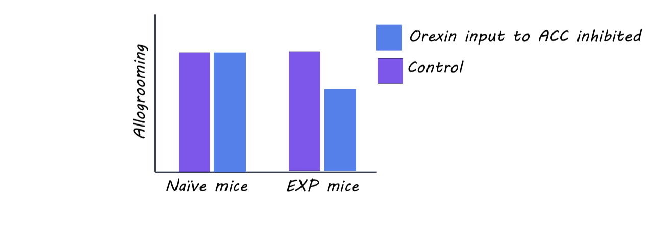
The authors evaluated empathy’s effects on prosocial behavior using an observational fear-conditioning paradigm (one mouse watches another receive a foot shock) combined with a consolation assay measuring allogrooming (the observer mouse grooming the foot-shocked mouse). They tested two paradigms: an experience-dependent observer (EXP), which had previously received a foot shock, and a naïve observer with no prior fear experience. They measured vicarious freezing (the observer mouse freezes at the tone when the experimental mouse receives the foot shock) and allogrooming in both groups and evaluated the impact of consolation on the shocked mouse using an open-field test. To dissect the neuronal mechanisms involved in these behaviors, the authors recorded ACC theta oscillations (5–7 Hz) during behavior. They conducted fiber photometry recordings using a genetically encoded orexin sensor (OxLight1), and using optogenetics, they inhibited ACC-projecting orexigenic circuits in observing mice only during the observation period when the experimental mouse received a foot shock

What did they find?

Both naïve and EXP observers exhibited vicarious freezing during observation and increased allogrooming after foot shock reunion. However, EXP mice displayed stronger vicarious freezing and more allogrooming compared to naïve mice. Self-grooming increased after observing the foot shock, but did not differ between groups. Notably, emphatic-like behaviors required visual attention - an observer mouse looking away during the foot shock showed no effects. Additionally, the authors found that the increased allogrooming by EXP observers resulted in less anxiety-like behavior in the mice that received the foot shock. Together, these data suggest that shared experiences enhance prosocial behaviors but don’t alter self-directed care.

To dissect the mechanism behind these behaviors, the authors show that both naïve and EXP observers have increased 5- to 7- Hz theta oscillations in the ACC while mice are observing the foot shock and during allogrooming (i.e. empathy and prosocial behavior), but not self-grooming. Concordantly, there was a selective increase in orexin activity at the same time, but only in EXP mice, suggesting that orexin-dependent increases require shared experience rather than observation alone. Inhibiting orexin input to the ACC specifically during the observation period reduced theta power and allogrooming in EXP mice and had no effect in naïve mice. These findings suggest that orexinergic inputs drive ACC theta oscillations to modulate affective empathy and prosocial action when there is shared experience.

What's the impact?

This study found that orexin-dependent increases in ACC theta oscillations link affective empathy to prosocial comforting behaviors. It demonstrates that social behavior relies on specific neuromodulatory rhythms to translate emotional experience into action. Elucidation of this upstream mechanism can provide potential targets for treating disorders that lead to empathy deficits. 

Access the original scientific publication here.

Practice Does Not Always Make Perfect: How Reward Timing Impacts Learning

Post by Annika Matthiesen 

The takeaway

This study challenges the idea that we learn faster just by repeating something more often. Instead, it shows that learning depends on timing, and even when rewarded events are spaced far apart, overall learning progresses at the same pace with fewer experiences.

What's the science?

Most theories of reward learning in neuroscience argue that the learning rate is a flexible parameter with no fixed rule, and that dopamine signals whether something is better or worse than expected. However, psychology has shown that learning can improve when experiences are spread out over time, though no clear biological rule has been established.

This week in Nature Neuroscience, Burke and colleagues sought to understand how learning rates vary under different conditions.

How did they do it?

The authors trained several groups of mice to associate a sound cue with a drop of sugar water, varying the waiting period in between trials by tenfold. They measured learning by tracking anticipatory licking and monitored dopamine release in the nucleus accumbens, a key reward-related brain region, using a dopamine-sensitive virus and an implanted lens. Comparing across groups, they identified a relationship between reward spacing and learning and used mathematical models to predict both behavior and dopamine response. To test their mathematical model, they also tracked learning using partial reinforcement, giving rewards on only some trials to increase the time between rewards, keeping cue frequency the same. In addition, they examined reward omission trials, where an expected reward was withheld, to test whether dopamine signals changed when rewards were unexpectedly absent.

What did they find?

The researchers found that learning rate depends on the time between rewards, not just how often a cue and reward are paired. When rewards were spaced farther apart, animals learned more from each reward and needed fewer trials to form the association. However, the proposed mathematical learning rate prediction model did not fully hold at the most extreme condition, where rewards were separated by a very long interval. Even so, the researchers showed that the timing rule held under partial reinforcement, where rewards were delivered only some of the time, further confirming that learning scales with the interval between rewards. Additionally, dopamine signals followed the same pattern as behavior, and the emergence of dopamine responses to reward omission occurred much later than cue responses, challenging traditional reinforcement learning models. Overall, the findings reveal a fundamental timing-based rule that governs how the brain updates associations during reward learning.

What's the impact?

This study is the first to show that learning rate follows rules based on the time between cue–reward experiences, rather than simply increasing with repetition. In simpler terms, learning depends not just on practice, but on how that practice is timed. Practice alone does not make perfect, and carefully spaced timing may be key to more effective learning, reshaping how we think about education and habit formation.

Access the original scientific publication here. 

Distinct Neurobiological Signatures of Early-Stage Depression and Psychosis

Post by Shalana Atwell

The takeaway

Neurobiological signatures can distinguish early-stage depression and psychosis and may help uncover mechanistic pathways and guide targeted interventions. 

What's the science?

Immune system alterations have been repeatedly linked to mood and psychotic disorders. Previous research has identified distinct and overlapping inflammatory markers in depression and psychosis whose dysregulation is associated with changes in brain gray matter volume (GMV). However, we need a more holistic view of the anatomical changes and inflammatory markers that are related to distinct pathologies of depression and psychosis. Furthermore, the complex relationship between immune factors and GMV makes univariate approaches poorly suited to capture higher-order patterns. Recently, in JAMA Psychiatry, Popovic and colleagues aimed to determine if multivariate patterns linking peripheral inflammatory markers with whole-brain gray matter volume could (1) distinguish early-stage depression and psychosis and (2) reveal how clinical factors such as childhood trauma and cognition relate to these biological signatures. 

How did they do it?

The authors analyzed baseline data from individuals with recent-onset depression, recent-onset psychosis, clinical high-risk state of psychosis, and healthy controls. All groups were medication-naïve or minimally medicated to reduce the confounding effects of antipsychotics and antidepressants. Peripheral blood was assayed for a panel of inflammatory and related markers such as interleukin (IL), tumor necrosis factor-alpha (TNF- α), and C-reactive protein (CRP), to name a few. These protein readouts were combined with demographic and technical covariates (age, sex, BMI, study group, MRI image quality) into a ‘blood’ domain. The ‘brain’ domain was generated using voxelwise gray matter volume (GMV) maps from structural MRI, which were mapped onto anatomical and functional network atlases to interpret where in the brain the signatures were expressed. To identify multivariate brain-blood domain relationships, the authors utilized sparse partial least squares regression (SPLS), which generates latent variables (LVs) that maximize covariance between the blood markers and GMV voxels. Each LV consists of weight vectors for blood markers and GMV voxels. Individual scores were computed by projecting each person’s data onto these weight vectors, and the correlation between blood and brain scores quantified how strongly the LV captures shared variance. Next, the authors used machine learning (linear support vector machine classification – SVM-C) to test whether life history (e.g., childhood trauma), cognitive function, and medications could predict high vs low expression of the psychosis-related and depression-related signatures. 

What did they find?

The authors found two significant brain-blood signatures that indicated a separation of psychotic and depressive disorders. Those with clinical high-risk status had higher levels of CRP compared to recent-onset psychosis, while recent-onset psychosis was associated with higher age, BMI, and certain inflammatory proteins (IL-6 and TNF- α). Additionally, they found disruptions in GMV mainly in cortico-thalamo-cerebellar circuitry that supports sensory integration and salience attribution. These findings suggest a different immune profile in high-risk and psychosis groups, as well as circuit disruptions that might serve as a neurobiological marker for defining different states of psychosis. In the depression signature, recent-onset depression was associated with higher levels of a mix of pro- and anti-inflammatory proteins (IL-1RA, IL-4, S100B, IL-1β, IL-2, and BDNF) and reductions in GMV in limbic system structures, such as the hippocampus and amygdala, compared to healthy controls. These findings support a complex immune-inflammatory and compensatory response as well as limbic-cortical dysregulation as a core neurobiological feature of depression.  

What's the impact?

This study is one of the first large, minimally medicated, transdiagnostic investigations to show that early-stage depression and psychosis are associated with distinct multivariate immune-brain signatures. Furthermore, the authors demonstrate that these signatures are shaped by childhood trauma and cognition, supporting stage-specific differentiation of psychosis and depression, which could guide targeted early interventions.

Access the original scientific publication here.