Neural Signature of Recovery Following Traumatic Brain Injury

Post by Shalana Atwell

The takeaway

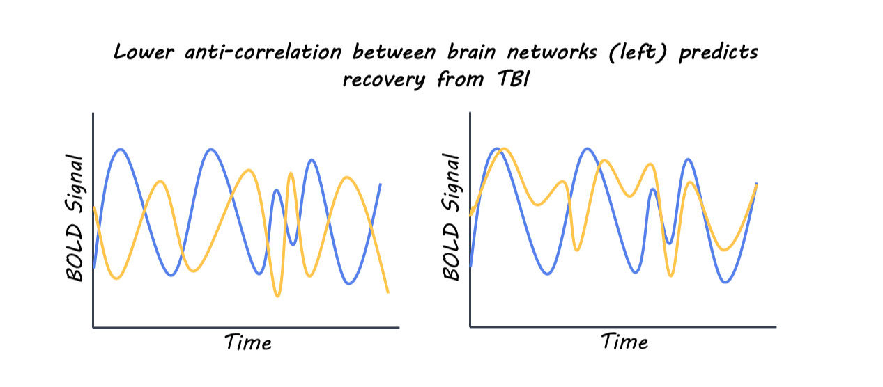
The extent of functional recovery from moderate to severe traumatic brain injury (TBI) in individual patients can be challenging to predict accurately. This research found that individuals with normal ‘on-off’ toggling (anticorrelation) between key brain networks following TBI are more likely to have a favorable functional recovery.

What's the science?

Current clinical models used to predict functional outcomes after TBI rely on age, extent of injury, motor scores, and the presence of other illnesses. Still, these predictive models do not account for neuroanatomical location or functional integrity of brain networks. Prior studies have used resting state functional magnetic resonance imaging (rs-fMRI) to measure functional connectivity (how brain regions communicate) in TBI studies. However, connectivity measures were inconsistent, and no model derived from fMRI data has been able to predict functional outcomes following TBI across independent datasets. Recently, in PNAS, Snider and colleagues investigated whether rs-fMRI can predict functional outcomes 6 months following injury across independent studies, as well as interrogated whether the predictive capacity of these measures is on par with or exceeds current prognostic tools for TBI.

How did they do it?

The authors pooled data from three TBI studies where each participant had completed multiple brain scans [MRI, diffusion tensor imaging, and rs-fMRI] and a 6-month follow-up after injury that assessed functional dependence [Glasgow Outcome Scale–Extended]. Brain scans measured structure (MRI), as well as the strength of connections between different brain regions by analyzing correlations in their activity over time (rs-fMRI) and organization and structural integrity of white matter tracts (diffusion tensor imaging). They cleaned and standardized MRI [CONN toolbox] and diffusion tensor imaging data to correct for excessive motion, technical differences across studies, and poor scan quality. 

Using one dataset, a model [logistic regression] tested the ability of 435 pairs of connected brain regions to discriminate favorable vs unfavorable functional TBI outcomes. These pairs were validated using two-fold cross-validation and repeated testing to ensure results were not due to chance. The best-performing connections were combined into a predictive model and then tested on a separate dataset, confirming generalizability. They utilized brain connectivity patterns to predict recovery outcomes following TBI. Finally, the authors investigated whether adding MRI-based measures matched or improved predictions beyond the standard clinical scoring systems.

What did they find?

Using their model, they found that the strongest predictor of 6-month recovery was the functional anticorrelation (one network turns off when the other turns on) between the salience network’s left anterior insula and the default mode network’s right parietal region. Patients with more normal anticorrelation between networks following TBI were more likely to have better recovery at 6 months following injury. Adding two more connections—the frontoparietal network’s right lateral prefrontal cortex to the visual network and the language network’s right frontal gyrus to the default mode network’s medial prefrontal cortex—created a three-connection model that improved performance and predicted recovery with high accuracy across independent data sets. Incorporating two additional connections created a more accurate prognostic model. Finally, they combined the three-connections identified from rs-fMRI with standard clinical scoring systems, which yielded a significantly improved outcome prediction, showing higher sensitivity and specificity for identifying patients likely to recover. This highlights the possibility of augmenting predictive TBI scoring models with rs-fMRI data to refine treatment planning.

What's the impact?

This study identifies preservation of specific resting-state anti-correlations between brain networks as a strong marker of functional recovery following moderate to severe TBI. Changes in brain connectivity patterns after injury could be incorporated into clinical prognostic tools and possibly leveraged as a therapeutic target. 


Access the original scientific publication here.

Brain Stages of Aging Across the Lifespan

Post by Lila Metko 

The takeaway

Topology in neuroscience is the study of the motifs that arise when observing how neural connections are interrelated and arranged. This research revealed that there are five major phases of topological development, with a significant topological turning point occurring between each phase at approximately the ages of 9, 32, 66, and 83 years old. 

What's the science?

Many scientists who study the development of the human brain look for linear trends in the developmental trajectory, or differences between age groups. To date, there are few papers that look at development outside of the confines of linear relationships. Recently, in Nature Communications, Mousley and colleagues analyzed brain imaging data from a large sample of individuals aged zero to 90 and found that rather than smooth trends in topographical organization, the human brain undergoes major age-specific turning points in topography. 

How did they do it?

The study included nine separate data sets, so the first step was to harmonize the data to ensure the differences between the data sets, such as the brain scanners and data acquisition protocols used, did not create nuisance variation. A double harmonization method was used to harmonize the data across atlas and study. Next, the authors used UMAP (Uniform Manifold Approximation and Projection), a manifold learning method, to project high-dimensional data onto a lower-dimensional space. Doing this makes highly complex data easier to understand while still maintaining its intrinsic structure. They mathematically analyzed the lower-dimensional manifold to find topological turning points within the data set. Finally, to understand the topological characteristics of these turning points, they did a principal components analysis of the data with eleven different topological metrics. These topological metrics measured how efficient communication was throughout a network, how many non-overlapping, communicating nodes can be found within a network, and to what extent different nodes are particularly important for network function. 

What did they find?

The major topological turning points were characterized by differences in three properties: segregation, integration, and centrality. Segregation is defined as the partitioning of the network into subgroups, integration measures the ease of communication across the network, and centrality is network communication’s dependency on a few nodes that are particularly important for network function. Segregation measures loaded most heavily onto principal component 1 (PC1), and integration measures onto PC2. Both segregation and centrality load most heavily onto PC3. Significant shifts in PC 1 and 2 (network subdivision and network communication ease) are found at roughly the ages of 9 and 32. At roughly the age of 66, there are significant shifts in segregation, integration, and centrality (network subdivision, network communication ease, and dependence on central nodes). At around age 83, there is a significant shift in integration, or network communication. The two epochs with the biggest differences in trajectories were epochs three and four, suggesting that topological change is particularly dissimilar in the years before around age 66 and after around age 66. 

What's the impact?

This study is the first to examine a large dataset with the goal of finding true qualitative transitions in brain topology, not just age group comparisons or linear trends. This paper allows us to see aging as unique stages rather than just progression and decline. Moreover, understanding the impact of therapeutics on different brain aging stages can allow for a more targeted approach.


Access the original scientific publication here.

Rugby Players Show Signs of Neurodegeneration in the Brain

Post by Anastasia Sares

The takeaway

This study reveals that former rugby players have elevated blood levels of a protein called Tau, which is associated with neurodegeneration, underscoring the risks of participating in contact sports where sub-concussive impacts are common.

What's the science?

Rugby is a high-contact sport where players can expect to experience head impacts regularly. Previous research has shown that players of high-contact sports have an increased risk of dementia, specifically Chronic Traumatic Encephalopathy (CTE) – a neurodegenerative disease first made famous in boxers, which can include mood swings, aggression, and memory problems. Studies on the brain tissue of players who have died with CTE show changes in the level of dementia-related molecules, specifically a protein called Tau. This is consistent with the idea that the head impacts and concussions suffered by players may trigger processes of neurodegeneration. However, it is difficult to track these subtle processes in people who are still alive but whose concussions or injuries are far in the past. 

This week in Brain, Graham and colleagues were able to show changes in dementia-related molecules in a large sample of ex-rugby players based on a blood sample, along with MRI data showing decreased brain volume and disrupted connectivity.

How did they do it?

Participants included 200 rugby players and 33 healthy controls, on average in their forties, along with a sample of older adults from a separate study, including 69 people with late-onset Alzheimer’s disease and their age-matched healthy controls. The authors obtained blood samples from the participants and used precise immunosorbent assays to detect specific proteins. An assay like this uses synthesized immune proteins (antibodies) that normally bind to foreign invading substances. By engineering the antibodies to bind to different proteins instead, they can capture these proteins and analyze the quantity of any molecule they choose. In this case, they were interested in dementia-related molecules such as amyloid beta and phosphorylated tau217, as well as other brain trauma indicators (plasma neurofilament light and glial fibrillary acidic protein). The participants were also scanned with MRI so that the size and shape of different brain regions could be estimated.

What did they find?

The rugby players had elevated levels of one key molecule involved in neurodegeneration: p-tau217. This increase in p-tau217 was associated with greater odds of traumatic encephalopathy syndrome (the clinical/behavioral symptoms of CTE). Alzheimer’s patients, on the other hand, had elevated levels of all molecules, even above the levels of the rugby players. 

MRI scans showed that players had lower brain volume in certain areas, like the frontal and cingulate cortex and the hippocampus—areas involved in executive function, emotional regulation, and memory. These changes in brain volume were related to the amount of time spent playing professionally. Finally, greater amounts of p-tau217 in the blood were related to a smaller volume in the hippocampus, a center of emotion and memory formation. It is important to note that these rugby players were recruited by self-referral, and one of the reasons for self-referral could include cognitive concerns. So, the results in this sample may be more extreme than for all rugby players.

What's the impact?

This study shows that rugby players coming in for cognitive concerns do indeed have elevated levels of tau protein, and that this is correlated with structural brain changes that could be part of CTE. Given their risk, it is important to monitor rugby players for signs of cognitive decline, and the methods used in this study are a useful step in developing this monitoring capability.

Access the original scientific publication here.