Brain Similarity Predicts Whether Strangers Become Friends

Post by Natalia Ladyka-Wojcik

The takeaway

Friendships are more likely to form and last between people who already share similar brain patterns in response to the world, even before they meet. These pre-existing brain similarities, beyond factors such as physical proximity or demographics, predict who grows closer over time, suggesting that deeper interpersonal compatibilities shape enduring social relationships. 

What's the science?

Humans are thought to form social networks based on their resemblance to one another in demographics, behaviors, and preferences, a phenomenon known as homophily. Yet, past attempts to link social network closeness with inter-individual similarities in self-report measures of personality traits have yielded inconsistent findings. More recent research suggests that friends share similar feelings and thoughts about the world, which are mirrored by similarities in brain-based measures. However, because these studies are cross-sectional, they cannot determine whether brain similarity predicts a future friendship or instead emerges as people become friends and influence one another through shared experiences and environments. This week in Nature Human Behavior, Shen and colleagues aimed to test whether pre-existing similarities in neural responses to naturalistic movies among strangers could predict who would later become closer in a social network.

How did they do it?

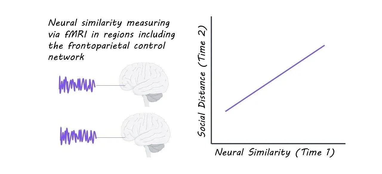
To determine whether similarities in brain activity predict who will later become friends, the authors recorded brain responses in a cohort of incoming graduate university students while they underwent fMRI scanning and watched 14 naturalistic movie clips. At later time points during the school year, the student cohort completed surveys reporting on their friendships, which allowed the authors to map out their social network. For each unique pair of participants, the authors calculated the correlation between their brain response time series at the start of the study in different brain regions, providing a measure of neural similarity, and the students’ social network positions two and eight months later, focusing on whether people who became friends had more similar brain activity than those who did not. The researchers also tested whether demographic factors like age, academic background, or shared interests predicted friendships more strongly, and whether enjoyment of the movies themselves could account for brain similarities.

What did they find?

The study found that people who later became friends, or who grew closer over time, tended to have more similar brain responses when watching the same movies before they had met. This effect was strongest in key brain regions of the frontoparietal control network, which has been linked to processing emotions, decision-making, and attention. Importantly, these similarities could not be fully explained by whether people simply enjoyed the movies or found them interesting. Whereas shared demographic factors like age accounted for some of the resemblance in brain responses between people, they did not explain the broader pattern observed in friendships that grew closer over time across the social network. Taken together, the findings suggest that friendship is shaped not only by shared backgrounds or interests, but also by deeper similarities in how people’s brains process the world around them.

What's the impact?

This study is the first to show that over time, strangers who exhibit similar brain responses for interpreting, attending to, and emotionally processing the external world are more likely to become friends and increase their social closeness. 

Access the original scientific publication here.

Expression of Motor Neuron Embryonic Factors Increases Resilience to ALS Pathology

Post by Amanda Engstrom

The takeaway

As neurons mature, they become less resilient to insults, which impacts the progression of age-dependent neurodegenerative diseases. Re-expression of embryonic factors in postnatal neurons reactivates aspects of a younger gene expression profile and slows degeneration.  

What's the science?

For many neurodegenerative diseases, aging is a major risk factor. This could be due to the loss of resilience in mature neurons compared to young neurons. Amyotrophic lateral sclerosis (ALS) is a progressive adult-onset degenerative disease specifically affecting motor neurons with no known cure. This week in Nature Neuroscience, Lowry, Patel and colleagues hypothesize that re-expression of embryonic motor neuron transcription factors, ISL1 and LHX3, in adult motor neurons could reactivate their “young” neuron state, increasing their resistance to the negative effects of ALS-causing mutations. 

How did they do it?

To investigate the effect of postnatal expression of ISL1 and LHX3 (both transcription factors downregulated after birth), the authors performed post-natal Day 0 (P0) mouse injections of adeno-associated viruses (AAVs) expressing a transgene for either Isl1 or Lhx3 driven by ChatE (an enhancer for the Chat gene) so both proteins would be expressed in motor neurons continuously throughout adulthood. This resulted in 90% of CHAT expressing motor neurons in the adult lumbar spinal cord also expressing ISL1 and LHX3, though their expression did slowly decline over time. They performed single-nucleus multiome RNA and ATAC sequencing on motor neurons isolated from the mouse spinal cord, allowing them to compare the transcriptional changes and correlated changes in chromatin accessibility at the single-cell level. Lastly, the authors re-expressed ISL1 and LHX3 by P0 injection in the SOD1G93A ALS mouse model and assessed disease-relevant phenotypes.

What did they find?

Re-expression of ISL1 and LHX3 led to significant increases in expression of MNX1, a key embryonic target that is typically downregulated after birth. Using single-nucleus RNA analysis of the motor neurons, the authors identified three clusters corresponding to alpha motor neurons, gamma motor neurons, and type 3 motor neurons. Despite ISL and LHX3 expression in all three clusters, only alpha motor neurons and type 3 motor neurons had differential gene expression. Further, the differentially expressed genes identified in these two subtypes had little overlap. These data suggest that re-expression of ISL1 and LHX3 is not only specific to motor neurons, but also selective among motor neuron subtypes, and the effect is unique to each subtype. The changes in chromatin accessibility were also distinct between alpha motor neurons and type 3 motor neurons. However, in both cases, the top motifs enriched in upregulated peaks were Lhx3 motifs, similar to their accessibility during motor neuron development. To determine which genes were being altered, the authors compared the differentially expressed genes to the normal temporal expression profile of alpha and type 3 motor neurons. In both subtypes, there was an upregulation of genes normally expressed embryonically and early postnatally, while downregulated genes were most highly expressed in typical mature motor neurons. Taken together, these data suggest that despite the cell-type-specific differences in gene expression changes, the global effect of re-expression of ISL1 and LHX3 results in a less mature state in both alpha and type 3 motor neurons.

Two histological hallmarks of disease pathology in ALS mouse models are large, round aggregates of SQSTM1 round bodies and SOD1 vacuoles. The percentage of both SQSTM1 round bodies and SOD1 pathology was reduced in motor neurons with re-expression of ISL1 and LHX3 compared to those without. Treatment did not alter overall survival in ALS mice, but did delay the onset of tremors and increased the total number of CHAT+ motor neurons. The proportion of ISL1+ LHX3+ cells was increased at late stage timepoints, suggesting that sustained expression of ISL1 and LHX3 can preserve the health CHAT+ motor neurons. 

What's the impact?

This study is the first to show that re-expression of ISL1 and LHX3 at an early postnatal stage can revert mature motor neurons to a younger state and reduce neuronal phenotypes in an ALS mouse model. This study provides a foundation for more targeted and biologically relevant reprogramming of mature cell types, increasing their resilience against age-dependent neurogenerative diseases. 

Access the original scientific publication here 

Predicting Dementia in Veterans With a Brain Injury

Post by Anastasia Sares

The takeaway

In this study, the authors used health data from thousands of veterans to build a risk model for dementia and death after sustaining a traumatic brain injury (TBI). This is important for the care of veterans specifically, as well as our understanding of the long-term consequences of traumatic brain injury.

What's the science?

Longitudinal studies, where data is collected over many years, are critical to establishing the long-term health effects of different life experiences, such as brain injury. However, these kinds of studies are few and far between because running them is complex and expensive. Another way to assess long-term health outcomes is to search in medical archives or records and use that information to try and predict a person’s health over time. In other words, past and present medical data are used to create a model of health risks that can be used going forward. The model can tell us about how likely it is that a similar person will develop a health problem in the future.

Previously, models have been developed for the general population showing that traumatic brain injury (TBI) increases the risk of both death and dementia in the following years. However, there are certain populations where this risk may be higher or lower. For example, veterans may have combat-related experiences that could exacerbate the effects of TBI.

This week in Neurology, Barnes and colleagues developed a model to predict the risk of death and dementia after TBI, based on over 100,000 medical records. They focused on veterans and included an assessment about combat-related experience to understand how these factors influence the risk of death and dementia.

How did they do it?

The authors were granted access to a medical database containing information from medical visits of many veterans. For their sample, they specifically targeted older people (over 55), who had a TBI diagnosis between 2001 and 2019 (with no dementia at that time), and had at least one follow-up visit. They gathered demographic information as well as two key variables related to military service: whether the person had served in a theater of combat operations, and whether they had previously had an injury caused or worsened by their active service. As for outcomes, the authors divided the participants into 3 groups: people who died within 5 years of the incident, people who developed dementia in that same period, or people who survived that period without death or dementia.

What did they find?

Of all the participants with TBI, 11% were later diagnosed with dementia, and 19% later died. The authors reported the hazard ratio to show how different factors influenced this statistic: how much more or less likely a person was to develop dementia or die. A hazard ratio of 1 means that there was no influence on the rate of dementia or death; a number above 1 indicates that these negative outcomes were more likely, and a number below 1 indicates they were less likely. As might be expected, age was a significant risk factor, with the hazard ratio increasing each decade of life, starting at 1.4 and climbing to 13.1. Having other conditions like Parkinson’s elevated the risk as well, with a hazard ratio of 3. Other physical and mental health conditions also increased the risk, with the hazard ratio between 1 and 2, depending on the condition. Older age and psychosis contributed more to the risk for dementia, while physical health issues and hospitalizations contributed more to the risk for death. The model performed fairly well in veterans with a service connection, veterans with combat service, and those with neither, indicating that these predictions generalize to a variety of TBI cases.

What's the impact?

The model developed in this study can be used to predict the risk of veterans developing dementia or other health complications in the future. This can help clinicians to be vigilant and suggest preventive care measures for those most at risk.

Access the original scientific publication here.