How Word Processing and Facial Recognition Adapt After Stroke

Post by Amanda Engstrom

The takeaway

Within the brain, facial recognition is lateralized to the right, and word processing is lateralized to the left. In individuals who suffer an early stroke in the left hemisphere, both word processing and facial recognition occur on the right without any impairment to facial recognition ability.

What's the science?

In the neurotypical human brain, the fusiform face area (FFA), a region within the ventro-occipital temporal cortex (vOTC), responds preferentially to faces. Face processing is right-lateralized, meaning the right hemisphere of the FFA is more active and dominant than the left. Conversely, language and visual word form processing are left-lateralized in a region called the visual word form area (VWFA) within the left VOTC. It has been hypothesized that these two lateralization patterns are linked: as children learn to read, the VWFA establishes itself in the left vOTC, either displacing or blocking the expansion of facial processing there, causing face processing to rely more heavily on the right vOTC. While correlations between literacy and face lateralization support this idea, evidence of direct competition between word and face processing remains mixed. This week in the Journal of Neuroscience, Seydell-Greenwald and colleagues used functional MRI (fMRI) to test whether face processing is altered in individuals who suffered a left-hemisphere perinatal stroke (LHPS) and consequently developed right-lateralized language and word processing. 

How did they do it?

The authors performed fMRI on 15 individuals with LHPS (mean age 19 years) who had previously established right-hemisphere language dominance and right-lateralized VWFA and compared them to 14 neurotypical individuals (mean age 16 years) with typical left-hemisphere language dominance. Both groups were fluent readers with comparable language comprehension scores. Participants completed a visual localizer task in which they viewed rapid sequences of images from three categories: faces, places, and word forms, allowing the authors to map and compare the lateralization of face and word form processing across groups. Participants then completed the Cambridge Face Memory Test, a standardized assessment of unfamiliar face recognition, requiring them to learn six faces and then identify them across different viewing angles and conditions. Together, these two tasks allowed the authors to assess both the neural lateralization of face processing and its behavioral consequences in individuals with LHPS relative to unaffected control participants. 

What did they find?

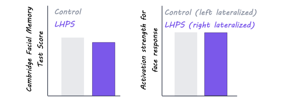
Control participants showed the typical pattern of left-lateralization for word forms and right-lateralization for faces, while the LHPS group was right-lateralized for both. This indicates that when word processing reorganizes to the right hemisphere after an early stroke, face processing does not compensate by shifting to the left. Instead, both functions are supported by the same hemisphere. There were no significant differences in the strength, spatial extent, or peak activation of face responses between the two groups, and both groups showed a positive correlation between face lateralization and word form lateralization, meaning stronger right-lateralization for words was associated with stronger right-lateralization for faces. Lastly, LHPS participants performed slightly but not significantly worse than controls on the Cambridge Face Memory Test, and both groups scored within normal range. Together, these findings suggest that sharing the right vOTC with word form processing does not meaningfully compromise the neural representation of faces or the ability to recognize them. 
What's the impact?

This study was the first to show that although the neurotypical brain delineates facial processing and visual word processing to either the right or left hemisphere, respectively, patients who have experienced early stroke to the left hemisphere can process both faces and written words together in the right hemisphere without any meaningful cost to either. This challenges the long-standing idea that the typical opposite-hemisphere arrangement of face and word processing is driven by competition. These findings provide insight into the key drivers of lateralizing specific neuronal pathways and demonstrate the remarkable capacity of the developing brain to reorganize after injury. 
Access the original scientific publication here.

Can Brain Stimulation Help Jump-Start Awareness?

Post by Annika Matthiesen

The takeaway

Stimulating specific brain regions may help “jump-start” brain activity in patients who have limited awareness and disrupted brain communication. While the effects were modest, the study suggests this noninvasive approach could help support recovery of awareness and responsiveness.

What’s the science?

People with severe disorders of consciousness have limited awareness and responsiveness because communication between different parts of the brain can be disrupted. Currently, there are very few effective treatments to improve wakefulness or recovery in these patients. This week in NeuroImage, Sangare and colleagues aim to improve brain activity and awareness in patients with limited wakefulness through brain stimulation.

How did they do it?

The authors studied 18 patients with disorders of consciousness. The authors used brain stimulation on the frontal cortex, an area at the front of the brain involved in higher-level thinking and control. Before and during treatment, the researchers measured how patients responded to sounds, sights, movement, and attempts to communicate. They also used EEG to record the brain’s electrical activity through sensors placed on the scalp, to track changes in brain function. Patients received stimulation or no stimulation (control), and the authors compared their responses before and after treatment. Six months later, the researchers followed up again to see whether any changes lasted over time.

What did they find?

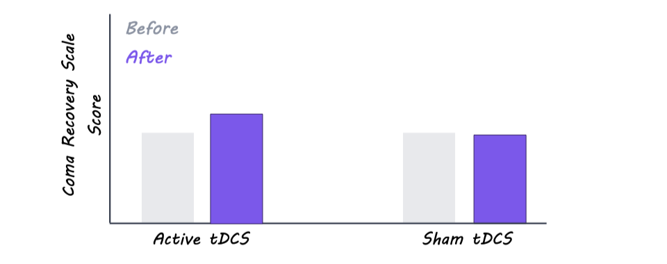
The researchers found some signs that the brain stimulation may have helped improve patients’ responsiveness and awareness, as the treatment group showed slightly greater behavioral improvement than the control group after stimulation. However, they did not observe changes in the overall level of consciousness. EEG results showed more active and complex brain activity in certain regions after treatment, which may suggest stronger communication between different parts of the brain. Overall, stimulation increased electrical activity across seven areas throughout the brain, although these changes did not consistently lead to major improvements in consciousness or behavior. Together, the findings suggest the stimulation may influence brain function, but larger studies are needed to determine whether it can produce meaningful recovery for patients with severe disorders of consciousness.

What’s the impact?

This study found that brain stimulation may help improve brain activity and responsiveness in patients with limited wakefulness by strengthening communication across different parts of the brain. If future studies confirm these findings, noninvasive brain stimulation could offer a safe way to improve awareness and brain communication in people with a wide array of serious neurological conditions.

Access the original scientific publication here.

Changing the Direction of Traveling Brain Waves Influences Attention and Memory

Post by Shalana Atwell

The takeaway

Neural information spreads across the cortex through brain oscillation patterns called “traveling waves.” Altering the direction of these traveling waves using traveling-wave transcranial alternating current stimulation (twtACS) influences cognitive performance.

What's the science?

Cortical “traveling waves” are rhythmic patterns of brain activity that move across the surface of the cortex, like ripples spreading across a pond, and they have been observed across species and during many different cognitive tasks. Cognitive dysfunction is a hallmark of many neurodegenerative diseases, psychiatric disorders, and aging populations, and previous research has shown that standard tACS can enhance certain aspects of human cognition. While tACS can synchronize brain rhythms, its effect is confined to one location and cannot generate dynamic patterns of neural activity. This week in PNAS, Lee and colleagues created and validated a stimulation method to impose a directionally controlled traveling electric field across the cortex and demonstrated how the wave's directionality influences specific cognitive tasks.

How did they do it?

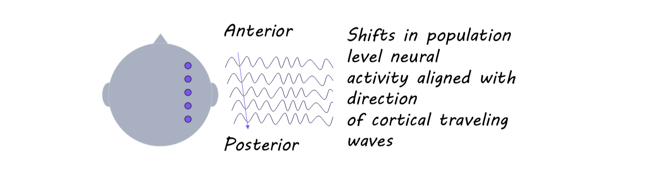
The authors used computer models of human head and brain anatomy to calculate how currents from multiple scalp electrodes would flow through the brain and then optimized the phases of the injected sinusoidal currents so that the location of the maximal electric field (the “peaks”) would sweep across the cortex over time (backward: from anterior to posterior), forming a traveling phase gradient. The team applied twtACS to two human patients with electrocorticography (ECoG) electrodes implanted for clinical reasons and showed that the measured phase at cortical electrodes closely matched the target pattern from the simulations and exhibited clear traveling phase gradients in the backward direction.

A nonhuman primate was implanted with a dense grid of intracortical microelectrodes spanning frontal to motor cortex, allowing measurement of multiunit activity (MUA; pooled local spiking) across space. The animal underwent four consecutive conditions at 10 Hz: baseline (no stimulation), standard tACS (same phase everywhere), forward twtACS (from posterior to anterior), and backward twtACS. For each condition, the authors computed the preferred phase of spiking relative to the stimulation cycle at each electrode. They confirmed that neural population activity aligned with the propagation of twtACS-induced electric fields.

Healthy participants received 10 Hz twtACS targeted to right frontal and parietal regions, with conditions optimized to produce either forward or backward traveling fields. During 20 minutes of stimulation, participants performed two tasks in fixed order: (1) A visual attention task where they covertly attended to flickering disks on the left or right, reporting whether a brief target appeared in the attended disk. (2) An episodic memory task where they encoded 60 images during stimulation, then, after a 10-minute rest, performed an old/new recognition test.

What did they find?

In ECoG patients, the phase of the recorded electric fields across electrodes showed robust gradients, confirming that twtACS can impose direction-specific electric fields across the human cortex that mimic observed cortical traveling waves.

Compared to tACS and baseline, twtACS produced systemic shifts in the preferred phase of spiking along the anterior-posterior axis. The spatial phase gradients in the MUA closely matched those of the electric field, indicating that population spike timing aligned with the propagating twtACS-induced field.

In the visual attention task, performance differed between forward and backward stimulation and depended on whether participants attended to the left or right visual field. Specifically, performance accuracy was higher with backward twtACS when participants attended to the right visual field. In the memory task, accuracy was higher under forward twtACS conditions. Together, these tasks indicate that the directionality of the imposed wave was critical to performance, and they are broadly consistent with previous work linking forward waves to feedforward sensory processing and memory encoding and backward waves to top-down control and spatial attention.

What's the impact?

This study provides evidence that manipulating the direction of traveling cortical waves can actively shape cognition. These findings support the idea that the direction of traveling waves is closely related to task demands, with different directions favoring feedforward vs feedback processing. This approach could eventually provide new therapeutic strategies for cognitive dysfunction in aging and psychiatric or neurological disorders that are thought to involve disrupted large-scale brain coordination.

Access the original scientific publication here.