
A Mega-Analysis of the Effects of Psychedelics on Brain Networks

Post by Anastasia Sares

The takeaway

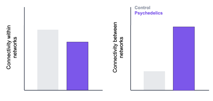
In a re-analysis of data from 11 different MRI studies, researchers found that psychedelics increase connectivity between many different brain networks, but only slightly decrease connectivity within networks. This analysis synthesizes and harmonizes findings in a field where research to date has been inconsistent and contradictory.

What’s the science?

Psychedelics are a class of compounds that primarily target serotonin receptors in the brain, changing neuronal activity and connectivity (see a previous BrainPost for more information on how these drugs work on a molecular level). There are currently many clinical trials looking at their potential therapeutic benefits for neuropsychiatric disorders like depression. Alongside this, researchers are trying to understand how exactly psychedelics act in the brain.

While we are beginning to understand the effect of psychedelics on brain networks, the picture is not consistent from study to study. Each psychedelic compound is a little bit different, and sample sizes in this kind of research tend to be small, so each brain network study has somewhat different findings—in fact, some papers report opposite results. Recently, in Nature Medicine, Girn and colleagues performed a “mega-analysis” from 11 different magnetic resonance imaging (MRI) datasets from studies on psychedelics to try to settle some of these debates and uncover the general effects of psychedelics on brain function.

How did they do it?

One common way of studying brain function that has been applied to many research topics, including psychedelics, is called resting-state functional MRI (or rs-fMRI). In regular functional MRI (fMRI), researchers measure the blood flow in the brain while a person lies in the scanner doing some kind of task: seeing images, hearing sounds, or thinking about certain things. As neurons use glucose and oxygen, the blood vessels in the brain open and increase the flow to supply them with more, and this can be tracked with the fMRI signal. However, in rs-fMRI, this signal is measured while people lie in the scanner doing nothing in particular—in other words, they are “at rest.” The brain is still active at rest, and a lot can be learned from studying this kind of data.

The authors collected and re-analyzed data from multiple laboratories that had scanned people under the effects of psychedelics using rs-fMRI. Most of these studies were randomized controlled trials with some kind of psychedelic versus a placebo. They didn’t just compile the results: instead, they started from scratch with the raw data and re-analyzed it with their own pipeline. MRI analysis is complex, and there is room for a lot of variation in processing methods, so by re-analyzing the data themselves, they hoped to iron out some of those inconsistencies. The type of analysis they conducted looked at functional connectivity, which tracks the fMRI signal to see which parts are correlated in their activity over time (and are therefore probably working together). They were primarily interested in testing one finding from previous studies: that psychedelics decrease connectivity within regions that usually work together, and they increase connectivity between regions that are usually distinct.

What did they find?

The main claim the researchers were testing was partially confirmed: compared to participants with a placebo, people who were under the influence of psychedelics showed increased connectivity between networks, especially general networks having to do with attention, self-reflection, sensory processing, and executive function. On the other hand, while they did see somewhat decreased connectivity within brain networks, these effects were much less robust. This amounts to more “cross-talk” between regions and only slightly less “internal chatter.” However, the one study using ayahuasca showed a completely different pattern: there was an overall decrease in connectivity between most brain areas. Since there was only one study on ayahuasca that had only 9 participants and lacked a placebo condition, more research will be needed to understand its effects. Overall, these results align better with some theories about how psychedelics work (like the subcortical connectivity account) while providing less support for others (like the ‘network disintegration’ account).
What’s the impact?

As clinical trials progress and potential therapies are identified, this research helps us understand the mechanisms behind those therapies and the mental health conditions they could treat. Knowing about the brain mechanisms of psychedelics might help us to better predict who could benefit from their use (personalized medicine), identify potential side effects, and develop sensible regulations.
Access the original scientific publication here.

How Does the Brain Create Social Network Maps?

Post by Amanda Engstrom

The takeaway

The brain constructs an internal map of social networks, even for relationships never directly observed. New research shows that the hippocampus and entorhinal cortex encode these maps, enabling us to infer and navigate complex real-world social connections.

What's the science?

The brain's ability to navigate complex social networks, like tracking how individuals are connected within a broader group, is fundamental to human social life. Neurons in the medial temporal lobe (MTL) are known to encode cognitive maps of physical space, and converging evidence suggests the MTL may similarly represent abstract relational structures; however, how the brain encodes large-scale, real-world social networks remains unknown. This week in PNAS, Teoh and colleagues combined computational modeling with fMRI and a longitudinal study of a real-world social network to investigate how activity in the hippocampus and entorhinal cortex scales to represent naturalistic social structures.

How did they do it?

To uncover how social network structure is encoded across individuals, the authors recruited undergraduates within a real-world social network and asked them to identify their friends within that network at multiple timepoints, generating a ground-truth friendship map (defined as mutual recognition between two individuals). Participants were asked to judge the relationships between other network members in a pairwise manner, including pairs they had never directly interacted with, to assess participants' ability to infer unseen social connections. The authors then tested four computational models of network representation to determine which best explained participants' behavioral patterns. These models varied in their relational complexity, the weight they gave to connections between individuals, the shortest path distance between two people, and the structural complexity of those paths.

To identify the brain regions supporting these representations, the authors used functional MRI (fMRI) to measure MTL activity while participants viewed photographs of community members and judged whether each person belonged to their network. Regions of interest included both the left and right hemispheres of the anterior hippocampus (aHC), posterior hippocampus (pHC), and the entorhinal cortex (EC). To link brain activity patterns to the computational models, they applied representational similarity analysis (RSA), a method that asks whether the pattern of similarity across brain responses mirrors the structure predicted by a given model of representation.

Finally, to assess whether these neural maps translate into functional social reasoning, the authors administered an Information Flow task where participants were asked to determine how information would travel from one participant across the network; a measure of how well participants could navigate their social network to trace indirect connections. Responses were linked to individual neural representations to determine how MTL activity patterns relate to real-world social inference.

What did they find?

Participants demonstrated structured, non-random judgments about unseen relationships, and their accuracy decreased with social distance: participants were most accurate when judging pairs close to their own friendships, with performance declining as the path distance between pairs increased. This suggests that participants do not simply recall direct ties but actively infer indirect connections using a structured internal representation of the network. Of the computational models tested, the Katz communicability model, which captures integration across multiple indirect paths between individuals, rather than just the shortest or most direct route, provided the best fit to participants' behavioral data. This indicates that people represent their social networks as distributed, multi-path structures rather than simple maps of direct connections.

At the neural level, the RSA revealed that the right EC encoded an abstract map of multistep network connections, while the right aHC may encode a more veridical representation of directly observed social ties. This regional dissociation suggests the MTL supports social network representation through at least two complementary encoding mechanisms, paralleling its known role in spatial navigation. 

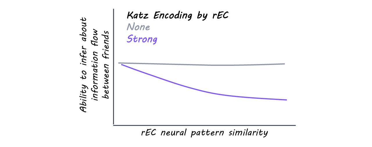
Finally, participants again relied on a Katz communicability-based strategy when reasoning about how news would spread through the network. Critically, when the right EC strongly encodes Katz communicability, the more strongly the right EC predicts task performance.

What's the impact?

This study is the first to demonstrate that the human brain encodes large-scale, real-world social networks as structured cognitive maps, within the MTL. These findings extend the MTL's known role in spatial navigation into the domain of complex social cognition. This work lays the foundation for investigating how social cognitive maps are updated over time, and how their disruption may contribute to deficits seen across neurological and psychiatric conditions.

Access the original scientific publication here

How Do Pain Pathways Drive the Placebo Effect?

Post by Lila Metko

The takeaway

Placebo pain reduction is a phenomenon where prior experience or expectations suppress pain in response to the administration of an inactive treatment. Placebo reduction of pain involves input from multiple cortical regions to the brainstem, which gates brainstem endogenous opioid release, reducing the experience of pain. 

What's the science?

Placebo analgesia (reduction of pain) is well known for its ability to complicate experimental procedures. It can also be a useful phenomenon relevant to therapeutic development. For example, if systems involved in placebo analgesia are understood, clinicians may be able to provide treatments that deliberately engage them to provide pain relief. This week in Neuron, Livrizzi and colleagues reverse translate a human placebo conditioning paradigm to mice, and uncover cortex to brainstem connections that gate release of endogenous opioids to downstream pain-modulatory regions. 

How did they do it?

The authors used a conditioning paradigm where contextual cues were paired with either morphine + pain stimulus or saline injection (placebo) + pain stimulus. For the morphine conditioning, the idea is that in the absence of morphine, these contextual cues would trigger placebo analgesia in the mice. After conditioning, the placebo test included placing these conditioned mice in similar contexts to the morphine conditioning, but with a saline (placebo) injection to induce placebo analgesia. They measured pain in animals by how long it took them to remove their paw from a pain-inducing apparatus (withdrawal latency). The tools they used to manipulate and record pathways involved in placebo analgesia were chemogenetics to activate pathways and fiber photometry to record opioid signaling. The authors used TRAP2 mice - mice that have genetic modifications that allow for labelling and then selectively analyzing neurons involved in a certain behavior or process. In this case, that behavior was placebo analgesia. They also used an interesting approach called in-vivo drug uncaging, which allowed the release of an opioid receptor antagonist over a specific temporal window, in this case, the placebo analgesia test window. 

What did they find?

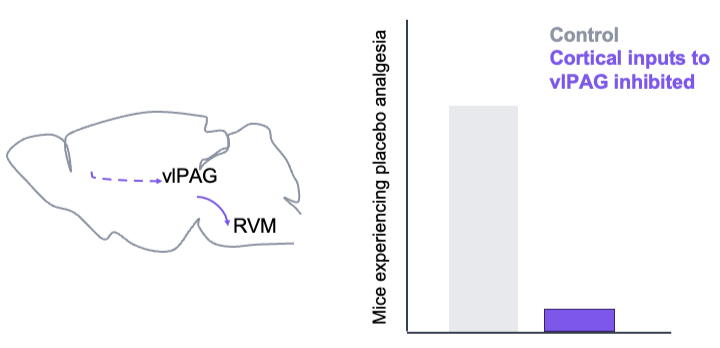
This study found that the vlPAG (ventrolateral periaqueductal grey), a brainstem region with glutamatergic neurons that activate to produce analgesia, is involved in placebo analgesia. They also found that neurons active in the vlPAC during placebo analgesia receive projections from neocortical and insular regions, while neurons in the rostroventral medulla (RVM) received projections from PAG analgesia neurons. After further experiments, they found that placebo analgesia was reduced when the neocortical regions, and not the anterior insular regions, were inhibited. Similar findings occurred when the PAG to RVM connections were inhibited, although both morphine and placebo nociception were altered in this case, not just placebo nociception. They additionally showed that placebo analgesia can be transferred between multiple pain modalities.

What's the impact?

This is the first research study to provide causal evidence of circuits involved in placebo analgesia. Importantly, it moves from correlational human evidence of cortex to brainstem circuits being involved in placebo analgesia to causational data using animal models. Understanding this circuit, especially its role in lasting analgesia after injury, opens up possibilities for future therapeutics. 

Access the original scientific publication here.