
How the Hippocampus Combines Place and Emotion in Memory

Post by Anastasia Sares

The takeaway

In this study, the authors show that different parts of the hippocampus, a memory formation structure in the brain, work together to bind environmental location and emotional information to form memories of reward or danger.

What's the science?

The hippocampus is a brain structure involved in memory formation. When an animal is navigating its environment, cells in the dorsal (“upper”) hippocampus called “place cells” fire when the animal is in different locations. Then, when an animal is sleeping, these cells fire again, in special bursts called “sharp-wave ripples.” This process reactivates other parts of the brain that were involved in the experience, and this is one way scientists think memories are consolidated and strengthened.

However, the hippocampus is not just for remembering where we’ve been. The ventral (“lower”) hippocampus has strong connections to the limbic system, a collection of deep brain structures that helps us process emotion. The limbic system includes the amygdala, which is involved in fear and other strong emotions, and the ventral tegmental area, which is involved in reward and motivation. This week in Nature Neuroscience, Morici and colleagues proposed that the ventral hippocampus is a key linking structure that helps us associate specific emotions with specific environments, driving behavior.

How did they do it?

The authors recorded brain activity from both the dorsal and ventral hippocampus of rats as they completed a navigation task, either to avoid danger (in the form of mild shocks) or to get a reward (in the form of a drink). They recorded this activity both as the animal was learning the task, as well as when the animal was sleeping that night and consolidating their memories from the day.

During learning, both the dorsal and ventral hippocampus were active, but the authors were specifically interested in whether they were working together or not. They used independent component analysis to identify patterns of coherent neural activity, which the authors called “assemblies.” They then tracked these assemblies during learning and during sleep.

What did they find?

During learning, the authors found 446 neural assemblies with coordinated activity. About 62% of these reflected activity mainly in the dorsal hippocampus, 14% reflected activity mainly in the ventral hippocampus, and 24% reflected activity in both regions (which they called “joint assemblies”). These joint assemblies looked very different between the reward and danger conditions, potentially indicating that the hippocampus was changing its internal connectivity depending on the emotional content of the experience. About half of the assemblies were associated with rewarding experiences, and half with dangerous experiences.
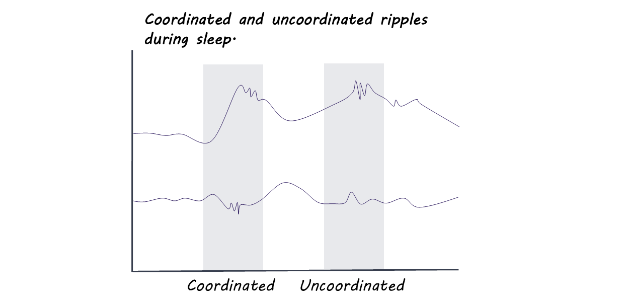
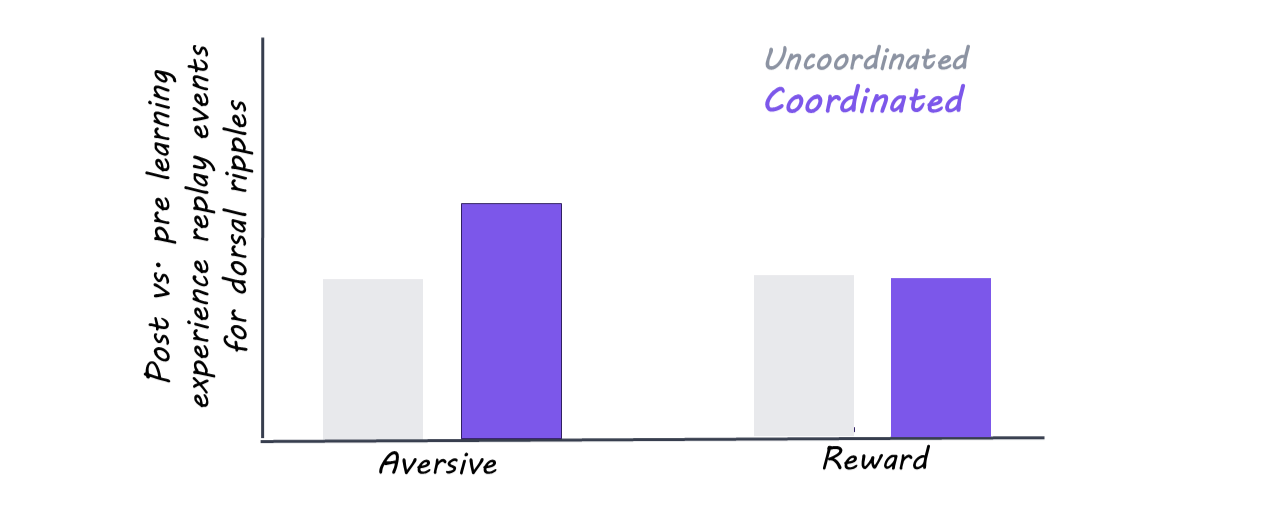
During sleep, the authors continued to track neural activity in both regions. Many of the ripples, those bursts of activity that help reactivate memories, were coordinated between the dorsal and ventral hippocampus. Depending on the experience of the day, different assemblies would be activated: specifically, on the days the animal had been learning from shocks, the cells in the ventral hippocampus responding to danger were more coordinated with place cells in the dorsal hippocampus. Again, this shows that the hippocampus may have different internal connectivity when linking a memory to reward or to danger.
What's the impact?

This work helps us understand the mechanism that links environmental location to emotional memories. It helps us tie what we know about the binding of memories to specific, anatomically connected parts of the brain.
Access the original scientific publication here.

How and When Does the Brain Show Resilience After Stress?

Post by Annika Matthiesen 

The takeaway

Resilience enables us to navigate challenges and recover from stress. Notably, resilience is not immediate; key neurophysiological changes emerge in a distinct window approximately one hour after stress exposure, revealing a delayed but dynamic process of recovery.

What's the science?

Stress is part of everyday life, whether it is a looming deadline or a never-ending list of chores. Humans are uniquely equipped to cope with these challenges, yet we vary widely in how well we adapt and recover from stress. This week in PNAS, Watanabe and colleagues set out to understand how the brain responds to acute stress by examining time-specific changes in neural activity and physiological responses following stress exposure.

How did they do it?

To explore how people respond to stress, the authors followed about 100 participants through a carefully timed experiment, beginning with self-reported assessments of their baseline stress levels. They measured both brain activity and physical stress responses, like heart rate, breathing, pupil size, and cortisol (a hormone released during stress). Brain activity was assessed using fMRI, which shows active brain areas, and EEG, which measures electrical brain signals. These measurements were taken before, immediately after, and up to 1.5 hours after the stress event to understand how responses change over time. The stress itself was induced using a cold pressor test, where participants placed their hand in ice-cold water for two minutes. By comparing responses across time and between individuals who were more stress-resilient vs. more stress-susceptible, the researchers were able to investigate when stress responses peak.

What did they find?

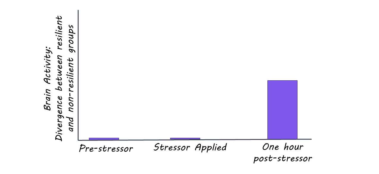
The researchers first found that self-reported stress levels did not clearly match immediate changes in brain and body activity, suggesting there is no simple biological pattern that separates more resilient from less resilient individuals right away. However, when they looked at brain activity over time, the most pronounced activity changes appeared about one hour after the stressor. At this point, two key brain networks showed opposite patterns, one becoming more active while the other became less active depending on individuals’ stress levels. This same one-hour window was confirmed by machine learning analyses as the most important time point for distinguishing resilience, highlighting that this time point after stress is especially dynamic in the context of stress response. Interestingly, EEG showed similar changes during this time in the same brain region, particularly in the prefrontal cortex (the part of the brain involved in decision-making and emotional regulation), suggesting this region may play a crucial role in stress recovery.

What's the impact?

This study is the first to show that the biggest differences between people who cope well with stress and those who don’t appear about one hour after a stressful event. In other words, resilience isn’t immediate; the brain takes time to shift into a recovery mode. There may be a critical window where the brain is most actively adapting to stress. Understanding this timing could help us better support mental health and develop treatments that target the right moment to improve recovery from stress.

Access the original scientific publication here.

Does Growing New Neurons Improve Memory?

Post by Rebecca Glisson

The takeaway

Recent studies have shown that humans can grow new neurons (neurogenesis) even as adults. Individuals with Alzheimer’s disease have much lower neurogenesis than normal, while older adults with high cognitive capacity have abnormally high neurogenesis.

What's the science?

It was long thought that adults do not grow new brain cells (neurogenesis), but more recently, we have come to understand that neurogenesis happens throughout our lives. Most studies use rodents as a model for human brains, which doesn’t yield enough insight into how neurogenesis occurs in adult humans. This week in Nature, Disouky and colleagues studied the brains of humans at all ages, with and without Alzheimer’s disease, to better understand the link between neurogenesis and cognitive functioning.

How did they do it?

To study neurogenesis in humans, the authors used the brains of deceased donors. The donors were groups of individuals at different ages and cognitive abilities, including young and older adults with normal cognitive ability, individuals who had or were starting to show signs of Alzheimer’s disease, as well as adults they called “SuperAgers”, or individuals who had high memory test scores. The authors identified neurogenesis from the genetic sequence present in cells in the hippocampus, a part of the brain involved in memory, using a method called RNA-seq. Cells that have just grown, immature cells, have a specific genetic sequence that the authors analyzed to determine how much neurogenesis had occurred.

What did they find?

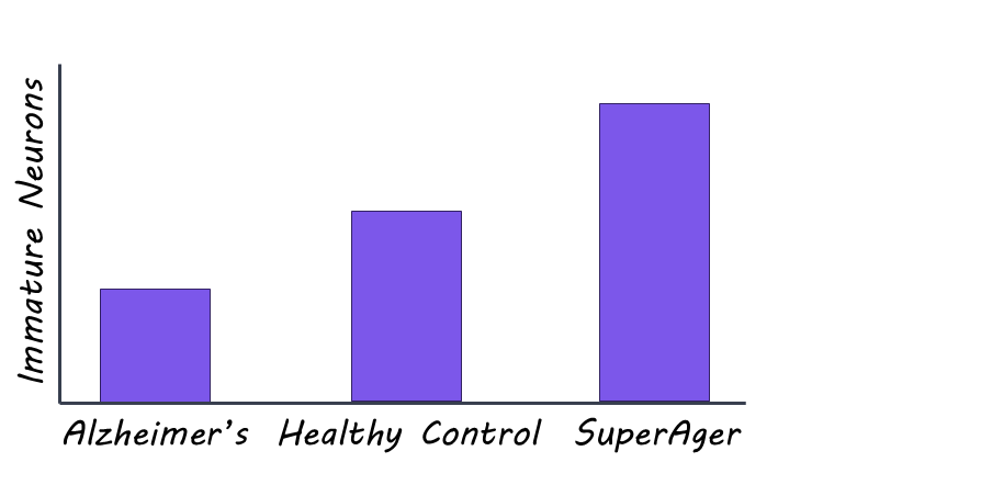
Both younger and older adults with healthy cognitive ability had high levels of neurogenesis in their hippocampi. In contrast, individuals with Alzheimer’s disease or symptoms of it had less neurogenesis and fewer immature neurons. This suggests that the slowing of neurogenesis may impact memory in diseases such as Alzheimer’s. One interesting result was that the “SuperAger” adults had significantly more neurogenesis and more immature neurons than any other group, including the healthy adults, suggesting that neurogenesis may help support healthy cognitive functioning as we age. 

What's the impact?

This study is the first to analyze the sequence of new immature neurons in humans and the first to link neurogenesis to cognitive function in individuals of different age groups and cognitive ability. Studies like these can help us better understand mechanisms of memory preservation or memory decline and how to develop better preventative care and treatment for those impacted.

Access the original scientific publication here.