Expression of Motor Neuron Embryonic Factors Increases Resilience to ALS Pathology

Post by Amanda Engstrom

The takeaway

As neurons mature, they become less resilient to insults, which impacts the progression of age-dependent neurodegenerative diseases. Re-expression of embryonic factors in postnatal neurons reactivates aspects of a younger gene expression profile and slows degeneration.  

What's the science?

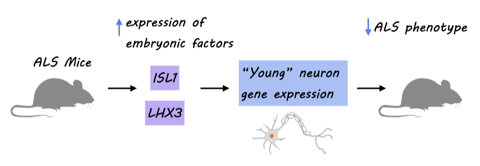
For many neurodegenerative diseases, aging is a major risk factor. This could be due to the loss of resilience in mature neurons compared to young neurons. Amyotrophic lateral sclerosis (ALS) is a progressive adult-onset degenerative disease specifically affecting motor neurons with no known cure. This week in Nature Neuroscience, Lowry, Patel and colleagues hypothesize that re-expression of embryonic motor neuron transcription factors, ISL1 and LHX3, in adult motor neurons could reactivate their “young” neuron state, increasing their resistance to the negative effects of ALS-causing mutations. 

How did they do it?

To investigate the effect of postnatal expression of ISL1 and LHX3 (both transcription factors downregulated after birth), the authors performed post-natal Day 0 (P0) mouse injections of adeno-associated viruses (AAVs) expressing a transgene for either Isl1 or Lhx3 driven by ChatE (an enhancer for the Chat gene) so both proteins would be expressed in motor neurons continuously throughout adulthood. This resulted in 90% of CHAT expressing motor neurons in the adult lumbar spinal cord also expressing ISL1 and LHX3, though their expression did slowly decline over time. They performed single-nucleus multiome RNA and ATAC sequencing on motor neurons isolated from the mouse spinal cord, allowing them to compare the transcriptional changes and correlated changes in chromatin accessibility at the single-cell level. Lastly, the authors re-expressed ISL1 and LHX3 by P0 injection in the SOD1G93A ALS mouse model and assessed disease-relevant phenotypes.

What did they find?

Re-expression of ISL1 and LHX3 led to significant increases in expression of MNX1, a key embryonic target that is typically downregulated after birth. Using single-nucleus RNA analysis of the motor neurons, the authors identified three clusters corresponding to alpha motor neurons, gamma motor neurons, and type 3 motor neurons. Despite ISL and LHX3 expression in all three clusters, only alpha motor neurons and type 3 motor neurons had differential gene expression. Further, the differentially expressed genes identified in these two subtypes had little overlap. These data suggest that re-expression of ISL1 and LHX3 is not only specific to motor neurons, but also selective among motor neuron subtypes, and the effect is unique to each subtype. The changes in chromatin accessibility were also distinct between alpha motor neurons and type 3 motor neurons. However, in both cases, the top motifs enriched in upregulated peaks were Lhx3 motifs, similar to their accessibility during motor neuron development. To determine which genes were being altered, the authors compared the differentially expressed genes to the normal temporal expression profile of alpha and type 3 motor neurons. In both subtypes, there was an upregulation of genes normally expressed embryonically and early postnatally, while downregulated genes were most highly expressed in typical mature motor neurons. Taken together, these data suggest that despite the cell-type-specific differences in gene expression changes, the global effect of re-expression of ISL1 and LHX3 results in a less mature state in both alpha and type 3 motor neurons.

Two histological hallmarks of disease pathology in ALS mouse models are large, round aggregates of SQSTM1 round bodies and SOD1 vacuoles. The percentage of both SQSTM1 round bodies and SOD1 pathology was reduced in motor neurons with re-expression of ISL1 and LHX3 compared to those without. Treatment did not alter overall survival in ALS mice, but did delay the onset of tremors and increased the total number of CHAT+ motor neurons. The proportion of ISL1+ LHX3+ cells was increased at late stage timepoints, suggesting that sustained expression of ISL1 and LHX3 can preserve the health CHAT+ motor neurons. 

What's the impact?

This study is the first to show that re-expression of ISL1 and LHX3 at an early postnatal stage can revert mature motor neurons to a younger state and reduce neuronal phenotypes in an ALS mouse model. This study provides a foundation for more targeted and biologically relevant reprogramming of mature cell types, increasing their resilience against age-dependent neurogenerative diseases. 

Access the original scientific publication here 

Predicting Dementia in Veterans With a Brain Injury

Post by Anastasia Sares

The takeaway

In this study, the authors used health data from thousands of veterans to build a risk model for dementia and death after sustaining a traumatic brain injury (TBI). This is important for the care of veterans specifically, as well as our understanding of the long-term consequences of traumatic brain injury.

What's the science?

Longitudinal studies, where data is collected over many years, are critical to establishing the long-term health effects of different life experiences, such as brain injury. However, these kinds of studies are few and far between because running them is complex and expensive. Another way to assess long-term health outcomes is to search in medical archives or records and use that information to try and predict a person’s health over time. In other words, past and present medical data are used to create a model of health risks that can be used going forward. The model can tell us about how likely it is that a similar person will develop a health problem in the future.

Previously, models have been developed for the general population showing that traumatic brain injury (TBI) increases the risk of both death and dementia in the following years. However, there are certain populations where this risk may be higher or lower. For example, veterans may have combat-related experiences that could exacerbate the effects of TBI.

This week in Neurology, Barnes and colleagues developed a model to predict the risk of death and dementia after TBI, based on over 100,000 medical records. They focused on veterans and included an assessment about combat-related experience to understand how these factors influence the risk of death and dementia.

How did they do it?

The authors were granted access to a medical database containing information from medical visits of many veterans. For their sample, they specifically targeted older people (over 55), who had a TBI diagnosis between 2001 and 2019 (with no dementia at that time), and had at least one follow-up visit. They gathered demographic information as well as two key variables related to military service: whether the person had served in a theater of combat operations, and whether they had previously had an injury caused or worsened by their active service. As for outcomes, the authors divided the participants into 3 groups: people who died within 5 years of the incident, people who developed dementia in that same period, or people who survived that period without death or dementia.

What did they find?

Of all the participants with TBI, 11% were later diagnosed with dementia, and 19% later died. The authors reported the hazard ratio to show how different factors influenced this statistic: how much more or less likely a person was to develop dementia or die. A hazard ratio of 1 means that there was no influence on the rate of dementia or death; a number above 1 indicates that these negative outcomes were more likely, and a number below 1 indicates they were less likely. As might be expected, age was a significant risk factor, with the hazard ratio increasing each decade of life, starting at 1.4 and climbing to 13.1. Having other conditions like Parkinson’s elevated the risk as well, with a hazard ratio of 3. Other physical and mental health conditions also increased the risk, with the hazard ratio between 1 and 2, depending on the condition. Older age and psychosis contributed more to the risk for dementia, while physical health issues and hospitalizations contributed more to the risk for death. The model performed fairly well in veterans with a service connection, veterans with combat service, and those with neither, indicating that these predictions generalize to a variety of TBI cases.

What's the impact?

The model developed in this study can be used to predict the risk of veterans developing dementia or other health complications in the future. This can help clinicians to be vigilant and suggest preventive care measures for those most at risk.

Access the original scientific publication here. 

Vascular and Immune Cell Disease Mechanisms in Neurological Disease

Post by Lila Metko

The takeaway

There is a link between cerebrovascular dysfunction (i.e., dysfunction in the blood vessels of the brain) and neurological diseases, yet how genetic variants in cerebrovascular cells influence the risk of disease is unknown. The authors developed a novel technology called MultiVINE-seq to understand how gene variants influence disease, and found distinct mechanisms associated with both cerebrovascular and neurological disease.

What's the science?

Over 90% of disease-associated genetic variants reside in non-coding regions of our genetic material. It is estimated that these disease-associated variants are active in a cell-specific manner. There is a clear relationship between cerebrovascular pathology and neurological diseases like Alzheimer’s disease; however, the genetic associations underlying these pathologies remain unclear. Currently, most of our knowledge on these genetic variants that influence disease risk is from investigating non-vascular cell types, due to the difficulty in recovering genetic material from vascular cell nuclei. This week in Neuron, Reid and colleagues developed a method for obtaining high-quality genomic data in vascular cells and integrated it with GWAS data to better understand how these genetic variants influence neurodegenerative disease mechanisms. 

How did they do it?

The authors processed prefrontal cortex samples from 30 post-mortem human brains. The samples were from individuals with conditions ranging from no cognitive impairment to dementia. The MultiVINE-seq processing required collagenase III, an enzyme that specifically digests collagen fibers, and loose-fit homogenization, a type of homogenization that reduces mechanical stress. From their output of genetic material, they determined which variants were in active regulatory elements by finding out which ones were in accessible chromatin regions, and overlapping snATAC-sequence data (which measures chromatin accessibility) with GWAS data. They correlated this information with pre-mRNA transcripts to determine which gene’s expression levels were most likely regulated by the variant-containing regulatory element. Finally, they grouped the genes for which variants likely affected each category of disease to see if there were any commonalities between genes in the same disease group. 

What did they find?

Variants associated with vascular diseases, such as stroke and aneurysm, were strongly associated with disruptions in extracellular matrix genes, which are responsible for the structural integrity of blood vessels in the brain. Thus, the vascular variants may contribute to a deterioration of the structural integrity of blood vessels, leading to leakage in the brain. Variants associated with Alzheimer’s disease were associated with proteins involved in the activation of immune cells and immune system signaling molecules. One Alzheimer’s Disease variant was specifically associated with regulating a protein, PDK2B, that is involved in the activation of T cells. T cells are a type of immune cell that destroys cells that contain pathogenic or foreign material. Further experiments showed that PDK2B and T cells were found near β-amyloid plaques. This suggests that this disease variant may weaken the brain’s immune response and ability to clear protein fragments, such as the material that builds up, forming amyloid plaques in the brain’s of people with Alzheimer’s disease.

What's the impact?

This study is the first to provide insight intot how disease-related non-coding variants in vascular and immune cells may contribute to neurodegenerative disease pathology. This is important because many neurological diseases are associated with deficits in neural vasculature or immune dysfunction. Having this information can equip scientists to better develop biomarkers or treatments for these disorders. 

Access the original scientific publication here.