
Neuronal Hyperactivity Recruits the Immune System to Eliminate Synapses
Post by Shalana Atwell
The takeaway
Neuronal hyperactivity in the adult brain can trigger local synapse loss by recruiting the immune protein complement component 1q (C1q), which initiates a cascade that flags and removes synapses at specific terminals. In mouse hippocampal circuits, C1q-dependent pruning requires nearby antibody-producing B cells that secrete immunoglobulins, especially IgM, to mark vulnerable synapses for removal.
What's the science?
Synapse loss is tightly linked to cognitive decline in diseases such as Alzheimer’s disease (AD). Prior work showed that microglia and C1q mediate synaptic pruning; however, the mechanisms by which specific synapses are tagged and whether adaptive immune cells play a role in this process have not been characterized. This week in Science, Crowley and colleagues asked whether changing neuronal activity is sufficient to drive C1q-dependent synapse loss in adult hippocampal circuits and how immune proteins contribute to activity-dependent pruning.
How did they do it?
The authors used chemogenetic tools called DREADDs (Designer Receptor Exclusively Activated by Designer Drugs) to bidirectionally control activity in defined hippocampal pathways. They injected an excitatory DREADD virus into the medial entorhinal cortex to selectively activate perforant pathway neurons that project to the dentate gyrus, and an inhibitory DREADD virus into the same pathway in an Alzheimer’s mouse model (J20 amyloid model) to dampen its hyperactivity. For five days, animals received daily injections of the designer drug clozapine-N-oxide (CNO) to specifically activate these engineered receptors, and the authors confirmed efficacy through in vivo neural recording (Neuropixels) and immediate early genes (e.g. c-FOS) related to neuronal activity. To further validate their findings, they used other models of hyperexcitability (kainic acid and subthreshold pentylenetetrazole) that induce seizure-like network activity and then measured C1q deposition and synapse density.
To uncover molecular changes, they applied multiple forms of spatial transcriptomics (Visium and Xenium) to hippocampal sections, focusing on differentially expressed genes and cell types around the dentate gyrus in activated versus control hemispheres. These analyses unexpectedly highlighted immunoglobulin genes, so the authors used RNAscope, immunohistochemistry, and high-resolution microscopy to visualize B-lineage cells and different antibody isotypes in cells adjacent to hyperactive circuits. Finally, they used several transgenic and pharmacological tools targeting B cells and antibodies (e.g. IgM knockout mice) to test whether secreted, antigen-specific immunoglobulins are required for activity-dependent C1q deposition and synapse loss.
What did they find?
Inducing hyperactivity in the perforant pathway in healthy mice produced a focal increase in C1q deposition in the activated projection zone and targeted loss of specific presynaptic terminals (VGLUT2-positive). This pattern suggests that local circuit activity can spatially direct where C1q accumulates and which synapses are selectively removed, rather than causing global synaptic damage. Inhibiting perforant pathway hyperactivity in J20 amyloid mice reduced C1q accumulation, decreased amyloid-β immunoreactivity, and partially restored postsynaptic markers, supporting the relationship between hyperactivity and C1q deposition in disease. In mice lacking a functional C1q protein (C1qa knockout), chemogenetic activation increased c-Fos expression but no longer produced VGLUT2 terminal loss, demonstrating that C1q itself is required for this form of activity-dependent presynaptic pruning.
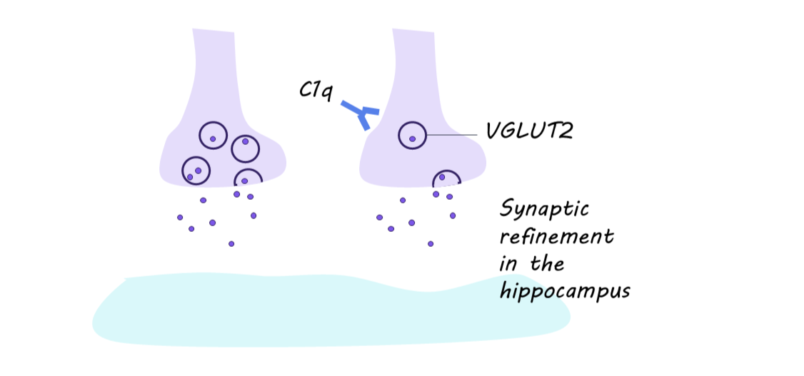
Spatial transcriptomics in activated wild-type brains revealed strongly upregulated immunoglobulin genes, indicating local antibody-secreting cells near the hyperactive pathway. RNAscope, spatial transcriptomics, and immunostaining identified B-lineage cells adjacent to activated hippocampal regions. Imaging revealed that IgM localized near C1q-positive VGLUT2 terminals, suggesting that locally produced IgM antibodies help recruit C1q to specific synapses during periods of increased activity. Functionally, mice lacking B cells or IgM, or with restricted B-cell receptor specificity (B cells all recognize the same kind of antigen), failed to show the typical activity-dependent C1q deposition and VGLUT2 loss after hyperactivity. This indicates that antigen-specific, secreted IgM from B-lineage cells is required to enable C1q-mediated synapse elimination in the perforant pathway.
What's the impact?
This study is the first to demonstrate that neuronal hyperactivity is sufficient to drive C1q-dependent synapse loss in the adult brain and that local B-lineage cells and their IgM antibodies are key enablers of this process. By revealing that adaptive immune cells can respond to circuit activity and tag synapses for C1q-mediated pruning, it broadens our understanding of how innate and adaptive immunity collaborate to sculpt adult neural circuits. Further, the work suggests that in disorders with early hippocampal hyperexcitability such as Alzheimer's disease, modulating neuronal activity or interrupting IgM-C1q interactions at synapses could be promising strategies to limit synapse loss and preserve cognitive function.