How Bilingual Brains Handle Grammar in Two Languages at Once

Post by Amanda Engstrom

The takeaway

The bilingual brain must compute different grammar rules for different languages at millisecond resolution. This computation relies on a shared brain network spanning the left frontal and temporal lobes, using flexible, reusable rules rather than a separate network for each language.

What's the science?

Human language requires the speaker to alter the pronunciation of words to account for changing information such as the plural form of an object, but the phonological rules for this are different across languages. This raises the question as to whether the brain utilizes language-specific neural systems or if it relies on a shared abstract mechanism that generalizes across linguistic contexts. Prior brain imaging work on this question has produced mixed results and has mostly examined language comprehension, leaving open whether the same holds true during the split-second demands of planning and producing speech. This week in Journal of Neuroscience, Chen and colleagues aimed to resolve this by using magnetoencephalography (MEG) to track millisecond-scale brain activity in Spanish/English bilinguals, using a task designed to separate the mental act of changing a word's grammatical form from other factors like the word's meaning or how it sounds, allowing them to pinpoint when and where this computation occurs in the brain as bilinguals produced singular and plural nouns in both languages.

How did they do it?

The authors recruited 23 highly proficient Spanish-English bilinguals to complete a phrase-completion task: participants saw a written noun (e.g., "boat"), then heard a spoken cue ("one," "two," or "say") prompting them to produce the singular form, plural form, or simply repeat the word aloud. The task varied language, whether grammatical change was needed, and whether that change altered the word's sound, all independently of one another.

While participants performed the task, the authors recorded brain activity using MEG, a non-invasive technique that detects magnetic fields from active neurons at a millisecond timescale, critical for capturing the split-second planning between hearing a cue and speaking. They also collected structural MRI scans and combined them with MEG data through source localization, which estimates where in the brain each signal originated using each participant's own anatomy. First, they compared brain activity when transforming versus simply repeating a word. Second, they trained a computer algorithm to recognize that activity pattern in one language (e.g., English) and then tested whether it could detect the same pattern in the other (Spanish). Successful detection across languages ("cross-decoding") would show that English and Spanish rely on a common neural code, rather than separate systems.

What did they find?

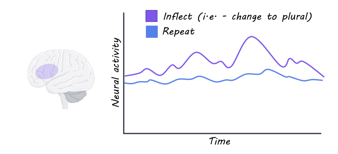
The authors found that transforming a word's grammatical form (e.g., "boat" to "boats") activated the left frontal and temporal regions much more strongly than simply repeating a word, even for words that don't change sound when pluralized (like "tuna/tuna"). This suggests that these brain regions carry out grammatical transformation as an abstract computation, rather than one tied to a specific sound pattern. Comparing English and Spanish separately, both languages activated nearly the same brain regions, with Spanish showing activity about 110 milliseconds earlier than English. A decoding algorithm trained to detect grammatical transformation in one language successfully detected it in the other, suggesting that English and Spanish rely on a shared brain mechanism for this computation rather than two separate systems, with the timing difference simply reflecting how quickly each language can be processed. Finally, this same pattern held even for words that don't exist in either language, and cognates (like "chocolate/chocolate") showed no added advantage over other real words. This suggests that the brain's grammar system is highly generalizable: it can apply grammatical rules to entirely novel words and doesn't depend on having a matching or overlapping vocabulary entry to work with.
What's the impact?

This study found that Spanish-English bilinguals engage in a shared left frontal-temporal network to process the grammatical transformation of pluralizing a word in both languages, suggesting the bilingual brain uses flexible, reusable rules rather than separate networks per language. These findings support a growing body of work showing that the brain favors efficient, generalizable abstract computations that can be reused across different contexts. Further, this study demonstrates how bilingualism can be used to interrogate general principles of neural organization. 
Access the original scientific publication here

Time Perception in Obsessive-Compulsive Disorder

Post by Anastasia Sares

The takeaway

In a preliminary study designed to understand how people with obsessive-compulsive disorder may differ in time processing, the authors found that while objective time perception and movement ability were intact, subjective time perception (including time management, time knowledge, and time experience) was significantly impacted.

What's the science?

Obsessive Compulsive Disorder (OCD) is a condition in which a person experiences recurring worries (obsessions) and enacts certain behaviors (compulsions) to try and manage these worries. OCD shares some features with depression (for example, some depression medications that act on serotonin can also be used to treat OCD). It additionally shares features with movement disorders such as Tourette’s, where differences in brain function cause involuntary actions that are difficult for the person to control. 

People with OCD can take longer to complete certain tasks and experience difficulties with time management. However, it is currently unclear what contributes to this. One possibility is that people with OCD have more difficulty controlling their movements, which means that doing things takes more time. Another possibility is that their objective time perception is altered—they might be less able to estimate time passing due to differences in time-keeping mechanisms in the brain. Finally, a third possibility is that people with OCD differ in their subjective experience of time, which could include difficulty making plans or realizing how much time they have spent on a task.

Recently, in an exploratory study published in European Archives of Psychiatry and Clinical Neuroscience, Mavrogiorgou and colleagues tested both objective and subjective time perception, along with movement ability and depression/anxiety symptoms, to better understand whether OCD uniquely contributes to altered time processing.

How did they do it?

In this exploratory study, the authors recruited 20 participants with OCD and 20 healthy participants of similar ages and genders. They ran them through a battery of tests, including questionnaires to establish the severity of OCD symptoms, questionnaires for depression and anxiety symptoms, measures of movement ability, experimental tests to measure more concrete/objective aspects of time perception, and questionnaires about subjective aspects of time perception.

The objective time perception tasks included flicker fusion frequency, which presents a blinking light that flickers at faster and faster frequency until the participant indicates that they perceive it to be stable and not flickering anymore—this measures the timing resolution of their visual perception. Another was the Chronotest, where participants are presented with a light stimulus and asked to estimate how long it was on, in seconds. They are also asked to reproduce different time intervals by turning a light on for that amount of time.

What did they find?

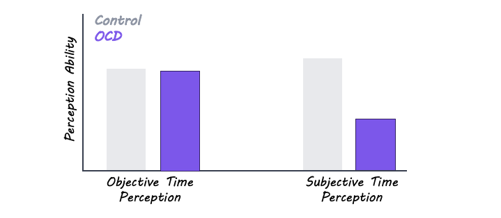
Participants with OCD were not significantly different from the healthy controls in their objective time perception, and there was only a slight trend for a difference in movement ability. In contrast, subjective time measures such as time knowledge, time experience, and time management were significantly impaired in OCD, and were correlated with OCD symptoms—in other words, those with more severe OCD had poorer subjective time perception. Subjective time perception was also correlated with depression scores, both in people with OCD and in the healthy control group, but some influence of OCD symptoms remained even after controlling for depression, anxiety, and movement issues, specifically on time management. It’s worth noting that subjective time perception was measured using a questionnaire, while the objective time measures were done using experiments, so self-perception had more of a chance to influence the results.

What's the impact?

Since this is an exploratory study, it should be treated as preliminary, but it has the potential to increase our knowledge of a lesser-known aspect of OCD. If these results hold, it means that there are time perception differences in OCD that are not related to movement difficulties or depression/anxiety symptoms.

Access the original scientific publication here. 

Using Large Language Models to Map Grammar in the Brain

Post by Anastasia Sares

The takeaway

This study shows that grammatical features are encoded by individual neurons in the brain, distinct from other characteristics of the speech signal. Grammar-sensitive neurons are distributed throughout the left hemisphere in a way that is hard to detect when averaging regional activity.

What's the science?

Since the time of Broca, scientists have developed more and more detailed maps of the language-processing areas of the human brain. They have used a variety of brain imaging techniques, including functional magnetic resonance imaging (fMRI), electroencephalography (EEG), and others. All of these techniques have been helpful, but still lack the precise detail of single-neuron recording that is done routinely in animals. The only time that electrodes are implanted in human brains is when they are already undergoing an operation, such as for epilepsy. During those times when the brain is exposed, researchers can gather precious single-neuron data.

One challenge about studying human language processing is the complexity of language itself. In everyday conversation, we are free to put together words in a myriad of forms and sentence structures. Most of the time, studies of language involve presenting pre-designed words, phrases, or sentences that have a feature we are interested in (for example, sentences with different relationships between the subject and the object). However, this eliminates the natural spontaneity of language production, and so we can’t be sure if the brain activity we observe is reflecting what our brains do daily.

Fortunately, the technology behind large language models (some of which are now commonly referred to as “AI,” though not all of them are on the same scale) has given us the ability to represent natural language production in a way that computers can digest, and we can identify interesting features without having to design awkward stimuli. This week in Nature, Cai and colleagues combined natural language processing and recordings from electrodes implanted in the brains of epilepsy patients to map human language production, including grammatical features.

How did they do it?

The authors had access to eight people who were undergoing surgery for epilepsy. At the time of their operations, the patients consented to have an array of electrodes temporarily implanted in their brains so that the researchers could gather data about the firing of individual neurons. Depending on the patient, these electrode arrays were located in different spots in the brain, so the researchers had a variety of brain regions represented.

After implantation, the researchers recorded brain activity through the electrodes while asking the participant questions and allowing them to respond freely. They recorded these conversations and applied natural language processing algorithms to classify each sample of speech. In addition to representing the individual words, this approach extracts both grammatical relationships and the hierarchical structure of phrases (see image below). By labeling these features, the authors could then look at which parts of the brain were selectively responding to parts of speech, hierarchical features, or specific conversation contexts.

What did they find?

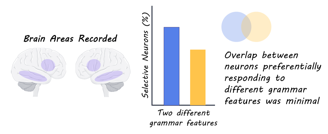
About 9% of neurons responded specifically to a word’s part of speech (noun, adjective, verb, etc). About 16% of neurons responded to constituency depth (how deep the word was in the nested hierarchy of the sentence), and so on. These neurons had very little overlap with each other, or with neurons that tracked other features of the speech (like the meaning of the word itself, or whether the pitch of the spoken word was high or low). Interestingly, these neurons that responded to different grammatical features were scattered throughout various brain regions, which may explain why it has been hard to pinpoint grammatical processing in the brain by summing up large amounts of activity. In addition, these grammar-selective neurons appeared in both hemispheres of the brain, but the most strongly predictive ones were in the left hemisphere, which lines up with decades of research confirming that language is processed on the left side of the brain (for most people).

What's the impact?

The results of this study inform debates about how the brain processes language. First, it supports the idea that certain high-level features of language can be coded by single neurons, rather than being only a result of multiple neurons firing in a pattern. These results also suggest that the popular but simplistic “modular” approach, where “region X is responsible for function Y,” doesn’t really work here: grammar-sensitive neurons are scattered throughout the left hemisphere.

Access the original scientific publication here.