A Device for Tracking Fluid Exchange in the Brain

Post by Lila Metko

The takeaway

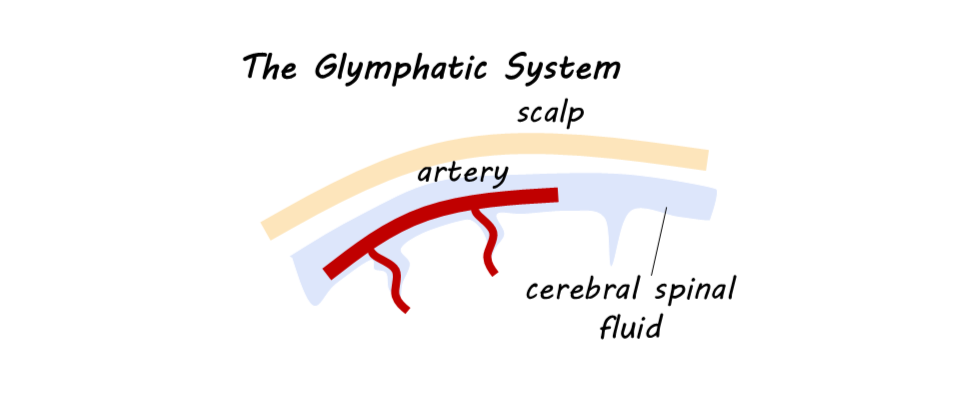
The glymphatic system is a fluid transfer and waste clearance system within the central nervous system (CNS) that supports the clearance of amyloid beta and tau proteins that accumulate in neurodegenerative disease. The authors developed a multielectrode patch, the first non-invasive method in humans to probe the activity levels of the glymphatic system during sleep.

What's the science?

Glymphatic function is a crucial fluid exchange process in the brain, facilitating the conversion of interstitial fluid into cerebrospinal fluid (CSF), which nourishes cells, regulates the volume transmission of neuromodulators, and is involved in the clearance of unwanted substances, such as amyloid beta and misfolded tau proteins. Glymphatic impairment in animal models has been shown to promote the development of amyloid beta and tau pathology. Additionally, glymphatic impairment is associated with aging, sleep deprivation, traumatic brain injury, and risk factors for neurodegenerative disease, including Alzheimer’s disease (AD). Glymphatic function has been shown to be more rapid during sleep. Currently, there is no non-invasive, temporally optimal device to monitor glymphatic function in humans. Recently, in Nature Biomedical Engineering, Dagum and colleagues describe a non-invasive device for monitoring glymphatic function in humans. 

How did they do it?

The authors conducted two complementary human studies in which participants wore the electrode patch device on the posterior area of the scalp and upper neck during one night of natural sleep and one night of sleep deprivation. The patch utilized electrical impedance spectroscopy (EIS) to measure the resistance to an electrical current applied to the tissue, thereby understanding the volume of fluid in the target region. A lower resistance score is indicative of higher glymphatic activity. The patch also monitored sleep stage using EEG, heart rate, and respiration using plethysmography, and an accelerometer for motion detection. In one of the studies, CE (contrast enhanced) MRI was used to monitor glymphatic function. Relationships between the variables were assessed using linear mixed-effects models that controlled for CE MRI contrast in brain areas other than the interstitial space, such as the blood vessels. 

What did they find?

The authors found an association between resistance in the tissue of interest (indicative of glymphatic function) and contrast enhancement. They found that lower resistance predicted greater contrast enhancement, indicating that resistance, the output of the EIS device, is a good proxy for glymphatic function. When they combined resistance with variation in heart rate and EEG powerband, two other outputs of the device, they found that it explained 74.8% of the variance in the model for the awake nights, indicating that combining the device’s outputs allows it to be an even better predictor of glymphatic function. Additionally, resistance decreased during sleep, which is consistent with previous findings that glymphatic function increases during sleep. Less time in REM sleep and more time in light sleep was associated with less CE MRI contrast, indicating that in lighter sleep stages, there is less glymphatic activity.  

What's the impact?

This device is the first to track glymphatic function in humans in a non-invasive and relatively time-resolved manner. This is critical to the field of neuroscience because of the glymphatic system’s association with clearing proteins involved in neurodegenerative disease, and its potential role in the control of volume transmission of neuromodulators. As a result, human glymphatic function will be able to be studied in more naturalistic environments. This device may also enable drug discovery by helping scientists understand the effect of certain drugs on glymphatic function. 

The Maintenance of Adult-Born Neuron Signaling Promotes Successful Aging

Post by Amanda Engstrom

The takeaway

Memory processing via adult-born neurons is essential for successful cognitive aging. A major distinction between people who are resilient and those vulnerable to cognitive decline lies, in part, in the maintenance of a network of long-lived adult-born neurons. 

What's the science?

Aging is frequently associated with cognitive decline; however, this decline varies among individuals - some individuals remain resilient while others are more vulnerable to the decline of memory functions. Memory formation relies on adult neurogenesis, the process of creating new neurons in the adult brain, but the role of long-lived adult-born neurons (ABNs) in cognitive resilience remains unclear. This week in Molecular Psychiatry, Blin and colleagues categorize aging animals as either resilient or vulnerable to cognitive decline, and examine their ABNs overall health and functionality. 

How did they do it?

To determine whether ABNs generated early in adult life contribute to preserved cognition, the authors labeled ABNs at 3 months of age in rats and assessed them at 8, 12, or 18 months of age. The rats were classified as either resilient or vulnerable to cognitive aging based on their performance in a behavioral memory task. Once characterized as resilient or vulnerable, the authors assessed the ABNs from both groups. The authors first assessed the survival and levels of senescent cells (a sign of cell arrest and inability to function) in the ABN population. Additionally, they used multiple retroviral vectors to label the ABN population and assess their dendritic morphology (GFP), glutamatergic post-synaptic density (PSD95-GFP), and mitochondrial network (MitoDsRed). Finally, they used optogenetic stimulation to artificially stimulate the ABNs. Rats were injected with ChannelRhodopsin-GFP at three months and underwent learning and memory testing at 12 and 20 months of age. The ABN population was activated by light during the learning phase to test if activating them at the later timepoints would increase the rat’s performance. 

What did they find?

The number of ABNs tagged at 3 months was the same in rats that were both resilient and vulnerable to cognitive aging. This was true for all three adult age groups (8, 12, and 18 months). The authors did detect senescent ABNs at all 3 ages, with an increased number of senescent cells at 18 months. However, resilient and vulnerable animals showed a similar number of senescent cells. Additionally, there was no difference in the dendritic morphology of ABNs in resilient and vulnerable rats. These data argue that the overall health of ABNs based on cell survival, entry into senescence, and gross morphology is not altered in rats vulnerable to cognitive aging. 

However, the authors did determine that rats vulnerable to cognitive aging progressively lost their glutamatergic inputs, indicated by a significant reduction in the labeling of postsynaptic density scaffolding protein, PSD95. The decrease of postsynaptic densities was observed at all ages in the inner molecular layer (IML) of the dendrite, but not in the middle or outer molecular layers. This suggests that the maintenance of proximal synaptic inputs (those closer to the soma or cell body) is especially important because these inputs are preserved only in resilient animals. Interestingly, the ABNs in vulnerable animals had a significant reduction of mitochondrial density, specifically in the IML at 8 months, but extended to the middle and outer layers in 18-month-old vulnerable animals. This suggests a progressive spread of mitochondrial dysfunction with aging in vulnerable animals. Optogenetic stimulation of ABNs improved the memory in all animals, and the memory of vulnerable rats improved to the level of non-stimulated resilient rats. This suggests that even when natural synaptic input is compromised in vulnerable rats, artificial stimulation can improve cognitive performance, indicating that ABNs can still function if properly engaged.

What's the impact?

This study found that long-lived ABNs play a role in cognitive aging. ABNs remain functionally viable in vulnerable animals and can transmit information when activated. Therefore, brain resilience relies, at least in part, on the preservation of the ABN integration into their neuronal network. This work highlights the potential therapeutic benefit of restoring the functionality of the ABN signaling network to improve cognitive functions in old age. 

Access the original scientific publication here.

How Do Motor Cortex Pathways Change in Aging?

Post by Rebecca Glisson

The takeaway

As we age, we sometimes lose our ability to move normally, which also significantly lowers our quality of life and capacity for independence. The motor cortex of the brain, which controls our movement, and the brain pathways descending from this area deteriorate in older adults, suggesting a focus on these areas could help us better treat neurodegenerative diseases.

What's the science?

Our movement is controlled by an area of the brain called the motor cortex, which can lose its function over time as we age. The motor cortex sends signals through two tracts, or pathways, of cells: the corticospinal tract (CST) and the corticostriatal tract (CStrT). This week in NeuroImage, Wen and colleagues investigated the changes in the CStrT that occur as people age, aiming better to understand the neural basis of movement-related neurodegenerative diseases.

How did they do it?

The authors wanted to study how aging affects several parts of the motor cortex and its pathways: its overall structure, how blood flows through this area, and the quality of the cells in these areas. To study these variables related to movement abilities, the authors used structural magnetic resonance imaging (MRI) to image and analyze the motor cortex in the brain. They also used diffusion MRI to analyze the pathways of the CST and CStrT. Linking this to movement, the authors measured participants’ motor function using endurance and locomotion walking tests and grip strength tests. The authors grouped participants into two age groups: the younger group, 36 to 65 years old, and the older group, 66 to 90 years old. 

What did they find?

While younger participants had normal brain structure, the authors found that older participants had less volume in the motor cortex and less blood flow to this region. The older group of participants also had significantly lower movement abilities involving locomotion, endurance, and strength. This suggests that the loss of certain movement abilities as we age is due to degeneration of the motor cortex. The authors also found that the CST and CStrT pathways were of significantly lower quality in the older group of participants and that this deterioration mediated the relationship between motor cortex atrophy and decline in motor function. Therefore, the CST and CStrT pathways are particularly important to movement function and are affected in aging.

What's the impact?

This study is the first to show that the changes in the motor cortex and its related pathways in the brain due to aging are directly related to a loss of movement functioning in older adults. This highlights the need to focus on these areas for studying movement diseases related to aging. Studies like these can help us detect neurodegenerative disorders earlier and develop better and more effective treatments.

Access the original scientific publication here.