Brain Volume in the Hippocampus is Influenced by Genetics

Post by Lani Cupo

The takeaway

Gray matter volume along the length of the hippocampus is influenced by genetic factors, which can be identified in living humans.

What's the science?

The volume of the hippocampus is considered to be about 80% “heritable”, meaning 80% of the variability of hippocampal volume is attributable to genetic variation. Measuring the volume of the entire hippocampus, however, neglects to consider that some regions of the hippocampus may be more susceptible to environmental influences than others. Examining the heritability of subfields, or regions within the hippocampus, may provide better estimates of whether the heritability of volume differs between regions. This week in NeuroImage, Pine and colleagues investigated the heritability of hippocampal subfields using magnetic resonance imaging (MRI) data, finding volume is influenced both by genetic factors general to the entire hippocampus, and specific to individual subfields. 

How did they do it? 

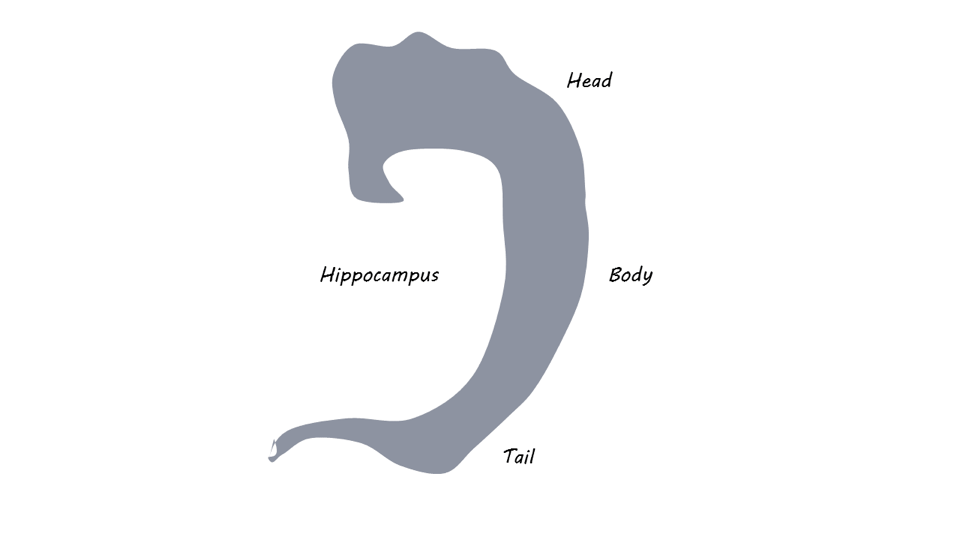
The authors used MRI from both children and adult twin pairs from three different datasets, including both dizygotic and monozygotic twins. They built biometric models to compare how highly correlated volumes were between monozygotic (single egg) and dizygotic (two eggs) twins. This method allowed them to detect differences in hippocampal subfield volume that could be attributed to sources of variance that are either genetic or environmental. For example, if there is variance in the hippocampal volumes of monozygotic twins (who begin life genetically identical) it can be attributed to an individual environmental influence (e.g. different levels of education). By examining how correlated hippocampal volumes were between monozygotic twins, compared to how shared hippocampal volumes were between dizygotic twins, the authors could estimate how much of the volume was heritable versus due to environmental influences. They compared the performance of different models to assess which one best fit their data before interpreting results. The hippocampal subfields examined were the head, body, and tail, dividing the hippocampus along its longitudinal axis (if you imagine the hippocampus as a c-shape uncurled). By including both males and females and both children and adults in this study, the authors were able to investigate how heritability by subfield differs across sexes and ages. 
What did they find?

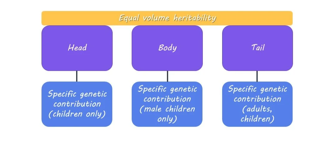
The authors didn’t find any differences in the heritability of volume across the different subfields. This suggests there isn’t a particular subfield where volume is more heritable than others, however, it is possible that using another division of the hippocampus or treating it as a gradient, rather than distinct regions, may reveal other subtle differences. 

Nevertheless, the authors did find evidence for subregion-specific genetic components, meaning different genes might contribute to the volume of different subfields. Subfield-specific genetic contribution was found to be both sex and age-dependent. In both sexes and in both children and adults, subfield-specific genetic contributions were found for the tail of the hippocampus. In contrast, subfield-specific genetic contributions were found for the head only in children (both sexes). Finally, subfield-specific contributions were found for male children only for the body of the hippocampus. The authors reported that these sex and age differences do not follow known systematic patterns, however, it is of interest that the genetic influence on hippocampal subfield volume is not constant across ages, and other properties of the hippocampus, such as white matter, may better reflect age-related changes. 
What's the impact?

These findings suggest the head, body, and tail may not show differences in heritability of gray matter volume, however, they show subfield-specific genetic contributions that differ across subfields by age and sex. The authors also demonstrate that noninvasive imaging can be used in living humans to estimate genetically based individual differences, which can set the stage for future population studies. 
Access the original scientific publication here

The Role of Dopamine in Integrating Costs, Benefits and Motivation

Post by Soumilee Chaudhuri

The takeaway

Eshel and colleagues found that dopamine release in the striatum is related to both the costs and benefits of a decision and that higher motivational states are associated with lower dopamine release. 

What's the science?

Dopamine (DA) release in two distinct regions of the brain: ventral and dorsal striatum, has been associated with the motivation underlying decision making. But there is insufficient evidence as to whether DA released from these regions impact cost, benefit and how this might be influenced by motivation. An article published in Neuron this week investigated the role of dopamine in cost, benefit and motivation  using mouse models and optogenetics to understand 1) how dopamine responds to variation in the cost and benefit of an action and 2) how these changes in dopamine release are associated with the animal’s motivation to act.

How did they do it?

The researchers measured motivation in mice to obtain a natural reward (sucrose) or an artificial reward (optogenetic stimulation of DA in the brain) while recording DA release in the ventral and dorsal striatum. This helped them study how DA dynamics are involved in reward and behavior. First, mice were trained to poke their nose to get a sweet reward (sucrose); thereafter, the number of pokes to get the same reward was increased every 10 minutes, which the authors considered to be the “cost” associated with the reward. The concentration and amount of sucrose quantity were constant within a session but varied with different sessions. The mice were analyzed based on their sensitivity to changes in reward and cost. In a separate group of mice, the researchers used optogenetics to directly stimulate the dopamine neurons in the brain and record & analyze its release; this was done to see the impact of dopamine release on motivation, as the mice were taught to poke for the optogenetic stimulation accompanied by light and sound cues.

What did they find?

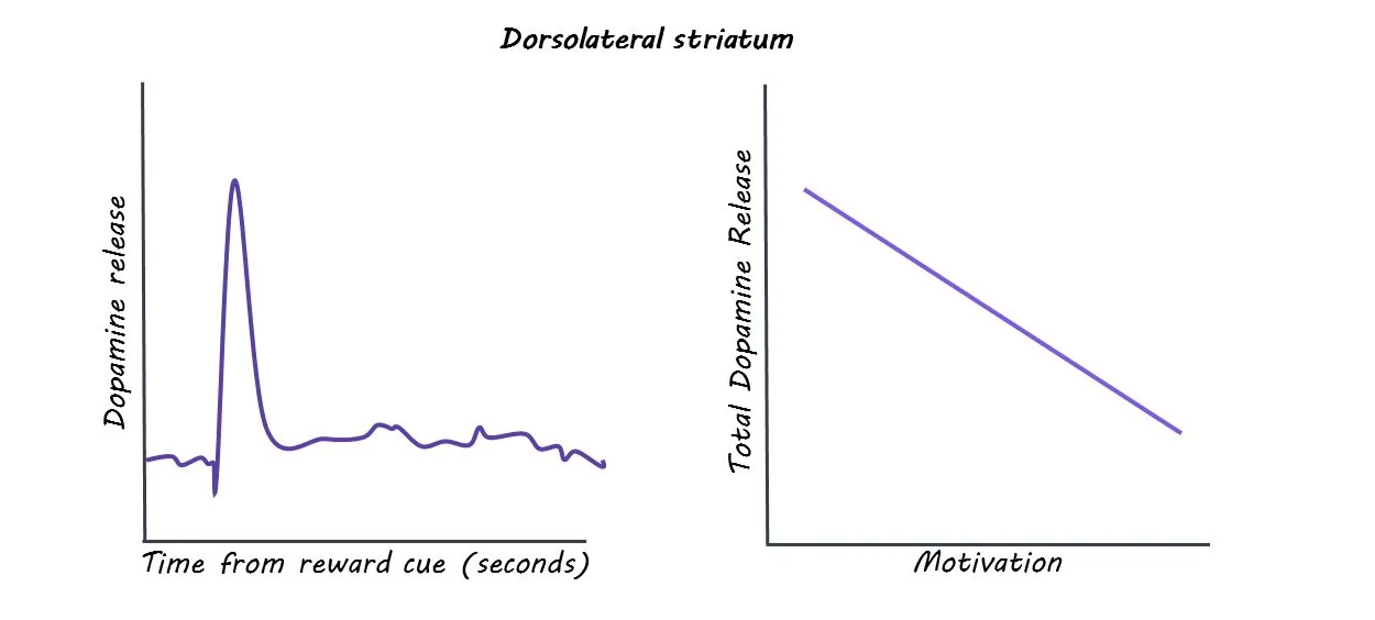
The researchers found that dopamine activity reflected both the size of the reward (i.e., benefit) as well as the effort that has already been made to achieve the reward (i.e., the cost). This was a key takeaway as it emphasized that DA was not preferentially associated with either benefit or cost associated with a task. The researchers also observed an unexpected and counterintuitive link between DA release and motivation: they found that highly motivated mice had lower dopamine release for a fixed reward and vice-versa. The authors speculate that this finding could be similar to what is observed in the cases of drug users, where high motivation is associated with low drug-induced dopamine release. Additionally, it was also found that optogenetics-induced DA stimulation was influenced by the environment, costs, rewards, as well as motivation. 

What's the impact?

This study shows that dopamine released in the brain’s striatum reflects the cost and benefit of a task, as well as the motivation underlying that task. Dopamine dynamics for each reward represents the size of the reward as well as the cost that’s already been paid for the task. The study also shows that dopamine release goes down in high-motivation states. Overall, the findings of this study are crucial to understanding the intricacies of the role of dopamine as a neuromodulator in the brain.

The Effects of Schizophrenia on Cognitive Mapping

Post by Christopher Chen 

The takeaway

We use cognitive maps to help make sense of the world, but these maps might be distorted and disorganized in people with schizophrenia (PScz). Researchers discovered that the reduced influence of semantic similarity and abnormal hippocampal ripples (i.e. transient bursts of high-frequency activity) in PScz contribute to cognitive disorganization found in PScz. 

What's the science?

Schizophrenia is a prevalent neuropsychiatric disorder causing conceptual disorganization, abstract reasoning difficulties, and language coherence issues. The presence of these symptoms, collectively known as "formal thought disorder," predicts impaired social functioning. Schizophrenia is also associated with abnormalities in association-guided cognition, where relationships between memories, concepts, or objects in the brain, known as "relational knowledge," are disrupted.

Recent studies suggest that the hippocampal–entorhinal cortex (HEC) encodes abstract relational knowledge. In rodents, researchers have shown that HEC cells encode spatial relationships during navigation of the environment and exhibit neural "replay" during rest. In humans, the HEC also encodes associations in abstract domains, supporting a domain-general "cognitive mapping" function that spans conceptual and semantic spaces. Furthermore, hippocampal ripple power, a neural output linked to cognitive map stabilization, is known to be disrupted in PScz. However, the impact of schizophrenia on cognitive maps as well as the neural signatures of these outputs are unknown. In a recent article in PNAS by Nour et al., researchers used a combination of verbal fluency analysis and brain imaging to help fill gaps in understanding the impact of schizophrenia on cognitive mapping. 

How did they do it?

To assess verbal fluency, 52 participants (26 PScz and 26 controls) were given 2 separate language-based tests, each 5 minutes long. For the first test, they were asked to name as many animals as they could in the allotted time (category fluency), while in the second test, they were asked to name as many words as they could that started with the letter "p" (letter fluency). 

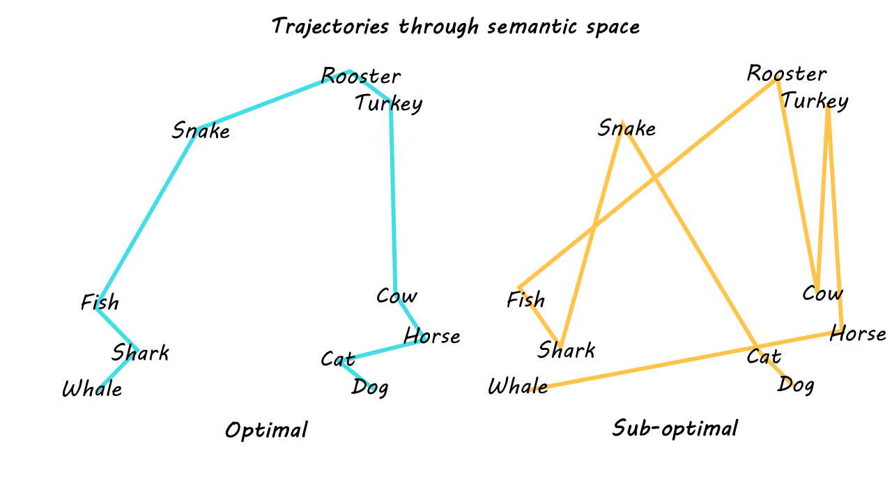
Researchers used several analysis tools, including a novel Natural Language Processing (NLP) machine learning approach, to measure semantic associations (i.e. similarities) between the words each patient provided. The researchers then measured how closely participants' word sequences approximated the “optimal path” of word sequences generated by mathematical algorithms, with greater divergences from the optimal path metrics suggesting “looser” conceptual organization. 

Researchers were also interested in investigating how neural signatures associated with verbal fluency manifested in PScz. To do so, researchers used a brain imaging technique called magnetoencephalography (MEG) to characterize brain activity in participants following verbal learned task sequences. Specifically, MEG data were collected from participants who had completed a sequence learning task and were resting quietly, to reveal whole-brain activity patterns of spontaneous neural replay of those learned task sequences.  

What did they find?

PScz showed less optimal word selection over time during the category fluency task, suggesting that their cognitive processes were more disorganized. Additionally, computational modeling demonstrated that the balance between category and letter-based information in task performance was notably influenced by the task goal (context). In other words, category-based (semantic) bias was seen in the animal-naming task while letter-based bias (similarity between words, letter by letter) was seen in the ‘p’ word task. The extent of this influence, referred to as "goal-induced semantic modulation," was significantly reduced in individuals with schizophrenia, again suggesting that PScz exhibits an impairment in how semantic information is used to guide the flow of thought. Furthermore, greater variability in goal-induced semantic modulation across participants correlated with greater difficulty with abstract conceptual reasoning, which is considered to be a “negative” symptom in schizophrenia. This suggests that the observed deficits in goal-induced semantic modulation may be linked to specific symptoms in schizophrenia, providing valuable insights into the cognitive mechanisms underlying the disorder.

As for brain imaging data, MEG traces showed that in both controls and PScz, participants with greater ripple power at the time of replay demonstrated a greater tendency to use semantic information for context-sensitive word selection. Essentially, greater ripple power was correlated with greater cognitive organization during verbal fluency behavior. Interestingly, the study did not find a correlation between goal-induced semantic modulation and a previously reported measure of spontaneous replay, providing valuable insights into the nuanced relationship between neural processes and semantic cognition in individuals with schizophrenia.

What's the impact?

This study provided preliminary support for the hypothesis that some symptoms of schizophrenia are linked to dysfunction in cognitive maps. Overall, these findings are some of the first to connect clinical features of schizophrenia to the neural processes underlying abnormalities in semantically guided cognition.