How Fixational Eye Movements are Related to Covert Attention

Post by Lina Teichmann

The takeaway

Microsaccades are small involuntary eye movements that occur when we are attending to peripheral things in our environment. These eye movements correlate with alpha-band activity (a particular frequency of brain activity) in the brain - a neural signature of spatial attention - but are not necessary for neural modulation to occur. 

What's the science?

When driving a car, we usually attend to what is right in front of us, but at the same time, we need to allocate some of our attentional resources to other things in our periphery like pedestrians or cyclists. This process is known as covert spatial attention and allows us to prioritize relevant information that is not currently in fixation. Covert attention is associated with directional biases in fixational eye movements or microsaccades, which are small jerk-like, involuntary eye movements. In the human brain, alpha activity has commonly been associated with spatial attention. This week in Nature Communication, Liu and colleagues examined if and how the commonly observed biases in microsaccades and the modulation of alpha activity in the brain are related when deploying covert attention.

How did they do it?

Participants completed a spatial memory task while their brain activity data was recorded with electroencephalography (EEG) and their eye movement data was recorded with an eye tracker. To compare the spatial modulation of the alpha activity, the researchers looked at lateralization (i.e., comparing the strength of alpha over the right and left posterior EEG sensors which is typically modulated by whether attention is deployed to the left or right visual field).

In the memory task, participants were asked to recall the orientation of one of two lines which were shown on right and the left of the screen. The researchers then measured whether (1) the presence and direction of eye movements during recall were indicative of performance and (2) whether lateralized alpha activity – a marker for spatial attention – is correlated with attention-directed microsaccades.

What did they find?

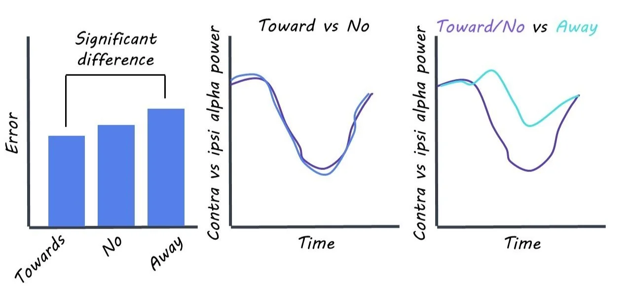
Overall performance was not dependent on microsaccades, as participants recalled the orientation of the line equally well regardless of the presence of an eye movement. However, when an eye movement was present, the direction was linked to performance, with eye movements towards the location of the to-be-recalled item resulting in better performance.

Looking at the neural data, the researchers found that there was clear lateralized alpha (a marker for spatial attention) activity when eye movements occurred towards the location of the to-be-recalled item. However, a highly similar pattern was observed when no eye movement was present, highlighting that eye microsaccades are not necessary for spatial attention. Considering only trials where a microsaccade occurred, the data showed that eye movements away from the location of the to-be-recalled item reduce the lateralized alpha modulation. This highlights that both markers for covert attention, microsaccades, and lateralized alpha-band activity are correlated, but that microsaccades are not necessary for spatial attention.
What's the impact?

Measuring and understanding covert attention is critical for gaining insights into how we navigate through our visual world. Liu and colleagues have found an elegant way to show that there is a correlational but not necessarily an obligatory link between microsaccades and lateralized alpha activity in the brain, which is frequently used as a marker of spatial attention. 
Access the original scientific publication here.

Using Artificial Neural Networks to Understand Emotion Recognition in Autistic Adults

Post by Negar Mazloum-Farzaghi

The takeaway

Compared to neurotypical adults, autistic adults have difficulty recognizing facial emotions. Machine learning helped to determine that neural activity in the inferior temporal cortex may explain these differences.

What's the science?

Autism spectrum disorder (ASD) is characterized by difficulty recognizing others’ moods and emotions from facial expressions. Previous research has found that the fusiform face area and the inferior temporal (IT) cortex are involved in facial recognition. Moreover, neural activity in the human amygdala has been associated with recognizing facial emotions. A critical question that remains to be answered is whether atypical facial emotion recognition in autistic adults can be explained by perceptual difficulties or by an atypical development and functioning of regions associated with facial emotional processing. Brain-mapped computational models can help us predict how facial emotion is represented across different brain regions in primates and how these representations are associated with performance on tasks involving facial emotion judgement. This week in The Journal of Neuroscience, Kar used artificial neural network models of primate vision to investigate neural and behavioral markers of emotion recognition in ASD.

How did they do it?

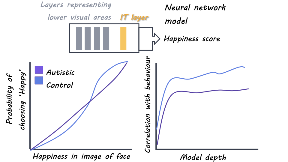
Kar began by analyzing behavioral and neural measurements conducted by Wang and Adolphs (2017) and Wang et al. (2017). During the behavioural task, Wang and Adolphs (2017) presented images of faces to neurotypical controls and high-functioning autistic adults. The participants were asked to make a judgment as to whether each face depicted happiness or fear. Wang and Adolphs (2017) found that compared to controls, autistic individuals had reduced specificity in facial emotional judgement.

To further investigate their findings using computational modeling, Kar trained artificial neural network models to perform the same task. The neural network models were comprised of layers of units that closely corresponded to the brain areas (i.e., IT cortex) and neurons in the primate ventral visual cortex. In order to test the accuracy of these models, Kar compared the facial emotional predictions they made with the behavioral measurements obtained from the neurotypical controls and autistic adults. Next, the author deleted different layers of the network in order to determine which layers of the network were responsible for discriminating between the behavior of the neurotypical controls and autistic adults.

In order to find out whether the network models could establish the IT cortex or the amygdala as the primary contributor to facial emotional processing, Kar also reanalyzed recordings obtained from electrodes implanted bilaterally in the amygdalas of patients with epilepsy by Wang et al. (2017). Similar to the task described above, participants were presented with images of faces and were asked to discriminate between two emotions; fear and happiness. Finally, to assess the efficiency of neural connections in autistic adults during facial emotional processing, the author looked at the synaptic strengths (weights) of the connections between the IT layer of the neural network models and its behavioral responses. Moreover, the author added various levels of noise to the activity of the IT layer in the neural network, to evaluate whether added noise would improve or weaken the match between judgments made by neural network models and by autistic adults.

What did they find?

Kar found that the artificial neural network models could accurately predict human facial emotion judgements at an image-by-image level. Interestingly, the network’s behavioral responses matched the patterns observed from the neurotypical control group more than those of the autistic adults. The author found that the greatest difference in the network’s ability to match the behavioral responses of controls versus autistic adults was based on the final and deepest layer of the network, which resembled the primate IT cortex. These results suggested that the neural activity in the primate IT cortex could play an essential role in abnormal facial emotion processing in autistic individuals. In terms of the role of the amygdala in facial emotional processing, the author found that, when controlling for the IT-cortex layer of the network models, the amygdala provided very little information during facial emotion recognition, suggesting that the IT cortex is the main driver of amygdala’s role in discriminating the emotion of a face.

Finally, the weights in the network models (the importance of different connections) were more strongly related to the neurotypical controls than the autistic adults, suggesting that the neural connections in the autistic adults were noisier and therefore different during facial recognition. Moreover, added noise increased the match between the network’s performance and that of the autistic adults, however, it did not improve the network’s similarity to the neurotypical controls. These findings suggested that atypical facial emotional processing in autistic individuals might be due to the presence of additional noise in their sensory representations.

What's the impact?

This study found that primate IT activity was a critical neural marker involved in a computational model of atypical facial emotion recognition in ASD. Overall, this study showed that artificial neural network models of vision were a useful tool in probing underlying neural and behavioral mechanisms involved in autism and that these neural models could be a promising tool in investigating other aspects of cognitive function.

Access the original scientific publication here.

Slow Oscillations Facilitate Brain-Wide Communication and Memory Consolidation During Sleep

Post by Lani Cupo

The takeaway

Coordinated waves of global brain activity play an important role in memory consolidation and information flow during non-rapid eye movement sleep, a sleep phase otherwise characterized by dampened activity.

What's the science?

Previous research shows that non-rapid eye movement sleep (NREM) is important in the consolidation of memory, possibly through waves of coordinated activity across the cortex called slow oscillations. However, evidence also suggests that NREM is characterized by a loss of network connectivity, which seems to be in opposition to the role of this sleep phase in memory consolidation. This week in PNAS, Niknazar and colleagues used electroencephalography (EEG) in healthy young adults to investigate the causal function of slow oscillations on the direction of information flow and on memory.

How did they do it?

The authors conducted a sleep experiment where 59 healthy young adults underwent EEG. Participants completed a memory test (word pair associations) before and after sleeping. During sleep, they wore an EEG cap measuring brain activity with electrodes at 32 different locations, or channels (10 for reference, 22 for recording). Data from the 22 channels were examined to identify the presence of slow oscillations, and data from 12 channels (three each in the frontal, parietal, temporal, and occipital lobes) were analyzed to establish the direction of information flow. Of these 12, data from four channels were considered as potential “sources” of activity, while all channels were considered potential recipients, or “sinks”. When patterns of slow oscillations were detected near the source channels, the authors used a mathematical model (generalized partial directed coherence) to compare waves of activity following the activation throughout the other electrodes. They examined how the magnitude of information (captured by the height of peaks) was impacted by the distance of the source to the origin of the slow oscillation, how much signal at sinks was impacted by the distance of the sink from the origin of the slow oscillation, and how much other factors impacted the flow of information. Then, the authors tested the hypothesis that global slow oscillations facilitate more information flow than local slow oscillations. Finally, they examined whether information flow associated with slow oscillations impacted memory performance on the post-sleep test.

What did they find?

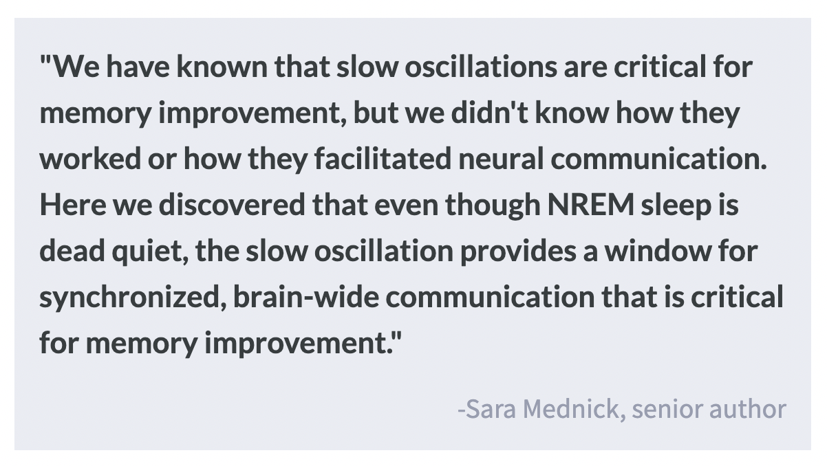
First, the authors found that NREM sleep is indeed a period where neural communication is very low. However, slow oscillations allow for widespread communication across the whole brain. During this burst of causal information flow, the position of the slow oscillation impacts the magnitude of information flow, with the source channel closer to the oscillation sending the greatest magnitude of information and sinks farthest from the oscillation receiving the greatest magnitude of information. For example, if a slow oscillation was detected in the frontal lobe, a source electrode in the frontal lobe would show more outgoing information compared with one in the parietal lobe, and a sink at the occipital lobe (far away) would receive more information than one at the central channel. This suggests that slow oscillations are promoting large waves of long-distance brain communication. Supporting this result, the authors found that global slow oscillations that propagate across more electrodes allow for greater information flow than localized ones.

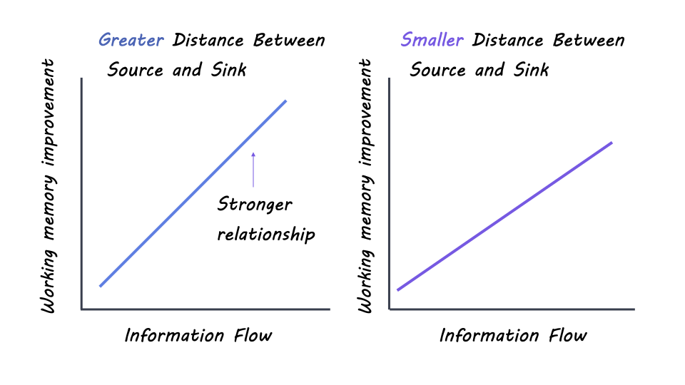
Finally, investigating the impact of information flow on hippocampal-dependent episodic memory, global slow oscillations also facilitated greater improvement, while local ones only facilitated improvement when the source electrode was close to the source of the oscillation and when there was greater distance between the source and sink pair. 

What's the impact?

These findings begin to reconcile disparate findings in the field—namely that NREM sleep is characterized by dampened activity, yet seems to underly the consolidation of episodic memory. This research provides evidence that bursts of long-range communication during slow oscillations allow memory consolidation to occur during NREM sleep.