The Impact of Sleep, Physical Activity, and Sedentary Behavior on Dementia Risk

Post by Leanna Kalinowski

The takeaway

Three modifiable daily behaviors –  sleep, physical activity, and sedentary behavior – independently and collectively impact dementia risk by influencing brain structure.

What's the science?

Dementia, defined as a loss of cognitive functioning that is severe enough to interfere with daily life, does not yet have an effective treatment despite its increased prevalence. Previous research has determined that three modifiable daily behaviors – sleep, physical activity, and sedentary behavior – are associated with the risk of developing dementia. Specifically, sleep deprivation and lack of physical activity are likely to exacerbate brain atrophy, defined as the loss of neurons in the brain. However, research on these associations is limited and based on relatively small numbers of study participants. This week in Molecular Psychiatry, Huang and colleagues investigated the impact of sleep duration, physical activity, and sedentary behavior on dementia risk and brain structure.

How did they do it?

The researchers used data from 431,924 participants in the UK Biobank study, which is a large-scale biomedical study that has been collecting data in the United Kingdom since 2006. At baseline, participants were asked several questions about sleep duration, physical activity, and sedentary behavior, and underwent structural MRI scans. All participants underwent a follow-up assessment an average of nine years after baseline.

To measure sleep duration, participants were asked how many hours of sleep they get per night and were divided into three categories: low sleep (0-6h), moderate (7h), and high sleep (8+h). To measure leisure-time physical activity, participants were asked to describe the frequency and duration of different activities undertaken over the previous four weeks. These activities were then converted into their metabolic equivalents (MET), which is the resting metabolic rate obtained during quiet sitting. After summing total MET for all physical activities, physical activity was divided into three categories: low (<400 MET/week), moderate (400-1200 MET/week), and high (>1200 MET/week). Finally, to measure sedentary behavior, participants were asked to report the number of hours spent watching TV and using a computer during a typical day. Sedentary behavior was then divided into three categories: low (0-2h/day), moderate (>2-4h/day), and high (>4h/day).

What did they find?

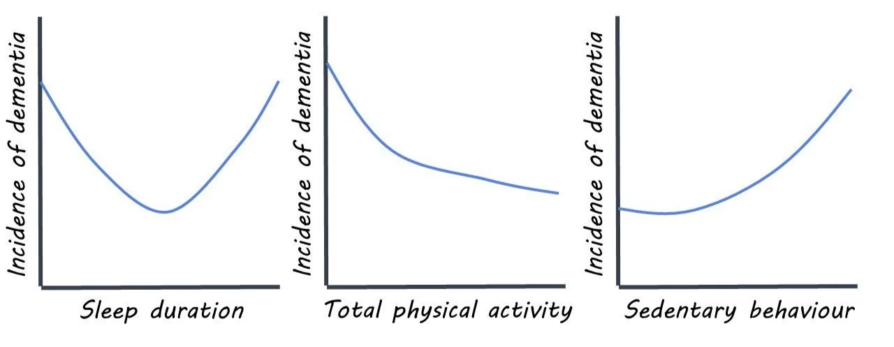
When measured independently, sleep, physical activity, and sedentary behavior were each associated with dementia risk. When compared to those in the moderate sleep category, both those in the low and high sleep categories demonstrated an increased dementia risk. In terms of physical activity, the risk of dementia dropped with every MET increase up until 1200 MET/week, above which there was little additional benefit of physical activity. As for sedentary behavior, there was no increased risk of dementia until the rate of sedentary behavior exceeded 3h/day, at which point the risk of dementia increased exponentially as the number of hours/day of sedentary behavior increased.

Taking each measurement together, the researchers found that a combination of moderate sleep, moderate-to-high physical activity, and low-to-moderate sedentary behavior showed the lowest risk of dementia. Structural MRI scans from these individuals showed larger cortical and subcortical grey matter volumes, suggesting that these activities impact dementia risk by affecting the structure of the brain.
What's the impact?

This study was the first of its kind to examine the independent and joint effects of sleep, physical activity, and sedentary behavior on the development of dementia. Findings from this study provide important insights into different risk factors for dementia and highlight the importance of changing these behaviors to reduce dementia risk.   
Access the original scientific publication here.

How New Neural Connections are Formed During Learning

Post by Shannon Kelly

The takeaway

New neural connections formed during learning tend to be active at the same time as other neural connections being used during learning and form in close proximity to them.

What's the science?

Previous research has shown that learning involves physical changes in connections between brain cells. These changes include the growth of new structures called “spines”, on neuronal dendrites which correspond to new connections with other neurons. However, the process by which learning causes the formation of these new spines is not yet well understood. This week in Nature Neuroscience, Hedrick and colleagues used microscopic brain imaging techniques to show how the activity of nearby connections affects the formation of new neural connections in the brains of mice while learning a new behavior.

How did they do it?

The authors measured the activity of existing dendritic spines and the growth of new spines on neurons in the primary motor cortex in adult mice over two weeks. During these two weeks, mice in the learning condition completed daily training in which they were given a reward for pressing a lever in response to hearing a specific noise. A control group of mice was rewarded regardless of whether they pressed the lever. Spine activity and growth were measured during training sessions using in vivo two-photon imaging, which uses fluorescent markers to provide high-resolution imaging of brain activity and structure at the level of individual brain cells, and even individual spines (~100 times smaller than cells). They examined the degree to which activity, location, and potentiation (the increase in strength of a neural connection with increased use) of existing spines affected the growth and maintenance of new spines. For a subset of the mice, they also used electron microscopy to examine mice brains after the final training session to study the structure of new connections developed during learning.

What did they find?

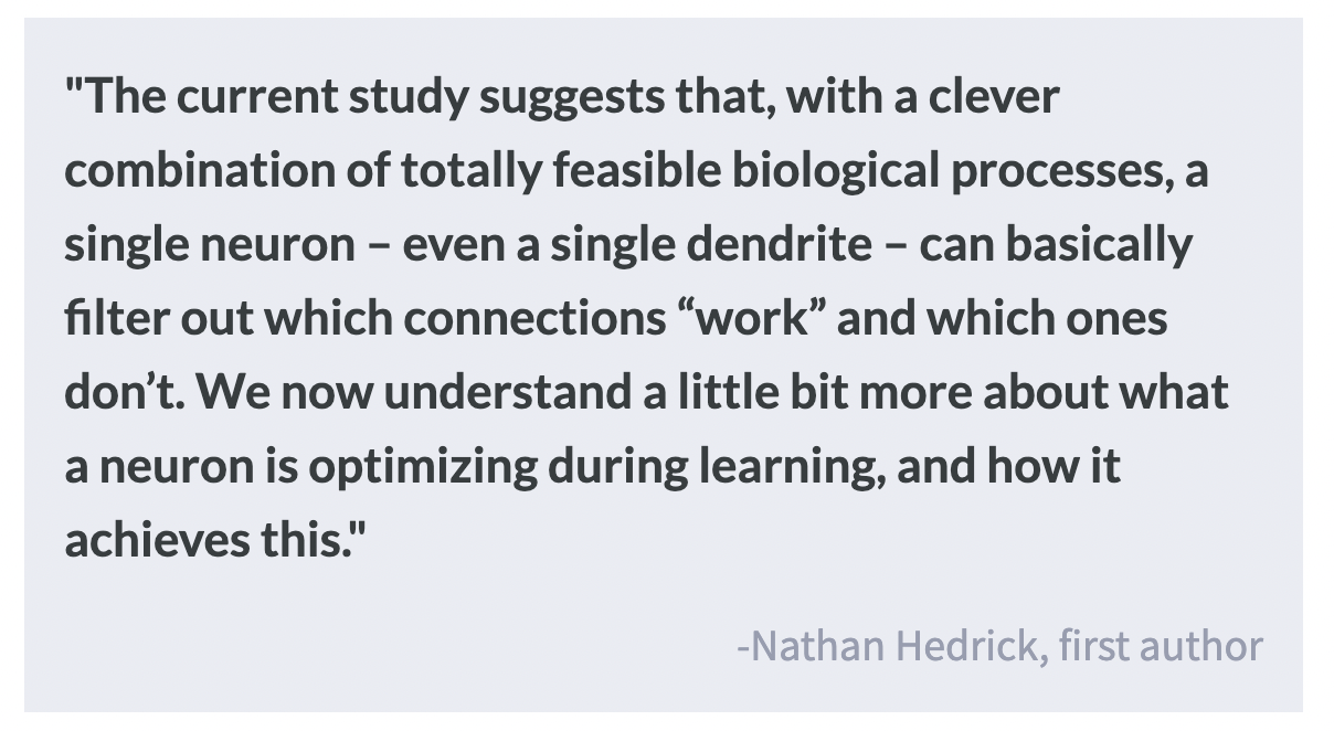
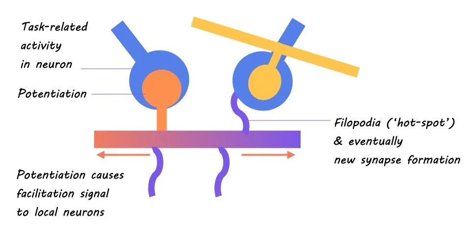
For mice in the learning condition, new spines formed more often near clusters of existing spines that were active during lever presses (movement-related spines) as well as near spines with high potentiation. These findings suggest that the location of new spine formation on the dendrite is guided by the activity and potentiation of pre-existing spines during performance of the behavior that is being learned. These new spines tended to be active at the same time as existing movement-related spines, and their combined activity was more prominent when mice showed the learned behavior pattern, suggesting that new spine development supports the performance of the learned behavior. Electron microscopy showed that filopodia (precursors of new spines) are more frequently located near fully developed new spines, suggesting that multiple new filopodia are produced physically close to each other in “hot spots”, and those that successfully connect to other neurons become new spines. Finally, new spines that tended to be active at the same time as nearby movement-related spines were more likely to remain throughout the study, while those whose activity was not related to that of movement-related spines disappeared by the final training sessions, indicating that new spines whose connections support learning are selectively maintained, likely by their synchronized activity with nearby movement-related spines.

What's the impact?

These results provide a model that describes how dendritic spines are developed and maintained during learning. This model fills a gap in what was previously known about the biology of learning by explaining the process by which the growth of new spines occurs and how it supports the learning of new behavior. Future research can build on these findings to describe the broader network that is developed through new spine growth to improve our understanding of how the aggregation of information from other brain regions supports learning.

Using Brain Lesions to Identify a Common Addiction Brain Circuit

Post by Megan McCullough

The takeaway

Brain lesions brought on by brain damage from events such as strokes have been known to interrupt addiction in some individuals with substance use disorders and even lead to remission. Lesion mapping shows that these regions connect to a specific brain circuit that can be used as a therapeutic target for the treatment of addiction. 

What's the science?

Substance use disorders (SUDs) are a leading cause of death in young populations. Neuromodulation of brain regions implicated in addiction —  a current therapy for addiction — involves delivering electrical stimuli to induce changes in specific neural circuits. However, there is a need to better identify therapeutic targets for neuromodulation. Previous research has shown that brain damage from strokes has led to addiction remission in some individuals with SUD. This week in Nature Medicine, Joutsa and colleagues aimed to link brain lesions that resulted in addiction remission to the human connectome in order to gain a better understanding of the specific brain regions involved in addiction remission.

How did they do it?

The authors examined data from 129 patients who were daily nicotine smokers at the time they acquired a brain lesion, with a subset of these patients entering addiction remission right after the lesion. Lesion locations within the patients entering addiction remission were analyzed to identify the specific brain regions connected with remission. The authors next tested if lesions within the addiction remission cohort mapped to a specific brain circuit rather than a single brain region. Utilizing lesion network mapping, connectivity patterns between lesion locations were then compared across groups: those entering remission versus non-quitters. Finally, the authors looked at which brain voxels [a location in three-dimensional space] have a similar connectivity profile to the profiles of the lesion locations that led to addiction disruption.

What did they find?

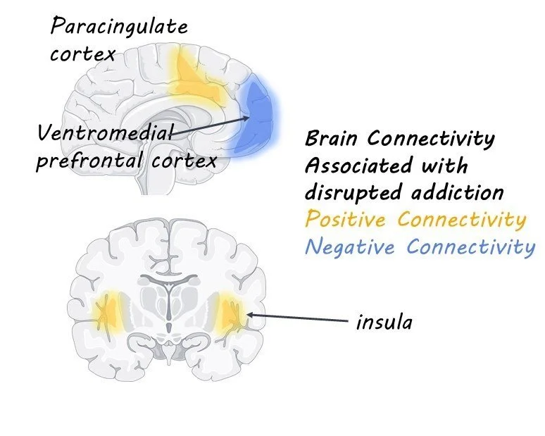
The authors found that lesions that led to a disruption in nicotine addiction occurred in multiple different brain regions. These data suggest that there is no one specific brain region implicated in addiction remission. However, the lesions all had the same pattern of connectivity to brain regions implicated in models of addiction. Brain regions that best matched the connectivity pattern of the lesions included the frontal operculum and paracingulate cortex. This study helps explain why some previous studies found that lesions to the insula are more likely to lead to remission while others have not replicated those results; lesions map to specific brain circuits, not specific regions.

What's the impact?

This study is the first to show that brain lesions that disrupt addiction map to specific brain circuits. This work has therapeutic potential, as neuromodulation techniques such as DBS and TMS may be most effective if they target brain regions with the same connectivity profile as the lesions that disrupted addiction in this study. This study provides targets for therapeutic neuromodulation, which has the potential to induce remission in those with SUDs.