Individual Differences and Similarities in How Pain is Represented in the Brain

Post by Lincoln Tracy

The takeaway

Brain regions receiving direct spinothalamic input display more consistent pain representations between different individuals. These regions have the potential to be used as targets for personalized clinical interventions.

What's the science?

Pain is a complex and multidimensional experience. Certain brain regions have consistently been reported to play a role in pain processing, while other regions have been less consistently linked. Researchers suspect pain arises from activity in a variety of brain pathways, which may differ from person to person. However, it is currently unclear as to which brain regions have more consistent versus variable representations of pain across individuals. This week in Nature Neuroscience, Kohoutová and colleagues used a personalized brain mapping approach to identify brain regions with high or low interindividual variability in their relationship to pain.

How did they do it?

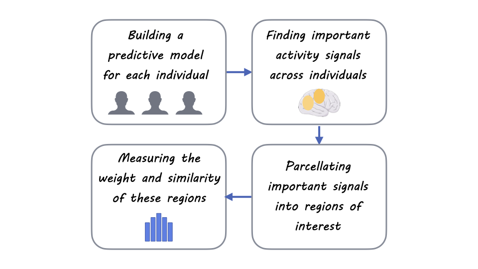
The authors obtained existing functional magnetic resonance imaging data from 404 individuals who had previously participated in one of 13 experimental pain studies across two independent laboratories. They then used these data to undertake personalized brain mapping of pain through predictive modeling. From these models, they sought to identify which brain regions were important for pain prediction. They also sought to explore how much variability each of the identified brain regions (either individually or as a collective) displayed in terms of pain prediction between individual participants. Finally, the authors validated their findings by replicating the analyses in a novel dataset of 124 individuals.
What did they find?

The authors identified 21 pain-predictive brain regions, including the anterior midcingulate cortex, the dorsolateral prefrontal cortex, and the cerebellum. When they compared the individual variability of these important regions, certain regions (e.g., the ventromedial prefrontal cortex) displayed high levels of variability between individuals, while other regions (e.g., the posterior midcingulate cortex) displayed lower levels of variance. This indicates substantial variability in the role of these regions in pain between individuals. Similar results were observed when considering the brain regions as a collective (i.e., multivariable analysis) rather than individually. The ventrolateral prefrontal cortex, the vermis, and the ventromedial prefrontal cortex showed the highest individual variability. In contrast, the posterior midcingulate cortex, the supplementary motor area, and the sensorimotor cortex were the most stable regions among individuals. Importantly, these findings were replicated when the analyses were repeated in a novel and independent dataset. Taken together, these findings indicate that the relationship between brain regions and pain perception at the individual level is more complicated than how they are usually summarized at the group level.  

What's the impact?

This study found that various brain regions display differing levels of variability in pain representation between individuals. These findings enhance our understanding of how individuals process pain differently. Knowing which brain regions contribute to pain processing at the individual level could assist treatment decision-making processes for people with chronic pain.
Access the original scientific publication here.

The Lateral Orbitofrontal Cortex Mediates Stereotyping

Post by Leanna Kalinowski

The takeaway

Actions towards other individuals are often driven by assumptions about what they are like based on their group membership (i.e., stereotypes). Researchers have identified the lateral orbitofrontal cortex as a key brain region that mediates the effect of stereotypes on social behavior. 

What's the science?

Stereotypes, defined as generalized beliefs about a category of people on the basis of gender, age, race, nationality, or occupation, often influence how individuals treat one another. It has previously been observed that stereotypes can be structured along two dimensions of trait perception: warmth, which is the degree to which people have good intentions toward others, and competence, which is the degree to which people are capable of acting on their intentions. Although the impact of stereotypes on social behavior is well-documented, it’s still unclear how traits inferred from social-group membership are represented in the brain, and how these neural representations guide social behavior. This week in PNAS, Kobayashi, and colleagues tested the impact of stereotypes on the distribution of resources in a social decision-making game.

How did they do it?

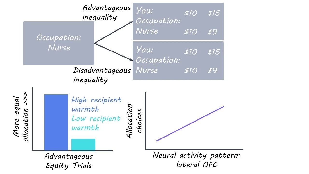
43 participants were asked to choose how to allocate monetary resources between themselves and a series of hypothetical recipients in a task called the Dictator Game. In each trial, the participants first viewed one piece of social-group information about a hypothetical recipient (e.g., “Occupation: Lawyer”). The researchers selected 20 social-group memberships to display, which were based on previous research that showed that these social-group memberships spanned a wide range of the warmth and competence dimensions. 

The participants were then shown two options to allocate the funds. Option one was an equal allocation of money between the participant and recipient, while option two was either (1) advantageous inequity, which was an unequal division of money that favored the participant, or (2) disadvantageous inequity, which was an unequal division of money that favored the recipient. Each participant completed 80 trials each while brain activity was measured using functional magnetic resonance imaging (fMRI).

What did they find?

In advantageous inequity trials (i.e., trials that favored the participant), participants were less likely to choose the unequal allocation of money when the recipient’s warmth was higher. Conversely, in disadvantageous inequity trials (i.e., trials that favored the hypothetical recipient), participants were less likely to choose the unequal allocation of money when the recipient’s competence was higher. Taken together, these results suggest that inferences about other people’s traits exert strong effects on social-decision making. 

Turning to the fMRI data, the researchers first conducted a representational similarity analysis, which looks for (1) brain regions where two recipients with similar traits display similar response patterns and (2) brain regions where two recipients with dissimilar traits display dissimilar response patterns. This analysis revealed that recipients’ warmth and competence are represented in the lateral orbitofrontal cortex (OFC), which has traditionally been associated with inference-based decision-making, along with two brain regions associated with mentalizing (i.e., temporoparietal junction & superior temporal sulcus). Finally, the researchers found that only activation of the lateral OFC predicted individual monetary allocation choices. Taken together, these results suggest that the trait representation in the lateral OFC contributes to allocation decisions.

What's the impact?

This study demonstrates that the effect of stereotypes on behavior is driven by inference-based decision-making processes in the lateral OFC. Future studies should investigate the role of the OFC in other types of inference-based decision-making, such as social projection and learning about traits from experience.

Access the original scientific publication here.

Reducing Neuroinflammation with Brain-Specific Gene Delivery

Post by Leanna Kalinowski

The takeaway

Acute central nervous system (CNS) trauma, which is present in neurological disorders such as multiple sclerosis, triggers the activation of immune cells in the brain causing inflammation. Researchers have developed a brain-specific gene delivery system for brain interleukin-2, which is effective in preventing and treating these neurological disorders in mouse models. 

What's the science?

Acute central nervous system (CNS) trauma is caused by diverse neurological injuries and illnesses, like traumatic brain injury (TBI), multiple sclerosis, or stroke. CNS trauma leads to the damage and loss of neurons, which can result in changes in cognition, sensorimotor function, and personality. Acute CNS trauma also triggers the activation of immune cells in the brain – like microglia and astrocytes – which leads to further negative effects.  

Brain interleukin-2 (IL-2) has been identified as a potential treatment option for acute CNS trauma. IL-2 works by supporting the survival and proliferation of regulatory T (Treg) cells, which have been shown to inhibit neuroinflammation. However, while IL-2 treatment shows promise, it is unclear whether such treatment can be localized to the brain rather than inducing unintended immune effects elsewhere in the body. This week in Nature Immunology, Yshii and colleagues (1) developed a new delivery system for brain-specific IL-2 treatment and (2) tested its effectiveness in preventing and treating models of acute CNS trauma.

How did they do it?

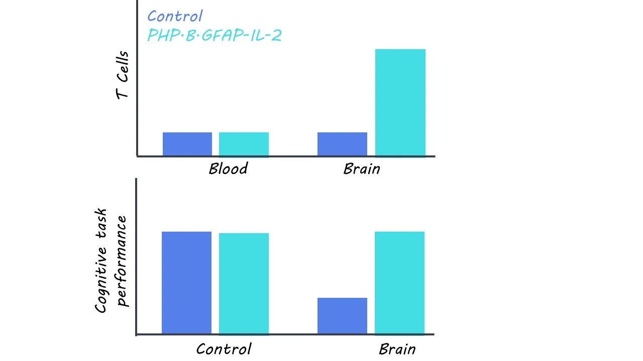
First, the researchers developed a gene delivery approach to increase IL-2 expression in astrocytes, which are a type of glial cell that responds to brain injury. To ensure that they specifically target astrocytes in the brain, the researchers first developed a vehicle for gene delivery by combining (1) a PHP.B-enhanced adeno-associated virus (AAV-PHP.B), which can be injected intravenously and delivers substances across the blood-brain barrier and (2) a modified GFAP promoter, which restricts gene expression to just astrocytes. Then, they incorporated the gene for IL-2 (PHP.B.GFAP-IL-2) into this virus, so that IL-2 is made by astrocytes in the brain, with subsequent measures of IL-2 and Treg cell production in the brain.

Then, they tested the effectiveness of PHP.B.GFAP-IL-2 in preventing acute CNS trauma in mouse models. Mice were first treated either with PHP.B.GFAP-IL-2 or a control virus (PHP.B) and then underwent a procedure to induce TBI. Fourteen days after TBI, neurological damage was assessed using MRI and histology, and behavior was assessed by running mice through two memory tests – Morris Water Maze and the Novel Object Recognition test. To examine other common sources of acute CNS trauma, this procedure was repeated for two other pathologies – ischemic stroke and multiple sclerosis.

Finally, to test the effectiveness of PHP.B.GFAP-IL-2 in curative mouse models of acute CNS trauma, they first subjected mice to acute CNS trauma (i.e., TBI, stroke, and multiple sclerosis). Then, they administered either PHP.B.GFAP-IL-2 or a control virus (PHP.B) and ran the same test battery as above.

What did they find?

First, the researchers found that PHP.B.GFAP-IL-2 was successful in delivering the IL-2 gene to the brain with expression in astrocytes. Over the course of fourteen days following administration, there was a threefold increase in IL-2 production. This was accompanied by an increase in Treg cell production, which was not observed outside of the brain and was not accompanied by any adverse symptoms.

Then, they found that pre-treatment of PHP.B.GFAP-IL-2 decreased the loss of cortical tissue and behavioral deficits in mice with TBI relative to those that received a control virus. Similar protective effects were observed in mouse models of ischemic stroke and multiple sclerosis. They also found that mice treated with PHP.B.GFAP-IL-2 after TBI also experienced lower levels of brain damage than control mice. This effect was mirrored in multiple sclerosis mice, but not in ischemic stroke mice, suggesting that the damaging effects of stroke are too rapid for this treatment to be effective before irreversible brain damage sets in.

What's the impact?

Taken together, the results from this study provide evidence for the effectiveness of brain-specific IL-2 gene delivery in preventing and treating acute CNS trauma. Future work is needed to determine whether such treatment is effective in humans with neurological injuries and diseases. Further, this study may also pave the way for the development of gene delivery therapies for other neurological disorders that require brain-specific delivery.