Brain Activity Associated with Positive Patient-Clinician Interaction

Post by D. Chloe Chung

What's the science?

Interaction between a patient and clinician can substantially impact therapeutic outcomes. For example, an unsatisfactory interaction can prevent patients from seeking medical care, but a positive patient-clinician interaction can actually lead to psychological relief of patients’ symptoms such as pain, amplifying benefits from the actual medical treatment. While it remains unclear what mechanisms underlie such phenomenon, several studies have emphasized the importance of synchronized behaviors and neural activity in social interactions such as bonding. This week in Science Advances, Ellingsen and colleagues used functional magnetic resonance imaging (fMRI) to demonstrate that social interaction between patients and clinicians can improve clinical outcomes by mirroring their behaviors and harmonizing neural activity in brain regions that regulate empathy.

How did they do it?

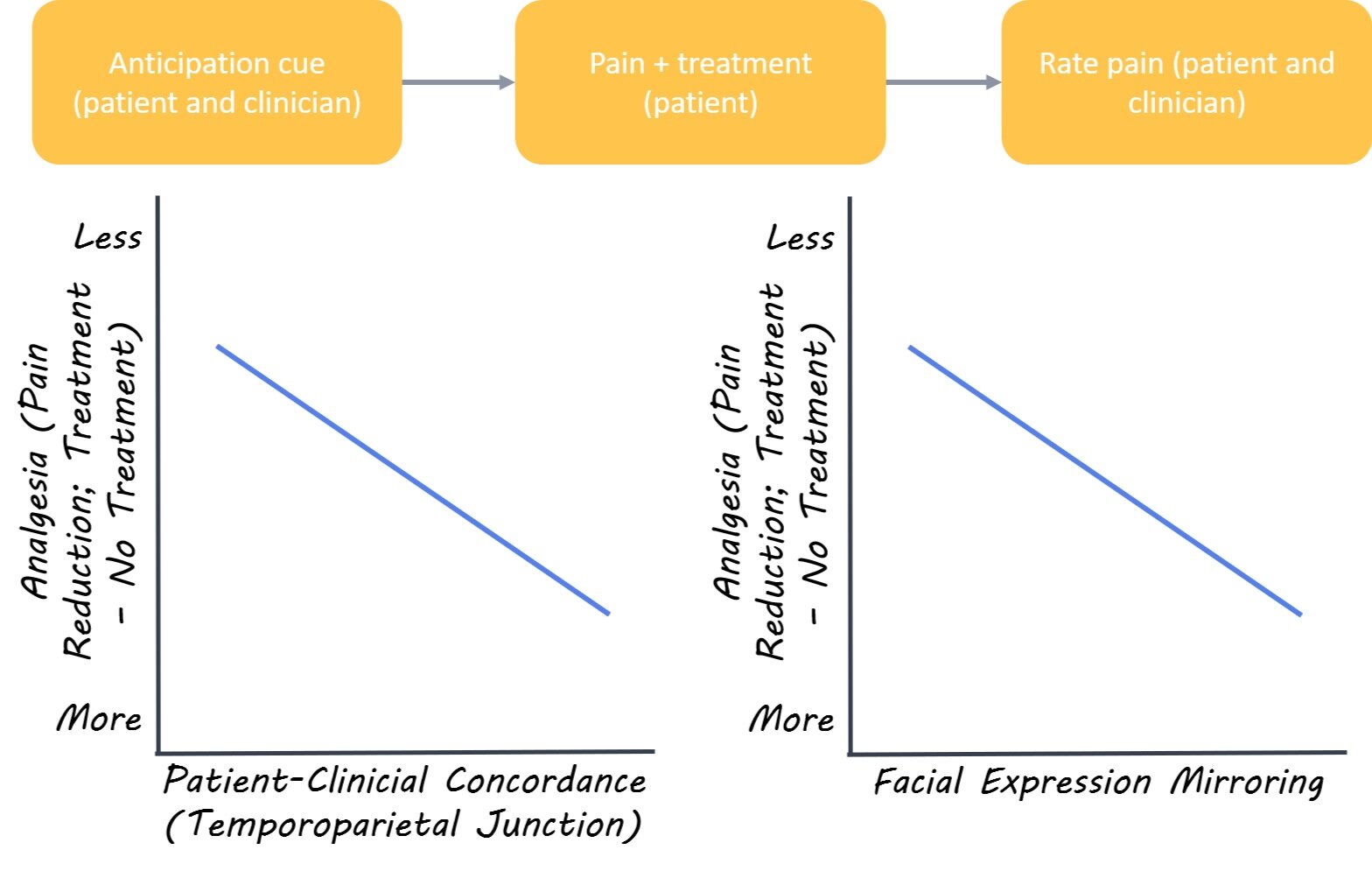
The authors recruited 23 fibromyalgia patients with chronic pain (all females) and 22 acupuncture clinicians (15 females) and matched them into 40 unique pairs. In the “clinical interaction” condition, the pair first established a rapport before the experiment (pain treatment) as the clinician gave consultation to the patient. In contrast, the pair in the “no interaction” control condition skipped the consultation and was only briefly introduced to each other before the experiment. On the day of the pain treatment, the patient and the clinician were placed in separate MRI scanners but were able to communicate with one another using facial expressions via video camera. Both participants received a visual cue to anticipate whether the patient would receive either a) treatment or b) no treatment following a moderate pressure pain evoked in the patient’s leg. Next, the clinician could press a button to give either real acupuncture treatment or a sham treatment to the patient’s leg. Both the clinician and patient were blind to whether a real treatment was given. After the trial, the pair were asked to rate the intensity of pain. The pair underwent 12 trials while their brain activity was scanned using fMRI

What did they find?

Both patients and clinicians reported less pain when the treatment was given, regardless of it being real or sham. Interestingly, video analysis of facial expressions revealed that patients tended to experience greater pain relief and stronger rapport with the clinician when the patients and the clinicians mirrored each other’s facial expressions. This suggests that social mirroring is linked to enhanced clinical outcomes. When the authors compared brain activity between groups they found that pairs from the “clinical interaction” group showed increased dynamic coupling of brain activity in the circuitry involved in social mirroring. In other words, the brain regions implicated in empathy were activated at the same time (i.e. concordant) in clinicians and patients if they already established a rapport prior to the pain treatment. Specifically, a brain area called the temporoparietal junction was activated at the same time in both pair members when they received an anticipation cue for treatment options. Upon further analysis, the authors found that concordance (co-activation between patient and clinician) of this brain region also led to greater pain relief via increased activity in a second brain region in the empathy neural circuitry that can also modulate pain.
Chloe+%281%29.jpg

What’s the impact?

This work sheds light on the neural circuitry involved in positive interactions between clinicians and patients. Further, this research demonstrates that positive patient-clinician interactions can improve clinical outcomes. The role of empathy in social interaction has been previously discussed in many studies, but this is the first to simultaneously monitor neural activity of two people who are interacting with each other in a clinical setting. Findings from this study may help guide clinicians in their interactions with patients to enhance clinical outcomes for patients.
ellingsen_quote_nov3.jpg

Ellingsen et al. Dynamic brain-to-brain concordance and behavioral mirroring as a mechanism of the patient-clinician interaction. Science Advances (2020). Access the original scientific publication here.

Axonal Injury Predicts Neurodegeneration After Traumatic Brain Injury

Post by Cody Walters 

What’s the science?

Data from non-human animal models of traumatic head trauma suggest that axonal damage can induce neuronal cell death. However, there is no direct evidence that axonal injury triggers progressive neurodegeneration in humans. This week in Brain, Graham and colleagues, examined the relationship between diffuse axonal injury and neuronal cell death in human subjects. 

How did they do it?

The authors imaged the brains of 55 patients (and 19 healthy control participants) with moderate-severe traumatic brain injury (TBI). Anatomical magnetic resonance imaging (MRI) was used to assess white/gray matter volume (via the Jacobian determinant [JD]) while diffusion tensor imaging (DTI; a type of MRI scan) was used to measure axonal integrity (via fractional anisotropy [FA]). Baseline brain volume and axonal integrity were established ~3.5 years after TBI, and participants received a follow-up scan ~1 year later.

What did they find?

The authors found that TBI participants had greater axonal injury at both baseline and follow-up scans relative to controls. This reduction in white matter integrity was most prominent in the corpus callosum, superior and inferior longitudinal fasciculus, external capsule, and posterior limb of the internal capsule. Similarly, the authors found that TBI subjects had lower gray and white matter volume across most brain regions at both baseline and follow-up scans relative to controls. Interestingly, the authors showed that the degree of axonal injury predicted the degree of brain atrophy a year later (with a positive correlation between FA scores and JD scores). Finally, the authors sought to determine the relationship between brain atrophy and cognitive impairment by administering a memory test. They found that performance on the memory test correlated with the degree of brain atrophy (with higher memory scores corresponding to less brain atrophy).

tbi_nov3.png

What’s the impact?

This study provides direct evidence that axonal injury following moderate-severe TBI predicts brain atrophy in human subjects. These data are clinically significant because they reveal a novel approach that has the potential to improve the assessment of neurodegenerative risk following TBI.

Graham et al. Diffuse axonal injury predicts neurodegeneration after moderate–severe traumatic brain injury. (2020). Access the original scientific publication here.
Political Views Bias Information Processing in the Brain

Post by Deborah Joye

What's the science?

With an important election fast approaching in the United States, it is clearer than ever that political polarization exists in our society. A powerful contributor to political polarization is motivated reasoning; when we make decisions based on emotional bias rather than evidence or facts. Motivated reasoning is made especially obvious when people with opposing beliefs are presented with identical information and their biased interpretation leads them to become even more entrenched in their original positions.

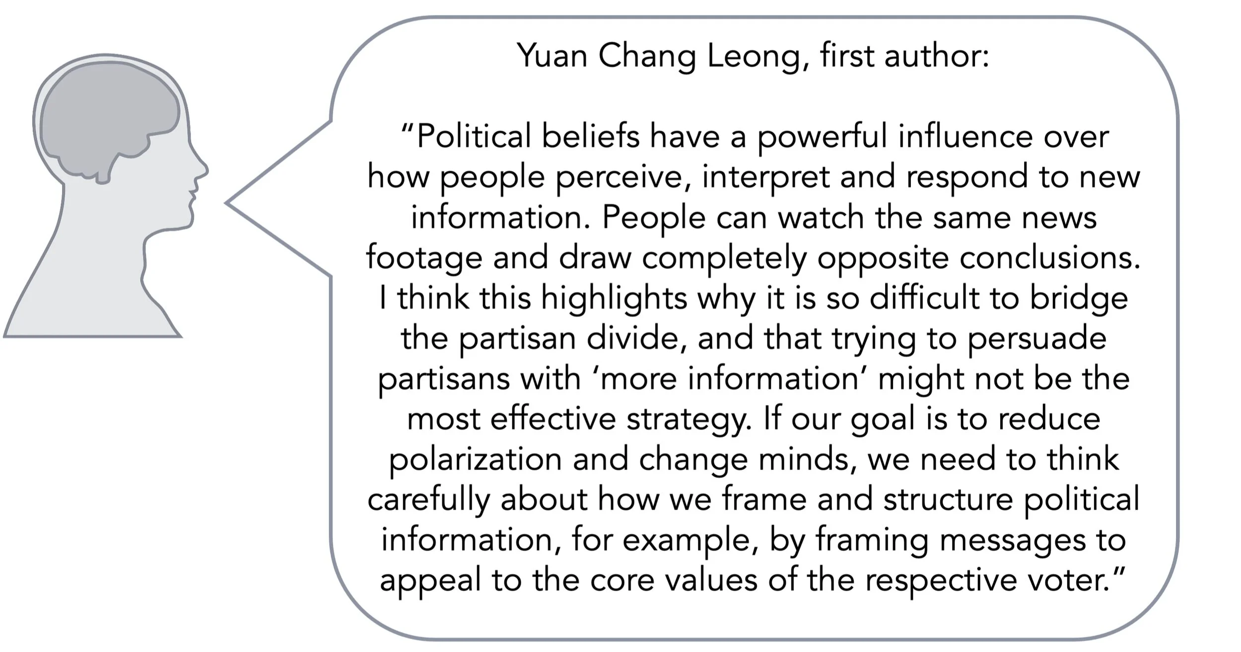
But how might identical information trigger such different outcomes, and can we see these different responses in the brain? It’s possible that our previously held beliefs change what we pay attention to when we absorb new information. For example, someone might view a political ad and only pay attention to information that supports their beliefs. It’s also possible that previously held beliefs change how people interpret incoming information. For example, one person may interpret actions as threatening, risky, or offensive where someone with different beliefs might not. This week in PNAS, Leong and colleagues demonstrate that when viewing political content,  conservative- and liberal-leaning people displayed differential activation of the dorsomedial prefrontal cortex which was intensified by moral-emotional and risk-related language.

How did they do it?

The authors scanned thirty-eight participants with functional magnetic resonance imaging (fMRI) while they viewed videos on immigration policies. Before scanning, participants indicated their support for each of the policies, and the authors calculated an overall “Immigration Attitude” to identify liberal- and conservative-leaning participants. Importantly, the authors controlled for age, sex, income, and education. The authors first asked if neural responses to the videos were different across participants. They calculated an intersubject correlation; a measure of how similar brain activity is across participants. This measure also allowed the authors to investigate which regions of the brain exhibited differential activity..

The authors next asked whether differential brain activity was specifically associated with certain content within the videos. The authors categorized words in the videos that were related only to emotions (e.g. fear, sad, etc.), related only to morals (e.g. ethics, etc.), or related to both morals and emotions (e.g. compassion, harm, etc.). The authors identified a total of 50 categories, including risk, religion, power, family, social, male, and female, among others. Finally, the authors asked whether brain activity that was more like either political group might predict the participant’s preference for supporting the position held by that group. To assess this, the authors averaged brain activity across both groups, creating a map of what the signal might look like for an “average” liberal or conservative. After each video, the authors asked each participant whether they’d be more or less likely to support that policy. The authors then assessed whether a particular participant’s brain activity during that video was more like that of the average conservative or the average liberal.

What did they find?

The authors found that overall neural responses to the political videos were shared across participants. Specifically, regions of the brain related to sensory processing displayed similar activity patterns regardless of the participant’s political leanings, suggesting that political attitudes do not alter sensory processing. However, activity patterns in the dorsomedial prefrontal cortex diverged between liberal and conservative participants. Previous research has demonstrated that this brain region is known to be active when participants are manipulated to have different interpretations of an ambiguous story. 

deb (1).png

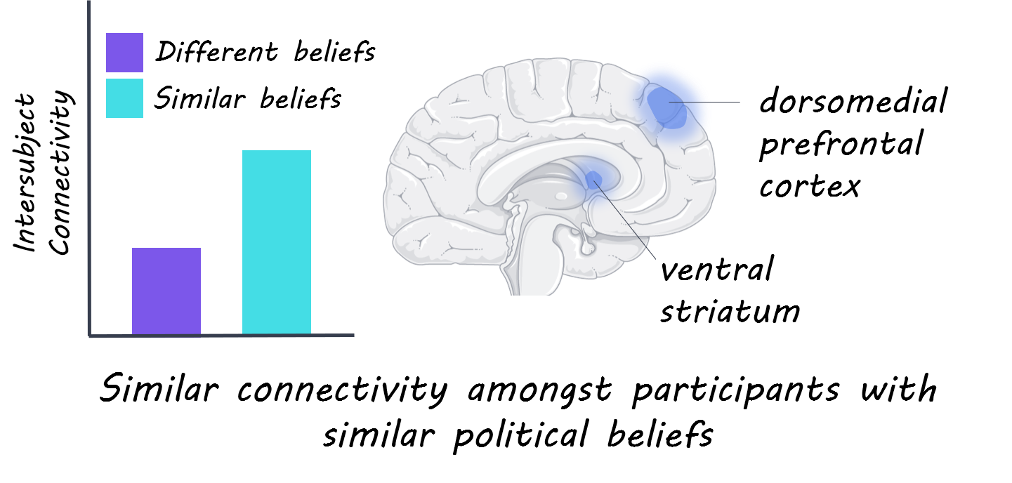
The authors also found that the difference in activity patterns in the dorsomedial prefrontal cortex was associated with the use of moral-emotional and risk-related language, suggesting that those categories of language are most likely to drive divergent neural responses between liberals and conservatives. The authors also found that connectivity between the ventral striatum (associated with emotional processing) and the dorsomedial prefrontal cortex was more similar for participants with similar political views. Lastly, the authors found that for each video, whether a participant’s neural activity was more like the average conservative or liberal predicted their attitude change. For example, a participant with brain activity patterns more like the average conservative was more likely to display attitude change further in the conservative direction.

What's the impact?

This study found that political attitudes can drive divergent neural processing of political information. One striking implication of these findings has to do with the “echo chamber” of social media, where online videos with moral-emotional content tend to spread faster within social circles sharing similar political beliefs. These findings suggest people with already formed political biases will respond to such political content with further attitude polarization. This study also demonstrates that content that features moral-emotional and risk-related language is most likely to be processed in this biased way. These findings have important implications for our political discourse moving forward and have implications for the type of language we use in a political context, as well as the impact this may have on an increasingly polarized society.

voting_quote_Oct27.jpg

Leong et al., Conservative and liberal attitudes drive polarized neural responses to political content, PNAS (2020). Access the original scientific publication here.