
Peripheral Macrophages Influence Disease Progression in Mouse Models of ALS

Post by Shireen Parimoo

What's the science?

Amyotrophic lateral sclerosis (ALS) is a neurodegenerative disorder that affects motor neurons and leads to loss of motor function, muscular atrophy, paralysis, and eventual death. Some cases of familial ALS are linked to the mutant SOD1 gene, which promotes motor neuron death. Motor neurons are surrounded by immune cells known as macrophages (peripheral nervous system) and microglia (central nervous system). Studies show that ALS mice without mutant SOD1 in their microglia live longer, but the precise impact of peripheral macrophages and microglia on motor neuron function and survival in ALS has not yet been identified. This week in Nature Neuroscience, Chiot and colleagues investigated the differential roles of microglia and peripheral macrophages in ALS disease progression.

How did they do it?

The authors used slow-progressing (slow-ALS) and fast-progressing (fast-ALS) mouse models of ALS that over-expressed different variants of the SOD1 gene. They used immunohistochemistry to characterize the morphology, number, location, and immunoreactivity (i.e., immune response) of peripheral macrophages and microglia at different stages of the disease throughout the disease course.

In the fast-ALS mice, the authors performed myeloablative conditioning and a bone marrow transplant to replace mutant peripheral macrophages with (i) control macrophages (without mutant SOD1), or (ii) macrophages with the neurotrophic over-expressing human SOD1 macrophages (non-mutant, wild-type) at the pre-symptomatic and disease onset stages. These macrophages also expressed the green fluorescent protein GFP+ for visualization. Finally, the authors performed an RNA sequencing analysis of microglia and macrophages in the mutant SOD1 and SOD1 wild-type ALS mice to better understand how the genetic profiles of these immune cells were modified in the wild-type mice.

What did they find?

In the fast-ALS mice, there was minimal infiltration of peripheral macrophages into the spinal cord, and only at the late symptomatic stage. In the slow-ALS mice, on the other hand, the number of peripheral macrophages increased at the early symptomatic stage and remained stable throughout, with a small increase at the end-stage of the disease. Nevertheless, the level of infiltrated cells at their peak was only 23% relative to the microglial cell population at the end-stage. This means that the infiltration of peripheral macrophages depends on disease duration but does not represent the major cell population in ALS spinal cords.
microglia_img_Oct27.png

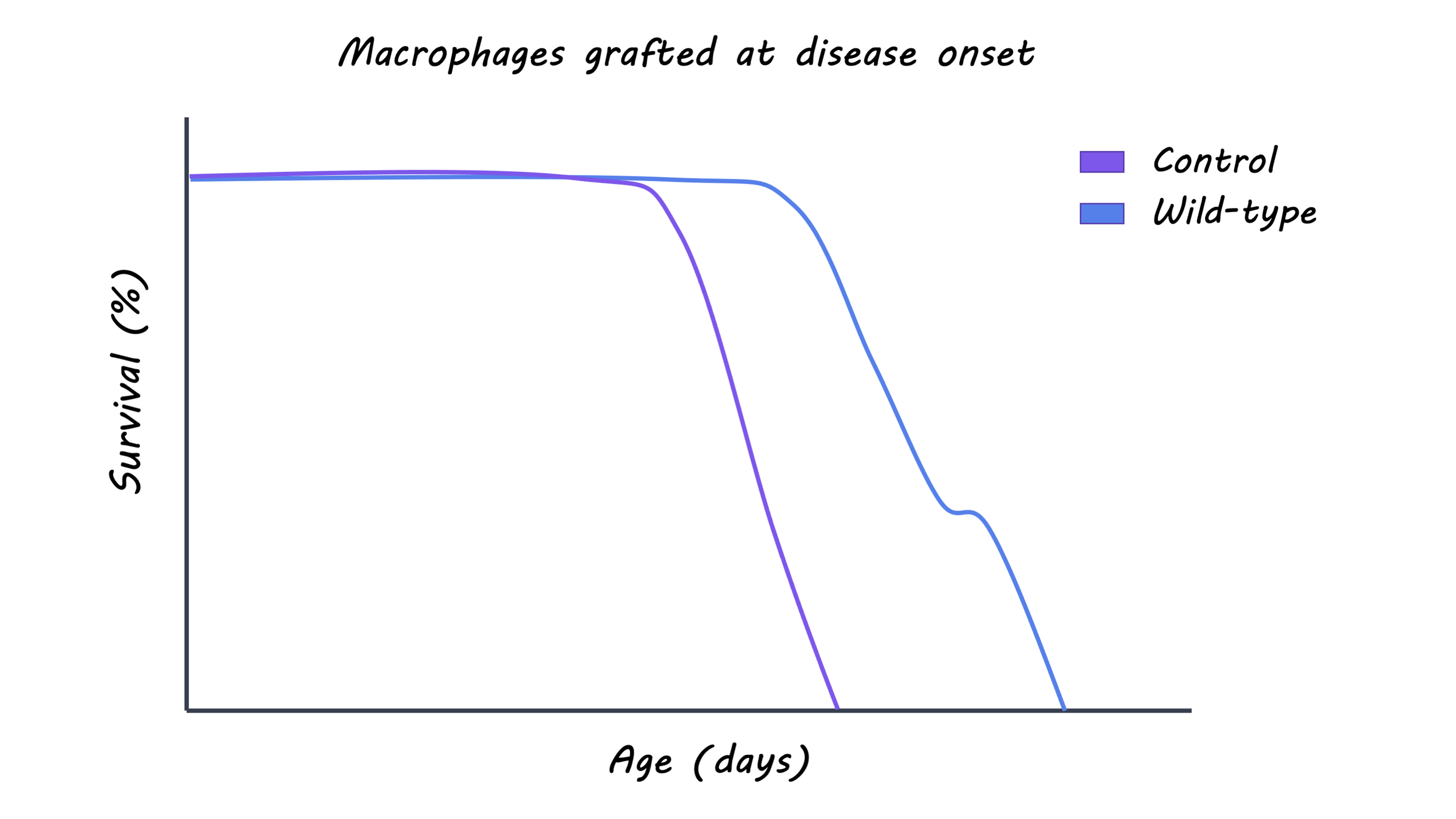
The neuroprotective effects of modifying peripheral macrophages depended on the disease stage. Specifically, replacing mutant macrophages with wild-type macrophages at the pre-symptomatic stage decreased immunoreactivity of the macrophages and microglia and delayed symptom onset, but did not increase survival. However, replacing the macrophages at disease onset additionally increased the survival of ALS mice. Macrophages and microglia also showed distinct gene expression profiles throughout the course of the disease in ALS mice. In the wild-type ALS mice that survived longer, there was a downregulation of inflammation-promoting genes in peripheral macrophages and an upregulation of synaptogenesis-promoting genes in microglia. These results highlight how modulating peripheral macrophages in mouse models of ALS can influence spinal cord microglia and impact survival by altering gene expression.

What's the impact?

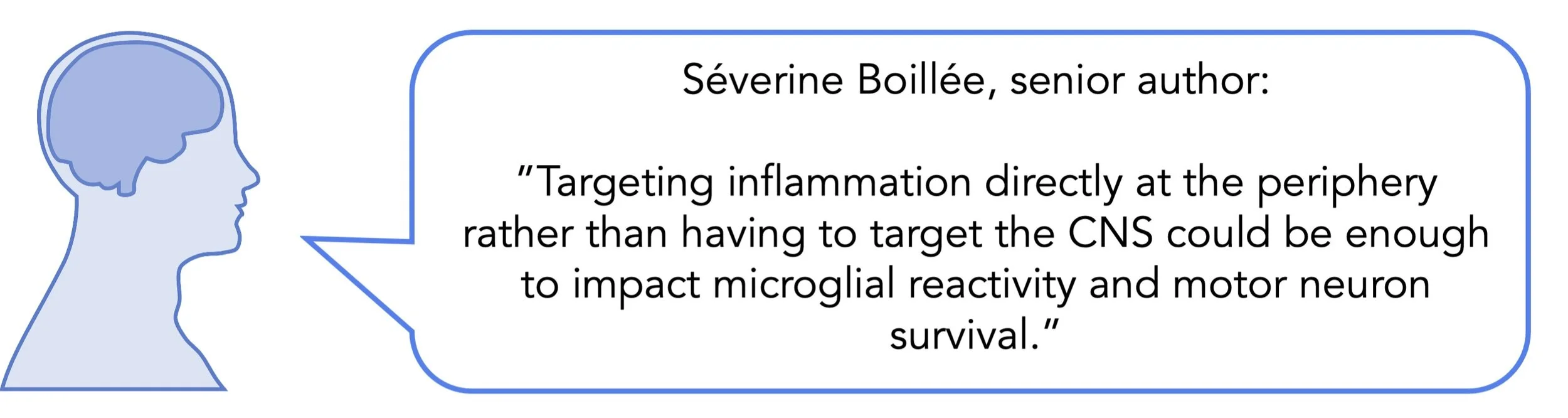
This is the first study to outline the independent contribution of peripheral macrophages and microglia to disease progression in mouse models of ALS. Additionally, this study demonstrates the beneficial effect of macrophage modification at disease onset on survival. The authors identified the role of inflammatory genes at different stages of ALS progression in central and peripheral immune cells, which has exciting implications for targeted interventions that could slow or potentially halt the course of disease progression.
microglia_quote_Oct27.jpg

Chiot et al. Modifying macrophages at the periphery has the capacity to change microglial reactivity and to extend ALS survival. Nature Neuroscience (2020). Access the original scientific publication here.
Brain Training: Do Computerized Games Improve Cognitive Ability?

Post by Flora Moujaes

What's the science?

In recent years, a billion-dollar industry has emerged.  It posits that you can enhance your cognitive ability, and even your IQ, simply by completing computerized games. But does science really support this claim? A limited number of studies have shown that training on a cognitive task can improve your performance on other tasks that recruit similar cognitive mechanisms; however many studies have failed to replicate these results. Even when training involves different tasks that engage multiple cognitive systems, research has shown that participants just get better at completing the specific tasks, rather than improving their general cognitive ability. This week in the Journal of Experimental Psychology, Stojanoski and colleagues conducted a large-scale online study, investigating whether brain-training tasks improve cognitive ability.

How did they do it?

The authors recruited 11,000 individuals online from a total of 145 countries. Over 1000 participants were active users of commercially available brain training programs including Luminosity, Peak, Elevate, Brian HQ and Neural Nation. The brain-training participants had used such programs for an average of 8.5 months. The authors assessed the participants’ general cognitive function using the Cambridge Brian Sciences online assessment battery, which measures cognitive skills such as working memory, verbal ability, reasoning, decision-making, and inhibitory control.

What did they find?

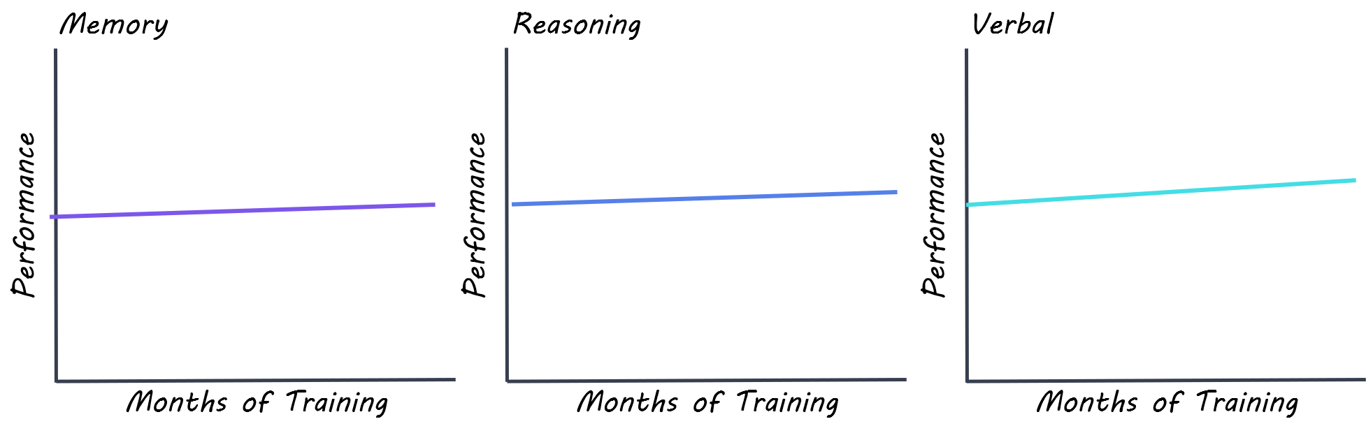
To see if brain training produces generalizable improvements in high-level cognition, the authors compared whether on average the 1009 participants with an active history of active brain training performed better than a demographically matched group who had no such history. The authors found that there was no difference in performance between active brain trainers and non-brain trainers, even when they compared non-brain trainers to those brain trainers who had been training for longest. They also examined whether the amount of time spent using brain training programs increased cognitive performance, but found that there was no relationship between self-reported length of time participants devoted to brain training and cognitive performance.

flora.png

What's the impact?

The findings of this study do not support computerized games as a way to improve cognitive performance. Given that a billion-dollar industry with over 70 million active users has been built on this premise, more research is needed into whether such brain-training programs are really worth people’s time and money. In particular, studies that employ a within-subject design or follow participants over time are needed in order to draw stronger conclusions.

A word of caution: The methodology in this study did not take into account individual improvements within-subjects (comparing each participant’s cognitive ability before and after brain training), and participants were not randomly assigned to receive brain training.

Stojanoski et al. Brain training habits are not associated with generalized benefits to cognition: An online study of over 1000 “brain trainers”. Journal of Experimental Psychology: General (2020). Access the original scientific publication here.

More Than a Watch: How Wearable Tech is Helping Advance Neuroscience

Post by Lincoln Tracy

What's the science?

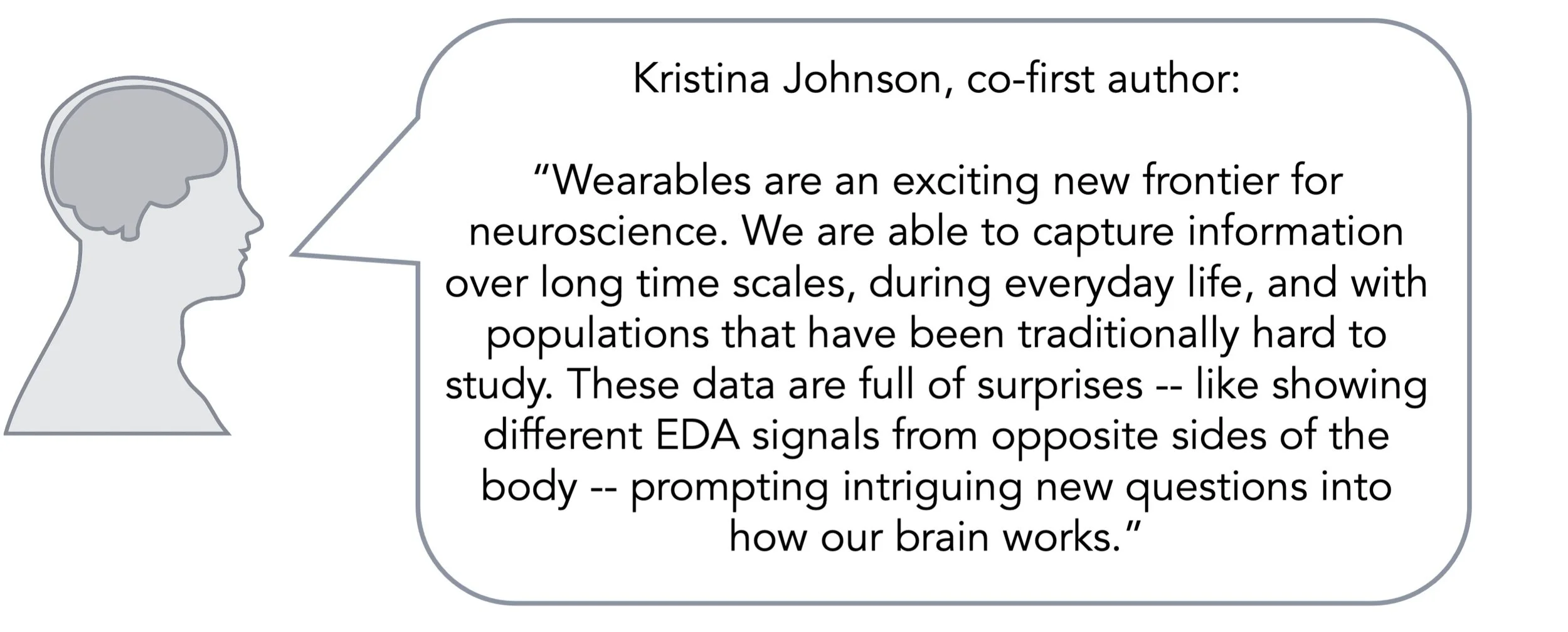
Wearable technology is a fast-moving and financially lucrative industry. The adoption of this technology is primarily being driven by smartwatches. These devices can measure a range of physiological signals including heart rate, blood oxygen levels, and sweat gland activation – all while looking sleek and stylish on your wrist. Smartwatches allow scientists to collect a wide range of long-term, real-time physiological data from large numbers of people at the same time. This week in Neuron, Johnson and Picard provide a broad overview of wearable technology and highlight its potential clinical applications.

What have we learned?

One type of smartwatch data is electrodermal activity, or EDA. EDA refers to changes in the electrical properties of our skin. When our sympathetic nervous system is activated, we begin to sweat. These increases in sympathetic activity can be measured by passing a small electric current across two electrodes on the surface of the skin and calculating the electrical conductance, giving us a measure of EDA. Measuring EDA allows scientists to examine purely sympathetic activity, as EDA does not have any known parasympathetic drivers.

However, EDA is more than a simple measure of arousal. One wider-ranging application of EDA is the detection and characterization of seizures, particularly generalized tonic-clonic seizures. The association between EDA and seizure activity has been identified from concurrent electroencephalography (EEG) and EDA recordings from patients having a seizure. The recordings show unusual electrical activity across all EEG channels during the seizure. But after the seizure stops, there is a flattening of EEG activity and a surge in EDA. Several studies in both children and adults have shown that measures of EDA correlate with the duration of the post-seizure suppression of EEG activity.

Why does it matter?

Correlations such as these allow for major advances in real-world diagnostic tests and interventions. For example, EEG suppression has been observed for all EEG-monitored cases of sudden unexpected death in epilepsy (SUDEP), the second leading cause of years of potential lives lost for neurological conditions. The exact mechanisms of SUDEP are unknown, but the risk of SUDEP is greatest with frequent seizures and being alone at the time of seizure. However, by applying machine learning to EDA and accelerometry data, life-threatening seizures can be detected in real-time with almost 100% sensitivity. Detecting the seizures before they end allows caregivers and loved ones to attend to the person seizing and provide aid, potentially preventing death.

What are the next steps?

Despite EDA being studied since the 19th century, the advent and popularity of wearable technology allow scientists to explore the role of this (and other) physiological signals in everyday life. The simplicity and practicality of smartwatches allow their use in vulnerable or underserved populations where typical neuroimaging technologies cannot be used. But it is important to note that wearable technology is not without its limitations. Many devices have their own proprietary algorithms, meaning that the underlying raw data is kept under strict lock and key. There are also issues surrounding the privacy, transparency, and ethical use of the data collected by these devices. These limitations aside, the addition of wearable technology to research kits and combining their use with existing neuroimaging techniques offer the possibility of a pipeline for the effective translation of basic science to new therapies, drug delivery, and personalized medicine.

Johnson_Oct20.jpg

Johnson and Picard. Advancing Neuroscience through Wearable Devices. Neuron (2020). Access the original scientific publication here.