Neuronal Computation Underlying Inference in the Brain

Post by Cody Walters

What’s the science?

Decisions often require that we infer relationships we have not directly experienced. While the ability to successfully perform inferential reasoning is critical for adaptation and survival, how the brain links multiple distinct events to guide behavior and accurately predict outcomes remains an open question. This month in Cell, Barron et al. used a cross-species approach to examine the neuronal computation underpinning inference in mice and humans. 

How did they do it?

The authors studied both humans and mice as they performed an inference task. The task involved an observational learning phase (pairing a tone with a visual cue), a conditioning phase (pairing a visual cue with an outcome), and an inference phase (during which the subjects were exposed to one of the tones from the observational learning phase and had to infer the outcome). Mice were shown visual cues consisting of two light configurations and received either sugar water (rewarding outcome) or water (neutral outcome). During the task, the authors recorded neural activity from the mice in the dorsal hippocampus using tetrodes. 

In humans, the task was performed in a virtual reality setting while participants were scanned with fMRI. The visual cues consisted of image patterns on a wall, and they received either coins with monetary value (rewarding outcome) or wood-chips with no monetary value (neutral outcome). The task, therefore, had two cue sets: the set of auditory and visual cues associated with a rewarding outcome and the set of auditory and visual cues associated with a non-rewarding outcome. Inference trials in which the subjects correctly inferred from the tone that they would be rewarded were called ‘correctly inferred trials.’

What did they find?

The authors found increased BOLD activity in the human hippocampus during correctly inferred trials. Using optogenetics in mice, the authors found that silencing activity in the mouse dorsal hippocampus during the inference phase (but not the conditioning phase) reduced reward-seeking behavior. These data suggest that the hippocampus plays an important role in inference. The authors then showed that the mouse hippocampus represents each of the task cues (i.e., the tones, visual stimuli, and outcomes) in a largely independent, non-overlapping manner. Next, the authors used representational similarity analysis (a method for determining how similar two neural representations are to one another) in both humans and mice to show that, on correctly inferred trials, the pattern of hippocampal activity during the tone predicted the pattern of hippocampal activity seen during that tone’s associated visual cue (from the observational learning phase). This implies that the tone elicited a prospective representation of that tone’s associated visual cue only when subjects correctly inferred the outcome of the trial.

cody+%281%29.jpg

Interestingly, the authors found that neither humans nor mice exhibited hippocampal representations of the outcome during tone presentation. In order to determine where in the brain the outcome was being represented, the authors looked for brain regions in human subjects that significantly encoded the outcome during the presentation of the auditory cue in the inference phase of the task. They found that the medial prefrontal cortex and the midbrain (a site containing reward-sensitive dopaminergic neurons) represented the inferred outcome. Finally, the authors showed that tone- and outcome-encoding cells in the hippocampus of mice were coactive during sharp wave-ripples. The authors further demonstrated that the ordering of these bursts of neuronal activity was temporally reversed (i.e., outcome-encoding cells fired before tone-encoding cells), a phenomenon known to occur during the replay of spatial trajectories nested within sharp wave-ripples. These results highlight sharp wave-ripples as a possible mechanism for bridging the gap between cues in inferring relationships.

What’s the impact?

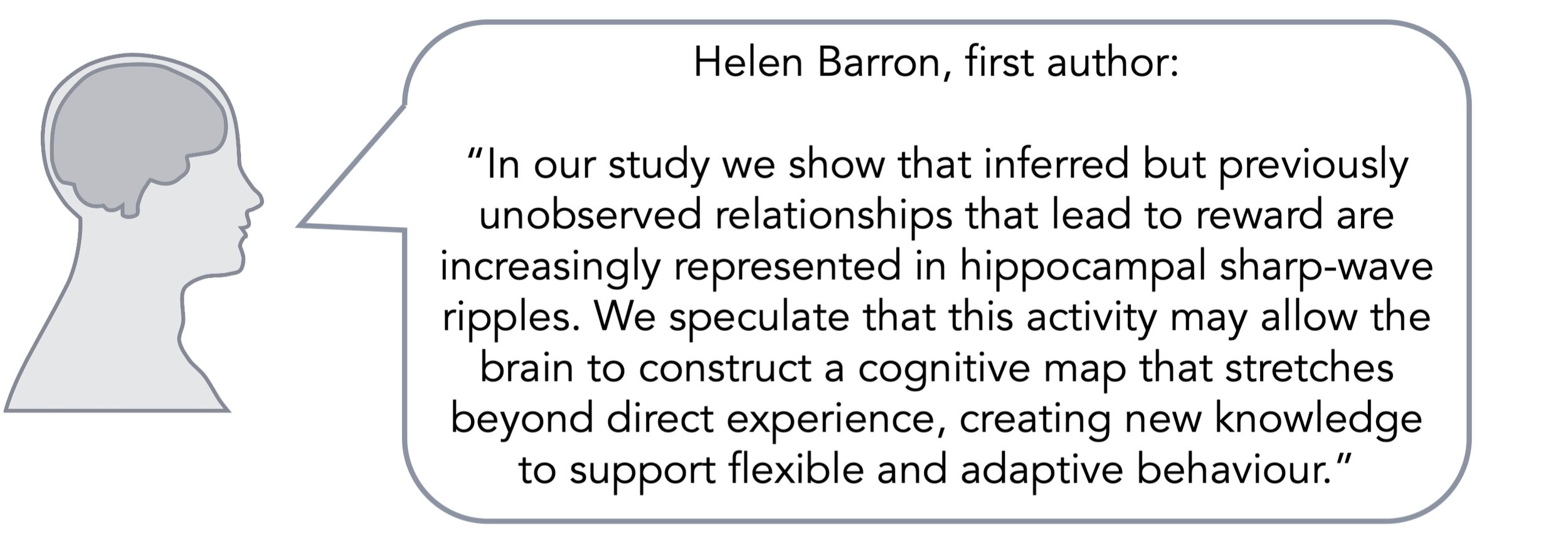
This study showed that 1) the hippocampus computes a prospective code that binds associated memories, 2) the midbrain and medial prefrontal cortex, but not the hippocampus, represent imagined outcomes during inference, and 3) that inferred relationships are represented during hippocampal sharp wave-ripples. These data outline the functional anatomy and neuronal computation that support inference in the mammalian brain.
barron-quote-oct6.jpg

Barron et al. Neuronal Computation Underlying Inferential Reasoning in Humans and Mice. Cell. (2020). Access the original scientific publication here.
Psilocybin-Assisted Psychotherapy in Complex Clinical Populations

Post by Flora Moujaes

What's the science?

Dubbed the ‘psychedelic renaissance’, a growing number of studies have emerged showing that psychedelic-assisted psychotherapy is beneficial for everything from treatment-resistant depression and PTSD to demoralization in terminally ill patients. Modern trials of psychedelic-assisted psychotherapy use carefully screened clinical populations, often excluding patients who have comorbid diagnoses where one or more disorders occur at the same time, or who have a history of psychosis. But what happens when psychedelic-assisted psychotherapy is utilized in more complex clinical populations? This week in EClinicalMedicine, Anderson and colleagues explored whether psilocybin-assisted psychotherapy is effective in treating distress in long-term AIDS survivors.

How did they do it?

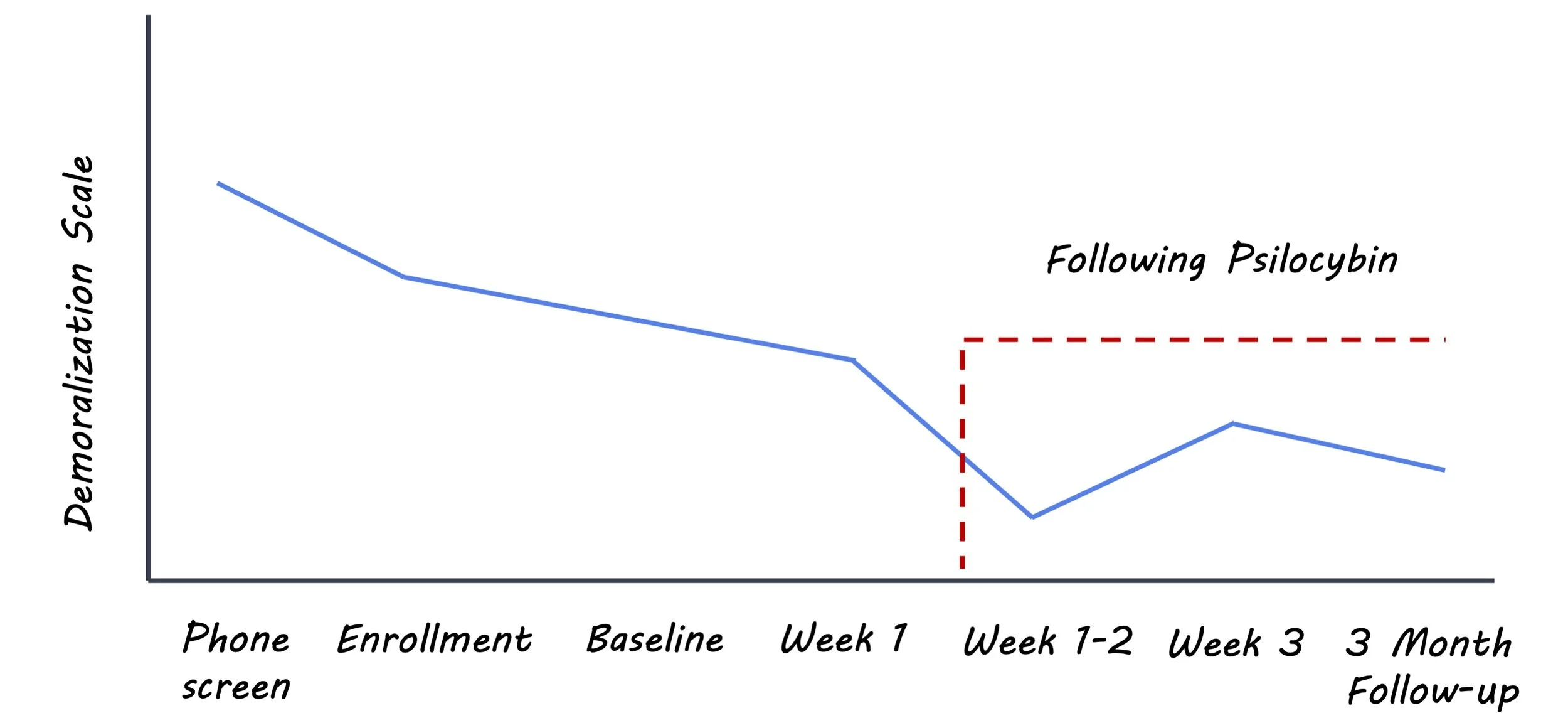
The researchers conducted an open-label pilot study where over the course of seven weeks participants underwent three hours of individual psychotherapy, 12-15 hours of group therapy, and one eight hour individual psilocybin administration. Eighteen older long-term male AIDS survivors took part in the study and were divided into three groups of six for the group sessions. This population was selected as it is known to have a higher degree of demoralization and loss. Participants received a moderate/high dose of psilocybin: 0.3-0.36 mg/kg. The main clinical outcome measure was change in demoralization from baseline at the end of the study and three months after the study had been completed.

The patients used in this study had a much higher rate of co-occurring psychiatric disorders than has been seen to date in the modern literature on psilocybin therapy. The patients had a range of psychiatric diagnoses including clinically active Borderline Personality Disorder (BPD), General Anxiety Disorder, Major Depressive Disorder, and Panic Disorder. They had also experienced a high degree of adverse life experiences.

What did they find?

Effect on Demoralization: Researchers found that there was a significant improvement in self-reported demoralization at both the end of the study, and three months after the study had been completed. In response to the feedback from the first group of six patients who partook in the study, the amount of post-drug group therapy visits was increased from four to six, suggesting that more complex clinical populations may need more support following the psilocybin session.

Fourteen out of 18 patients experienced expected moderate-to-severe adverse reactions to psilocybin including paranoia, transient thought disorder (e.g. dissociation with repeated self-talk), anxiety, nausea, and visual and tactile hallucinations. Twelve participants experienced moderate-severe high blood pressure, which seemed to co-occur with anxiety and in most cases self-resolved when clinicians calmed patients verbally. However, one participant who met the criteria for BPD was intermittently severely hypertensive for four hours, and his paranoia precluded the clinicians from offering oral medications to help lower his blood pressure. Finally, two participants experienced unexpected reactions to psilocybin: one participant experienced a post-traumatic stress flashback related to a previous sexual assault two days after receiving psilocybin, while another reported severe anxiety 10 days after receiving psilocybin and then had a lapse in his methamphetamine use and withdrew from the study.

Psilocybin_oct6.jpg

What's the impact?

Overall this study characterized the potential efficacy of psilocybin-assisted psychotherapy in treating distress in long-term AIDS survivors. However, given that moderate-severe side effects occurred in 14 out of 18 patients, this study highlights the importance of thorough screening, experienced clinicians, and providing ample therapeutic support after the substance session. This study also supports prior reports suggesting that patients with BPD traits may have more difficulty tolerating psilocybin. In summary, this study provides invaluable insights into the nuances of psychedelic-assisted psychotherapy in clinical populations. 

anderson-quote-oct6.jpg

Anderson et al. Psilocybin-assisted group therapy for demoralized older long-term AIDS survivor men: An open-label safety and feasibility pilot study. EClinicalMedicine (2020). Access the original scientific publication here.
REM Sleep is Necessary for Experience-Dependent Plasticity

Post by Elisa Guma

What's the science?

Sleep is an important physiological process. Many lines of evidence suggest that sleep is important for regulating neuronal plasticity during brain development and after learning. A prominent feature of experience-dependent synaptic plasticity is the pruning of existing synapses during development and adulthood, which is thought to contribute to changes in neuronal activity. This week in Nature Communications, Zhou and colleagues investigated the role of sleep in dendritic spine elimination in the developing visual cortex and frontal association cortex, respectively, to try to unravel the role of sleep in experience-dependent synapse elimination.

How did they do it?

To evaluate the effect of sleep and sleep deprivation on experience-dependent spine elimination the authors used transgenic mice expressing a yellow fluorescent protein in layer 5 pyramidal neurons of the cortex. Four week old mice were either monocularly deprived (MD) of light, wherein a shade cloth was used to cover one eye, or they were subjected to auditory fear conditioning (FC), wherein an unpleasant foot shock was paired with an auditory cue. Dendritic spines were imaged via a thinned cranial window in the visual cortex of MD awake mice and frontal association of awake FC mice, using transcranial two-photon microscopy. Sleep deprivation was induced by gentle handling, either over a four-hour period, or during specific sleep stages: rapid eye movement (REM) sleep and non-REM (NREM) sleep. Electroencephalography and electromyography recordings were taken to confirm that the gentle handling was inducing sleep deprivation, and to determine sleep stage. Imaging was performed over 4 hours after one session of sleep deprivation, and then again after a second session of sleep deprivation.

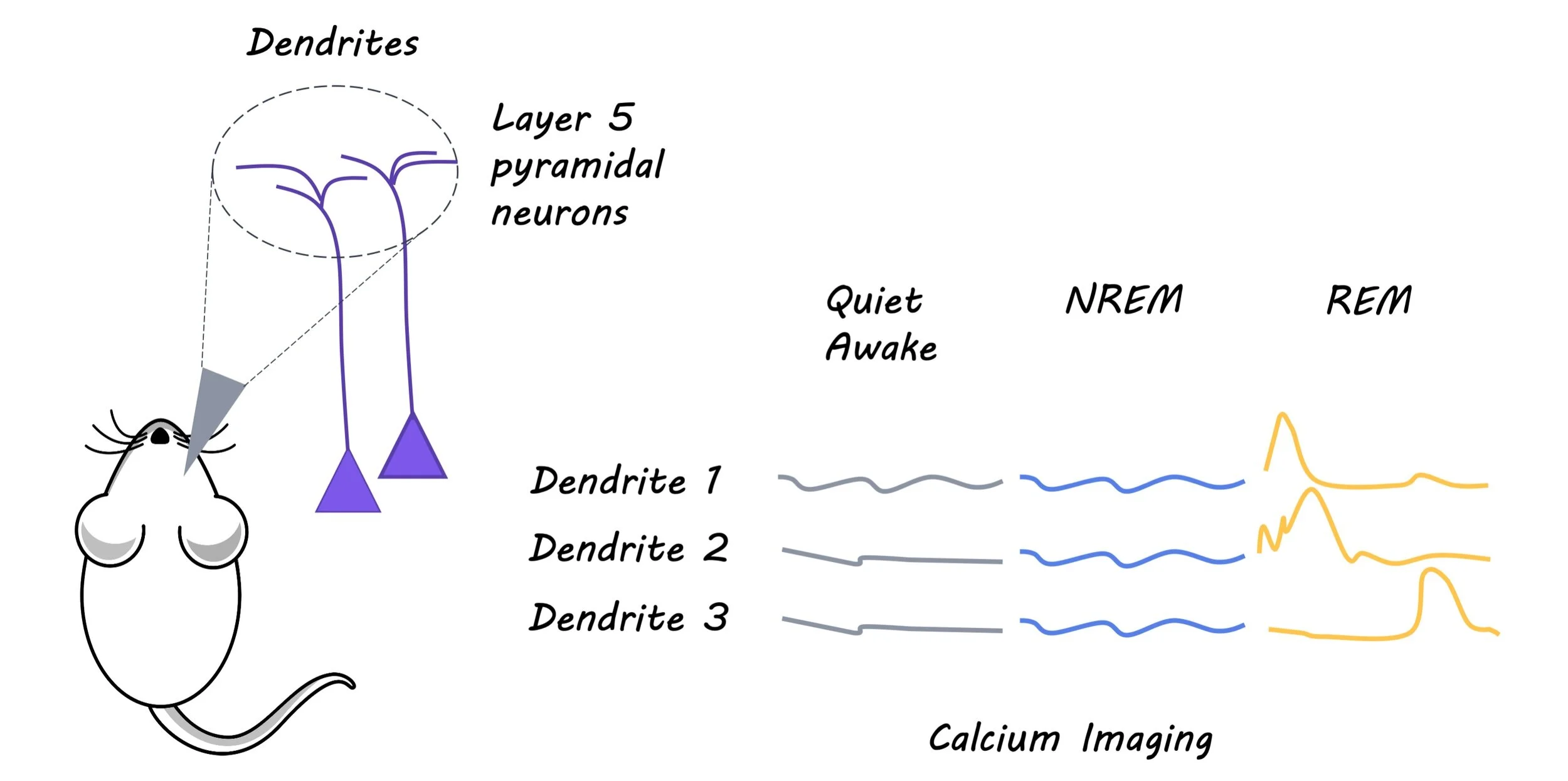
Next, the authors wanted to determine whether the change in rate of spine elimination was reflected in changes to neuronal activity. They measured the amount of intracellular calcium, indicative or neuronal activity, in the layer 5 pyramidal neurons of either the visual cortex for MD mice or the frontal association cortex for FC mice. Finally, to examine whether dendritic calcium spikes during REM sleep are important for dendritic spine elimination induced by MD or FC, they inhibited neuronal activity with an NMDA receptor antagonist at the beginning of REM sleep.

What did they find?

The authors observed an increase in dendritic spine elimination during a 24 hour imaging period in MD mice relative to mice that were not sleep deprived in the primary visual cortex, in line with previous findings. This is indicative of circuit remodeling during critical periods of development. Conversely, following FC, the authors observed an increased rate of spine elimination and formation in the frontal association cortex of FC mice, suggesting that FC induces rapid changes in dendritic spine remodeling. In the first 4 hours of recording following sleep deprivation, the authors observed no significant effect on spine elimination or formation for the MD group overall, and for REM and NREM sleep investigated separately.  However, they did find that following a second round of sleep deprivation, the rate of spine elimination and the rate of neuronal activity (as measured by calcium release) was decreased, with no changes to the rate of spine formation.

Interestingly, sleep deprivation, particularly in the REM phase, decreased the rate of freezing in response to the conditioned auditory cue in FC mice, as well as the rate of spine elimination and neural activity of layer 5 pyramidal neurons in the frontal association cortex. In both groups, the authors found that dendritic calcium spikes were higher during REM sleep. The blockade of these spikes via an NMDA receptor antagonist during REM sleep reduced dendritic spine elimination, indicative of the critical role that calcium spikes during REM sleep play in facilitating experience dependent dendritic spine elimination.

memory_image_sep29.jpg

What's the impact?

This study found that REM sleep is critical for experience-dependent synapse elimination and modulation of neuronal activity. The authors present compelling evidence for two different types of experience induced change, either via monocular deprivation or auditory fear conditioning, in two different brain regions. This further strengthens our understanding of the role that sleep plays in supporting plasticity in the brain during development and in response to learning. Future work may investigate the mechanisms underlying calcium spike-dependent spine elimination.

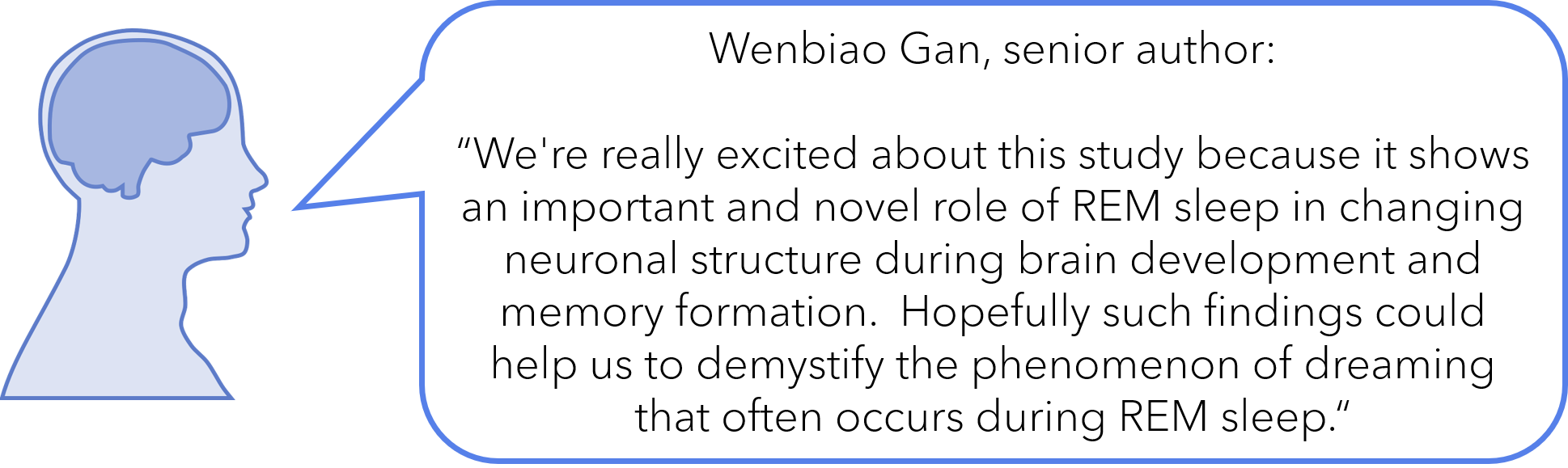
wenbiao.png

Zhou et al. REM sleep promotes experience-dependent dendritic spine elimination in the mouse cortex. Nature Communications (2020). Access the original scientific publication here.