Prediction of the Future Can Block Memory Formation

Post by Deborah Joye

What's the science?

Memory is a multifaceted system that helps us recall past events (episodic memory). This system can also use those memories to make predictions about the future (i.e. statistical learning). Remembering every unique detail about a past event may not be necessary, since those precise details are not likely to occur again in the exact same combination. Instead, it may be more efficient for our memory to generalize across common features within past events that can help us make predictions about the future. But these two functions of memory require different processes – episodic memory requires recall of specific memories linked to a particular time and space, while prediction requires synthesis of generalized features across many memories. Although these are very different processes, both have been linked to the hippocampus. How does the hippocampus mediate between these two potentially conflicting processes? This week in PNAS, Sherman and Turk-Browne demonstrate that when our memory uses statistical learning to predict what comes next, it prevents the current experience from being encoded into memory.

How did they do it?

For each experiment, the authors trained participants on a stream of images of outdoor scenes depicting distinct categories, such as beaches, mountains, fields, etc., then later tested their memory. All images were of 3 different overall types – category A images which were always followed by a category B image, and category X images which were randomly inserted and not preceded or followed by any particular type of image. Participants were not informed that images from category A would always be followed by category B, instead they learned about this association as they completed the task. This experimental design allowed the authors to assess how well participants remembered images they saw depending on whether the images were predictive (category A vs. category X) or predictable (category B vs. category X).

In the first experiment, the authors exposed participants to the image stream, then later had them view images and asked them to indicate whether they had seen that image before. To better evaluate the role of the hippocampus in this process, the authors next exposed participants to the image stream, then later asked them to identify whether they had seen that image before, and if so, what time they remembered seeing it. This additional timestamp question is a more detailed form of episodic memory recall that critically depends on the hippocampus. Finally, to see how brain activity changes during the memory task, the authors repeated their image stream test experiment while participants also underwent a functional magnetic resonance imaging (fMRI) scan.

What did they find?

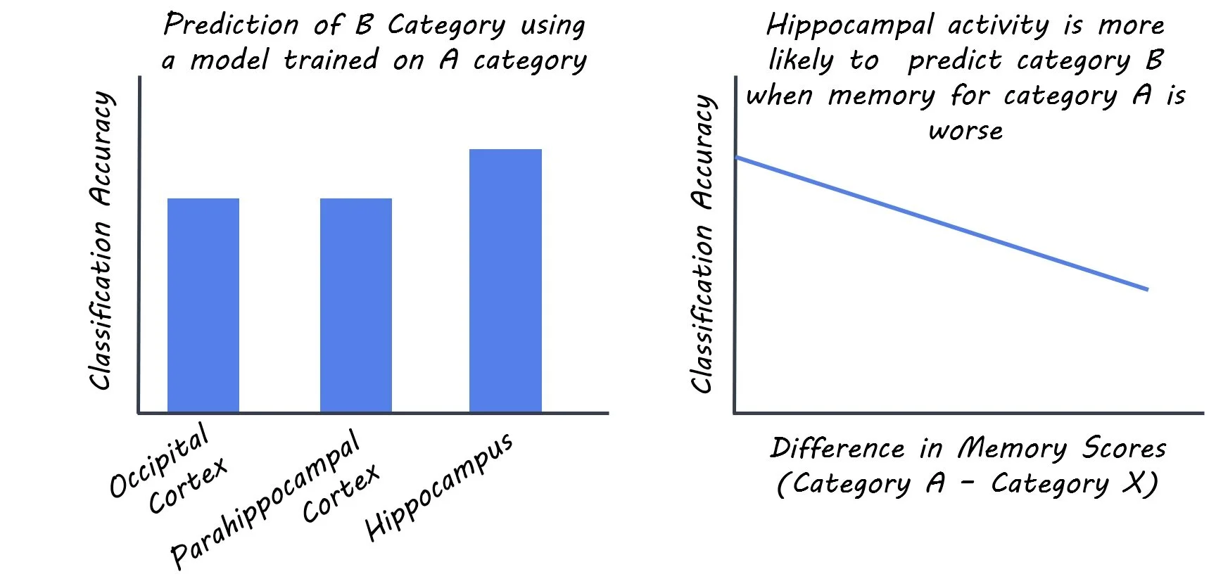
The authors first found that participants were worse at remembering images that predicted another image (category A) than they were at remembering control images (category X). Interestingly, participants remembered seeing predictable images (category B) about the same as control images, suggesting that the memory deficit was specific to images that allowed prediction of what came next. When the authors added the more difficult timestamp question, they found a similar result: participants were much worse at remembering when they had seen predictive images (category A) relative to control images. These findings suggest that when participants saw an image from category A, it may have triggered the hippocampus to retrieve a representation of the category B image that was to immediately follow and not the category A image itself. This prediction of the upcoming image may block the hippocampus from creating a new memory of the specific details of the current image the participant was seeing. Finally, when the authors had participants perform the original image stream task (without the timestamp question) while undergoing fMRI they found that hippocampal activity when viewing category A images looked as if it was viewing category B images (the predicted image). They also found that the better the hippocampus was at predicting category B images, the worse the participant was at recalling category A images (relative to category X images).
deb.jpg

What's the impact?

These findings demonstrate that when the hippocampus makes predictions from statistical learning, memory formation of the present is impaired. If statistical learning and memory encoding oppose one another, why does the hippocampus do both? One proposed idea is that hippocampal control of these processes allows them to regulate another. In an excellent analogy from the authors, “using one’s right foot to operate both the brake and gas pedals in a car serves as an anatomical constraint that forces one to either accelerate or decelerate, but not both at the same time.” Similarly, the hippocampus might likewise mediate between using already-formed predictive models to predict what’s next (“we already know what’s gonna happen, let’s GO”), and formation of new memories (“let’s pay attention closer here so we can remember this new situation”).
sherman_quote_sep29.jpg

Sherman & Turk-Browne, Statistical prediction of the future impairs episodic encoding of the present, PNAS (2020). Access the original scientific publication here.
The Occipital Cortex and Visual Memory Development

Post by Amanda McFarlan

What's the science?

The study of learning and memory commonly focuses on brain regions such as the hippocampus and the prefrontal cortex, however, recent studies have provided evidence to suggest that the occipital cortex may be involved in visual memory development. Indeed, age-related differences in cortical thickness indicate that the occipital cortex matures well into adolescence. Researchers postulate that synaptic pruning may occur throughout this maturation process as a way of enhancing visual processing. This week in Neuropsychologia, Yin and colleagues investigated whether the maturation of the occipital cortex played a role in enhancing memory formation of visual stimuli.

How did they do it?

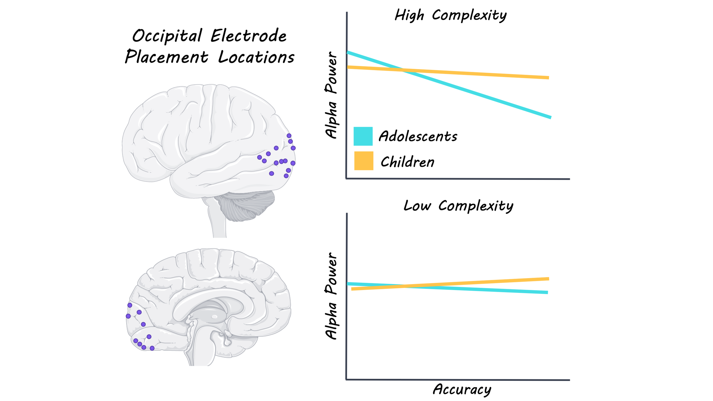
The authors used electrocorticographic recordings (electrodes placed directly on the surface of the brain) to measure brain activity in the occipital cortex in 24 children (6-13 years) and adolescents (13- 20 years) while performing a memory task. The memory task consisted of two parts: the “study” part and the “test” part. During the study part of the memory task, participants were presented with 40 scenic images of both high and low complexity (defined by the number of object categories in the image) and were asked to decide whether the image was of an indoor scene or an outdoor scene. Then, during the test part of the task, the participants were presented with the same 40 scenic images and 20 new scenic images. The participants were asked to classify the scenic images as “old” (ones they had previously studied) or “new” (ones they had not studied). The participants’ brain activity in the occipital cortex during the two parts of the memory task was recorded. To further investigate how visual processing in the occipital cortex might contribute to memory formation, the authors analyzed brain recordings during the study part, focusing on oscillations in the alpha range (7-14 Hz) since these are known to be important in visual processing. Specifically, they measured alpha power, which has been shown to decrease with increased neural activity and memory formation, and instantaneous alpha frequency which have been shown to increase with a higher temporal resolution of perception. 

What did they find?

The authors found that the recognition accuracy for high-complexity scenes was greater for adolescents compared to children, which suggests that memory formation for complex visual scenes improves during development. Additionally, decreased alpha power was found to be a predictor of recognition accuracy for high-complexity scenes in adolescents. Next, the authors determined that only high-complexity scenes were associated with both an age-related decrease in alpha power and an increase in instantaneous alpha frequency, suggesting that visual processing in the occipital cortex that is involved in memory formation improves in an age-dependent manner for complex scenic stimuli. Finally, the authors determined that there were no age-related differences in alpha power or frequency for scenes that were later incorrectly identified “new”, which suggests that age-related differences in alpha power and frequency may be predictive of memory formation, but not visual perception as a whole.

occipital_sept29.png

What’s the impact?

Using electrocorticographic recordings in children and adolescents, this study showed that developmental changes in the occipital cortex are associated with enhanced memory formation for complex scenic stimuli. The authors found that adolescents were better at recognizing high, but not low-complexity scenes compared to children. Additionally, they found that decreased alpha power was predictive of memory formation for high-complexity scenes in an age-dependent manner. In all, these findings highlight the importance of occipital cortex maturation in the enhancement of visual memory formation.

Ofen_quote_sep29.jpg

Yin et al. Direct brain recordings reveal occipital cortex involvement in memory development. Neuropsychologia (2020). Access the original scientific publication here.
Deactivation of Anterior Thalamic Nuclei Underlies Associative Memory Encoding

Post by Shireen Parimoo

What's the science?

Associative memory refers to memory for multiple pieces of information (e.g., a face and a name) and is a critical component of episodic memory. The involvement of regions in the medial temporal lobe (MTL) cortex – such as the hippocampus – in episodic memory has been well-established. The thalamus is a subcortical brain region made up of several nuclei that has structural connections to the MTL and is also implicated in the formation and retrieval of memories. However, the precise role of thalamic nuclei, particularly the anterior and mediodorsal nuclei, in associative memory is not clear. This week in Neuropsychologia, Geier and colleagues used functional magnetic resonance imaging (fMRI) to investigate the contribution of thalamic nuclei and their connectivity to the hippocampus and MTL regions in associative memory encoding and retrieval.

How did they do it?

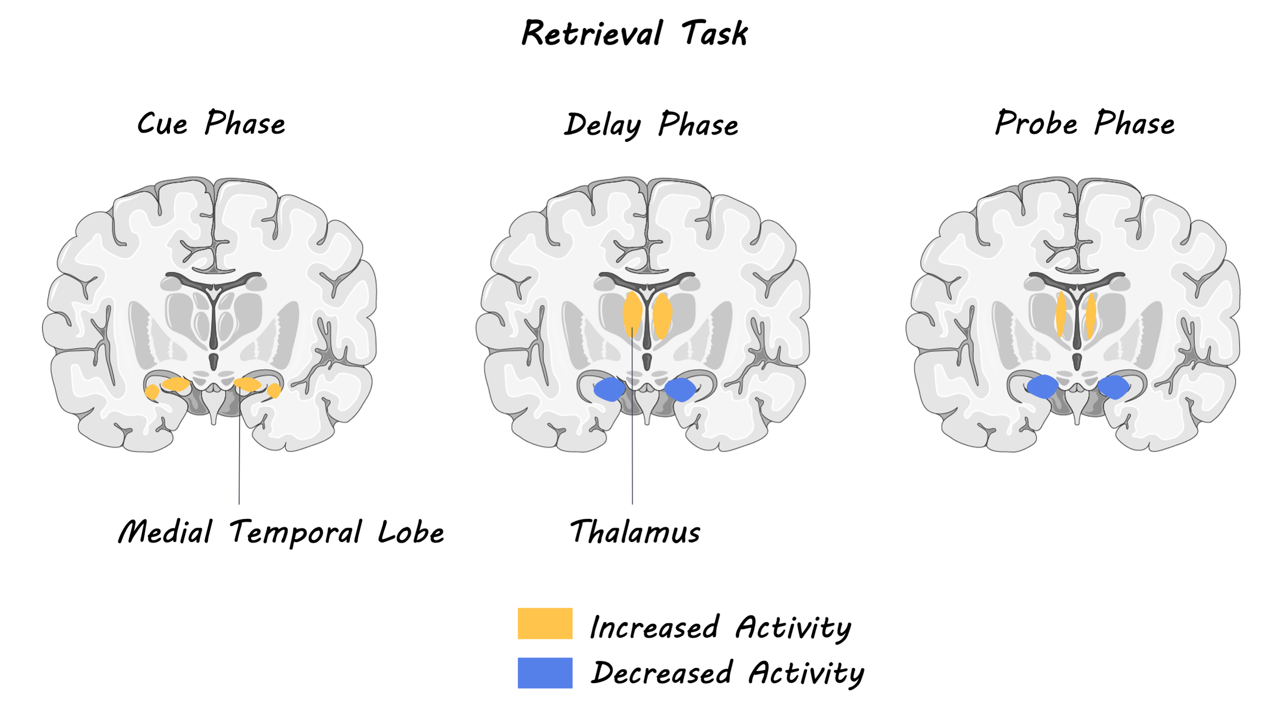
Twenty-seven young adults completed an associative memory task while undergoing fMRI scanning. In the encoding phase, they were shown a scene overlaid with a face, and participants made plausibility judgments about the face-scene pairs. The retrieval task consisted of three phases: (i) a cue phase in which a scene image was presented, followed by (ii) a delay phase, during which participants were instructed to mentally recall the face that was previously paired with the scene, and (iii) a probe phase in which three faces were presented with the scene and participants performed a memory judgment for the previously paired face.

The authors examined activity in the thalamus and MTL during memory encoding and the three memory retrieval phases. They also investigated how the time course of activation in each task phase was related to memory performance. That is, how did activity in the anterior and mediodorsal thalamic nuclei, as well as the anterior and posterior hippocampus, differ for correctly and incorrectly remembered faces? Finally, they assessed functional connectivity between thalamic nuclei and the MTL in associative memory and additionally compared the connectivity profiles of the anterior and mediodorsal thalamic nuclei to the MTL.

What did they find?

Thalamic activity significantly decreased during the encoding phase, whereas MTL regions like the hippocampus exhibited a nominal increase in activation. In particular, activation of the anterior hippocampus during memory encoding was associated with better face-scene memory performance. Conversely, reduced activation of the anterior thalamus at encoding was related to memory accuracy, while the mediodorsal thalamus showed little change across correct and incorrect trials. Interestingly, the opposite pattern of activation was observed at retrieval, with greater activation of the thalamus during the delay and probe phases but reduced delay-phase activation in the MTL. However, retrieval-phase activity in the hippocampus or thalamus was not significantly related to memory performance. These results indicate that the successful formation of associative memories is related to the increased involvement of the anterior hippocampus but deactivation of the anterior thalamus. Finally, the mediodorsal thalamus showed greater connectivity with the MTL compared to the anterior thalamus. However, differences in functional connectivity between the thalamic nuclei and the MTL were not related to memory performance.

encoding_img_sept22.png

What's the impact?

This study characterized the involvement of thalamic nuclei in memory and used neuroimaging to demonstrate the complementary role of the anterior thalamic nuclei in associative memory formation. Although patient and neuroimaging studies have shown that the thalamus is important for memory, we now know how specific nuclei in the thalamus are involved in the formation of associative memories, specifically. These results pave the way for future work that can further reveal how thalamic nuclei communicate with other regions of the brain to support memory encoding and retrieval.

memory_quote_sep22.jpg

Geier et al. The role of anterodorsal thalamus in associative memory encoding and retrieval. Neuropsychologia (2020). Access the original scientific publication here.