Inflammation Triggers Somatic Bassoon Protein Accumulation in Multiple Sclerosis

Post by Shireen Parimoo

What's the science?

Multiple sclerosis (MS) is a neuroimmunological disease characterized by myelin (insulation for neuronal axons) damage due to inflammation in the central nervous system. This damage impairs neuron function and can cause neuronal death, resulting in symptoms like fatigue, loss of coordination, and cognitive impairment. The molecular mechanism by which inflammation results in neuronal loss is not clear because isolating damaged neurons from neuronal tissue has been a methodological challenge, making it difficult to determine the involvement of various proteins in this process. This week in Nature Neuroscience, Schattling and colleagues used neuron-specific profiling of messenger RNA to investigate the gene expression profiles of different cell types in inflamed tissue from MS patients and model organisms with MS.

How did they do it?

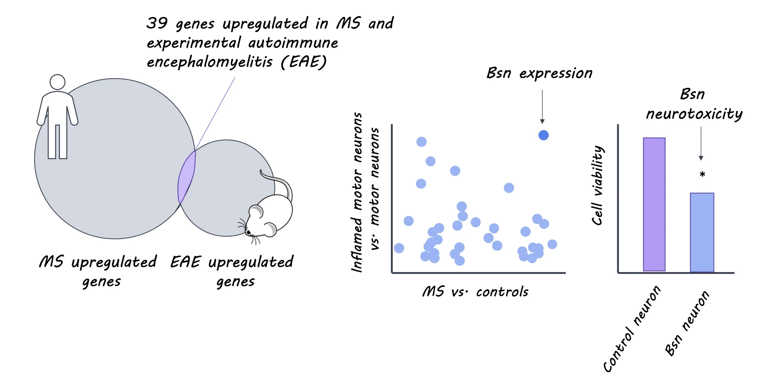
The authors first examined the genes expressed by neurons in inflamed spinal cord tissue. Mouse models of MS were created by inducing experimental autoimmune encephalomyelitis (EAE), which triggers inflammation and the demyelination of neurons, and results in partial or full paralysis. Translating ribosome affinity purification was used to isolate messenger RNA of different cell types in normal and inflamed cervical spinal tissue and gene set enrichment analysis was performed to determine whether certain genes were over- or under-expressed in those cell types. To identify which genes were up- and down-regulated in humans, they used a microarray dataset of MS patients to compare genes expressed in chronic MS plaques and in normal tissue. They then compared the overlap in genes expressed in both humans and mice using homology mapping, which is a method used to identify the genes shared by two different species. The Bassoon (Bsn) protein – a pre-synaptic scaffolding protein found in synaptic active zones – was highly expressed in inflamed motor neurons. The authors used immunohistochemistry to examine Bsn expression and localization in the mouse and human spinal cord. They further explored the cellular and behavioral effects of high and low Bsn expression and Bsn deletion (knockout) in mice and transgenic fruit flies. Cellular measures included energy metabolism, Bsn accumulation, and cellular survival and number. Behavioral measures included EAE symptomatic recovery in mice and climbing ability in flies. Finally, they investigated whether removing Bsn from neurons would rescue the adverse effects of inflammation and enhance neuronal survival by administering the ubiquitin carboxyl-terminal hydrolase 14 inhibitor (IU1), which allows proteins like Bsn to be degraded.

What did they find?

The authors identified 354 up-regulated and 448 down-regulated candidate genes in motor neurons found in inflamed mouse spinal cord tissue. The majority of the up-regulated genes were those involved in protein breakdown pathways, while the down-regulated genes were primarily those involved in energy metabolism, suggesting that the cellular energy metabolism process is impaired during inflammation. In humans, 11% of the up-regulated genes identified in chronic MS plaques overlapped with those observed in mice, including Bsn. There was a greater concentration of Bsn in spinal cord tissue obtained from EAE mice and MS patients than in wild-type mice and control participants. Interestingly, in these groups Bsn accumulated in the cell bodies of the inflamed motor neurons, even though it is typically located in the active zone of presynaptic neurons.

Increased Bsn expression in mouse cells was associated with Bsn accumulation in cell bodies, reduced energy metabolism, and reduced cellular survival. Similar to humans and mice, Bsn accumulation was observed in the neuronal cell bodies of transgenic flies, along with reduced climbing ability and increased mortality. On the other hand, mice with Bsn deletion (knockout mice) showed faster and better recovery from EAE symptoms compared to wild-type EAE mice. The knockout mice also had more axons and neurons in the spinal cord and fewer injured axons, but the same number of immune cells as wild-type mice. This suggests that the immune response to inflammation is not affected by knocking out the Bsn protein, and that Bsn likely plays a crucial role in determining neuronal fate. Consistent with this, administering IU1 facilitated Bsn degradation in neuronal cell bodies of mice and even restored pre-synaptic Bsn localization. Furthermore, mice that were administered IU1 had a larger number of axons and neurons, as well as a faster and better recovery trajectory than mice that were given a vehicle solution. These findings indicate that Bsn accumulation in the cell body might exacerbate the effects of inflammation by interfering with cellular energy metabolism and other intracellular processes.

Bsn_image_April30.jpg

What's the impact?

This study is the first to demonstrate that inflammation triggers Bassoon accumulation in neuronal cell bodies of both MS patients and model organisms. Importantly, both the cellular and symptomatic effects of inflammation can be partially reversed by promoting Bsn degradation. These findings have important implications for our understanding of the mechanisms underlying MS as well as potential targeted treatments for mitigating the symptoms of the disease.

Friese_quote_April30.jpg

Schattling et al. Bassoon proteinopathy drives neurodegeneration in multiple sclerosis. Nature Neuroscience (2019). Access the original scientific publication here.

An Oscillator Model Predicts Activity in Auditory Cortex in Response to Music

Post by Amanda McFarlan

What's the science?

Research has shown that when presented with an auditory stimulus, neural activity in the auditory cortex tracks rhythmic patterns in the stimulus. There are two distinct hypotheses that explain this phenomenon: the oscillatory hypothesis and the evoked hypothesis. The oscillatory hypothesis suggests that the auditory cortex has an intrinsic neural oscillator that will synchronize to an acoustic stimulus, as long as the frequency of the stimulus is within a range close to the oscillator’s resting frequency. Conversely, the evoked hypothesis suggests that the auditory cortex responds to each individual acoustic stimulus and shows evidence of rhythmic firing because the inputs it receives (i.e. music, speech, etc.) are rhythmic themselves. This week in the PNAS, Doelling and colleagues used computational models to study these neural behaviors and to uncover whether human auditory processing follows the oscillatory hypothesis or evoked hypothesis.

How did they do it?

The authors created two distinct computational models, an evoked model and an oscillatory model, based on the evoked and oscillatory hypotheses that describe the mechanisms of auditory neural processing. The evoked model was convolution based, while the oscillatory model was based on the Wilson-Cowan model of excitatory and inhibitory neural populations. Musical stimuli of varying frequencies (0.5 to 8 notes per second) from piano pieces were used as inputs for both models. To compare the outputs from both models at the different frequencies, the authors developed a phase concentration metric that analyzed the phase lag between the stimulus input and the model output in both models across stimulus rates/frequencies. Next, the authors used their phase concentration metric to analyze data from a previous study in which 27 participants listened to musical stimuli (the same stimuli used in the computational models) while undergoing magnetoencephalography (MEG) recordings. They used confidence intervals and Gaussian fitting to compare the participants’ data with their computational models. In a subsequent experiment, the authors aimed to reduce the effect of evoked responses by altering the musical stimuli such that the musical notes were either smoothed in their onset (resulting in a reduced evoked response) or characterized by a sharp attack (evoked response present). They had 12 new participants undergo MEG recordings while listening to these altered musical stimuli, and compared data from the participants’ recordings with their computational models.

What did they find?

The authors found that in their evoked computational model, the phase lag between the musical note stimulus and the model output increased as the frequency of the musical note increased, suggesting the phase lag is frequency dependent. The oscillatory computational model, however, was better able to keep up with the change in musical note frequencies, and displayed a relatively consistent phase lag between the stimulus and model output. Next, they used their phase concentration metric to analyze MEG data that was collected while participants listened to musical stimuli (the same stimuli used for the computational models). They determined that the mean phase concentration metric from the analyzed MEG data was better matched to that of the oscillatory model compared to the evoked model, suggesting that there may be an oscillatory mechanism in the auditory cortex. The authors reasoned that, although the oscillatory model was found to be a better predictor of MEG activity compared to the evoked model, the well-documented evidence for evoked responses in the literature suggested that the auditory cortex may use a combination of both evoked and oscillatory mechanisms to process external stimuli. To investigate the role of evoked responses, they analyzed MEG recordings from participants who were presented with the ‘smoothed’ or ‘sharp attack’ musical stimuli. They found that, similar to the first experiment, the oscillatory model was better than the evoked model at predicting the MEG activity when participants were presented with a sharp attack stimulus. Notably, they determined that when the evoked response was not present (when the smoothed stimulus was presented), the oscillatory model was an even better predictor of the MEG activity compared to the evoked model. These data suggest that the relative weights of oscillatory vs. evoked responses are shifted based on various stimulus features, including sharpness of the stimulus note onset.

PNASpaper.png

What's the impact?

This is the first study to show strong evidence of an oscillatory mechanism for processing neural inputs in human auditory cortex using MEG recordings and computational modelling. These findings provide insight into the underlying mechanisms by which the human auditory cortex integrates information. The techniques used in this study may be useful for studying other sensory brain regions to further explore the role of oscillatory activity in the brain.  

Doelling_quote_April30.jpg

Doelling et al. An oscillator model better predicts cortical entrainment to music. PNAS (2019). Access the original scientific publication here.

Restoration of Brain Circulation and Cellular Function after Death

Post by Deborah Joye

What's the science?

Interruption of blood flow to the brain can result in lasting damage within seconds, and “brain death” can occur within minutes. Unless blood flow is restored quickly, a series of progressive and irreversible mechanisms are thought to ultimately led to cell death and decomposition of the brain. The growing consensus has been that brain decomposition happens within a singular, narrowly defined time period after blood flow to the brain has stopped. But death of brain cells may occur in a much more gradual way than previously thought. This week in Nature, Vrselja and colleagues use a newly-developed device to demonstrate that cellular function and circulation can be successfully restored to the mammalian brain up to four hours after death.

How did they do it?

The authors developed a device called BrainEx, which allows them to remove a brain 4 hours after death and flush it continuously (also called perfusion) for 6 hours with a blood-like fluid that promotes recovery from lack of oxygen, prevents excess liquid accumulation, and provides the brain with the energy it needs to function on a cellular level. The researchers tested this device on brains of 6-8-month-old pigs from USDA-regulated food processing facilities. The researchers developed four conditions to compare with the BrainEx and the blood-like fluid: 1) perfusion with a control fluid; 2) perfusion with the specially designed blood-like fluid; 3) no perfusion and kept at room temperature for a total of 10 hours (the total interval of all brains after death – 4 hours after death plus 6 hours of perfusion) and 4) processed 1 hour after death with no perfusion. The authors then quantified circulatory and cellular health of the brain by measuring flow dynamics throughout the brains, relative size of neural landmarks, and integrity and functional properties of different cell types throughout the brain, including excitatory, inhibitory, and glial cells. The authors also investigated whether brains could mount immune responses by injecting brains with lipopolysaccharide (an agent that induces an immune response) and measuring inflammatory responses. Finally, the authors measured energy metabolism and electrical activity to determine whether their device could restore metabolic and electrophysiological activity across the whole brain.

deb (1).png

What did they find?

Overall, the researchers found that perfusion with blood-like fluid through the BrainEx device can restore and maintain circulation and cellular life to brains that have been “dead” for four hours. Compared to control groups, the authors observed that brains perfused with the blood-like fluid showed decreases in cell death and preserved neuroanatomical and cellular integrity. Vasculature and glia were also responsive to an agent known to cause immune responses in the brain, indicating a restoration of cellular function. The authors also observed spontaneous synaptic activity and active metabolism. It should be noted that while cellular function was restored, it should not be interpreted that normal brain function was restored. Global electrical activity and integrated brain function associated with awareness, perception, and other higher-order function was not observed.

What's the impact?

This is the first study to demonstrate that degradation of the brain after death is a much larger grey area than previously thought. Instead of rapid degradation within a short time after death, this study reveals that the brain undergoes a prolonged period of degradation. Further, it demonstrates that BrainEx perfusion with blood-like fluid up to four hours after death can restore cellular and circulatory function. This technology presents the exciting possibility of investigating how the brain recovers from large insults like oxygen and blood-flow deprivation.

Vrselja et al., Restoration of brain circulation and cellular functions hours post-mortem, Nature (2019). Access the original scientific publication here.