The Maintenance of Adult-Born Neuron Signaling Promotes Successful Aging

Post by Amanda Engstrom

The takeaway

Memory processing via adult-born neurons is essential for successful cognitive aging. A major distinction between people who are resilient and those vulnerable to cognitive decline lies, in part, in the maintenance of a network of long-lived adult-born neurons. 

What's the science?

Aging is frequently associated with cognitive decline; however, this decline varies among individuals - some individuals remain resilient while others are more vulnerable to the decline of memory functions. Memory formation relies on adult neurogenesis, the process of creating new neurons in the adult brain, but the role of long-lived adult-born neurons (ABNs) in cognitive resilience remains unclear. This week in Molecular Psychiatry, Blin and colleagues categorize aging animals as either resilient or vulnerable to cognitive decline, and examine their ABNs overall health and functionality. 

How did they do it?

To determine whether ABNs generated early in adult life contribute to preserved cognition, the authors labeled ABNs at 3 months of age in rats and assessed them at 8, 12, or 18 months of age. The rats were classified as either resilient or vulnerable to cognitive aging based on their performance in a behavioral memory task. Once characterized as resilient or vulnerable, the authors assessed the ABNs from both groups. The authors first assessed the survival and levels of senescent cells (a sign of cell arrest and inability to function) in the ABN population. Additionally, they used multiple retroviral vectors to label the ABN population and assess their dendritic morphology (GFP), glutamatergic post-synaptic density (PSD95-GFP), and mitochondrial network (MitoDsRed). Finally, they used optogenetic stimulation to artificially stimulate the ABNs. Rats were injected with ChannelRhodopsin-GFP at three months and underwent learning and memory testing at 12 and 20 months of age. The ABN population was activated by light during the learning phase to test if activating them at the later timepoints would increase the rat’s performance. 

What did they find?

The number of ABNs tagged at 3 months was the same in rats that were both resilient and vulnerable to cognitive aging. This was true for all three adult age groups (8, 12, and 18 months). The authors did detect senescent ABNs at all 3 ages, with an increased number of senescent cells at 18 months. However, resilient and vulnerable animals showed a similar number of senescent cells. Additionally, there was no difference in the dendritic morphology of ABNs in resilient and vulnerable rats. These data argue that the overall health of ABNs based on cell survival, entry into senescence, and gross morphology is not altered in rats vulnerable to cognitive aging. 

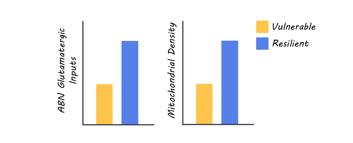
However, the authors did determine that rats vulnerable to cognitive aging progressively lost their glutamatergic inputs, indicated by a significant reduction in the labeling of postsynaptic density scaffolding protein, PSD95. The decrease of postsynaptic densities was observed at all ages in the inner molecular layer (IML) of the dendrite, but not in the middle or outer molecular layers. This suggests that the maintenance of proximal synaptic inputs (those closer to the soma or cell body) is especially important because these inputs are preserved only in resilient animals. Interestingly, the ABNs in vulnerable animals had a significant reduction of mitochondrial density, specifically in the IML at 8 months, but extended to the middle and outer layers in 18-month-old vulnerable animals. This suggests a progressive spread of mitochondrial dysfunction with aging in vulnerable animals. Optogenetic stimulation of ABNs improved the memory in all animals, and the memory of vulnerable rats improved to the level of non-stimulated resilient rats. This suggests that even when natural synaptic input is compromised in vulnerable rats, artificial stimulation can improve cognitive performance, indicating that ABNs can still function if properly engaged.

What's the impact?

This study found that long-lived ABNs play a role in cognitive aging. ABNs remain functionally viable in vulnerable animals and can transmit information when activated. Therefore, brain resilience relies, at least in part, on the preservation of the ABN integration into their neuronal network. This work highlights the potential therapeutic benefit of restoring the functionality of the ABN signaling network to improve cognitive functions in old age. 

Access the original scientific publication here.

How Do Motor Cortex Pathways Change in Aging?

Post by Rebecca Glisson

The takeaway

As we age, we sometimes lose our ability to move normally, which also significantly lowers our quality of life and capacity for independence. The motor cortex of the brain, which controls our movement, and the brain pathways descending from this area deteriorate in older adults, suggesting a focus on these areas could help us better treat neurodegenerative diseases.

What's the science?

Our movement is controlled by an area of the brain called the motor cortex, which can lose its function over time as we age. The motor cortex sends signals through two tracts, or pathways, of cells: the corticospinal tract (CST) and the corticostriatal tract (CStrT). This week in NeuroImage, Wen and colleagues investigated the changes in the CStrT that occur as people age, aiming better to understand the neural basis of movement-related neurodegenerative diseases.

How did they do it?

The authors wanted to study how aging affects several parts of the motor cortex and its pathways: its overall structure, how blood flows through this area, and the quality of the cells in these areas. To study these variables related to movement abilities, the authors used structural magnetic resonance imaging (MRI) to image and analyze the motor cortex in the brain. They also used diffusion MRI to analyze the pathways of the CST and CStrT. Linking this to movement, the authors measured participants’ motor function using endurance and locomotion walking tests and grip strength tests. The authors grouped participants into two age groups: the younger group, 36 to 65 years old, and the older group, 66 to 90 years old. 

What did they find?

While younger participants had normal brain structure, the authors found that older participants had less volume in the motor cortex and less blood flow to this region. The older group of participants also had significantly lower movement abilities involving locomotion, endurance, and strength. This suggests that the loss of certain movement abilities as we age is due to degeneration of the motor cortex. The authors also found that the CST and CStrT pathways were of significantly lower quality in the older group of participants and that this deterioration mediated the relationship between motor cortex atrophy and decline in motor function. Therefore, the CST and CStrT pathways are particularly important to movement function and are affected in aging.

What's the impact?

This study is the first to show that the changes in the motor cortex and its related pathways in the brain due to aging are directly related to a loss of movement functioning in older adults. This highlights the need to focus on these areas for studying movement diseases related to aging. Studies like these can help us detect neurodegenerative disorders earlier and develop better and more effective treatments.

Access the original scientific publication here. 

Cyclical Brain Rhythms Drive Key Cognitive Functions

Post by Soumilee Chaudhuri

The takeaway

Brain networks carry out day-to-day cognitive functions, such as focusing, remembering, and processing sensory information. The activity in these networks follows a cyclical pattern, with each activation supporting essential cognitive functions.

What's the science?

The human brain carries out numerous cognitive and bodily functions, but it has been unclear how these processes are coordinated over time. Previous studies using different forms of neuroimaging have observed directional relationships in network transitions; however, it remained unclear whether these asymmetries are part of a higher-level organization. This week in Nature Neuroscience, van Es and colleagues analyzed brain imaging data from five independent datasets and revealed that, while individual transitions appear noisy, they collectively form consistent cycles that repeat every 300–1,000 milliseconds, with each network having a preferred position within the cycle

How did they do it?

The study analyzed brain activity from over 800 participants across five Magnetoencephalography (MEG) datasets. Participants ranged in age from 19 to 88 years and included both males and females. MEG signals were mapped onto the brain’s cortex, divided into 38–78 regions depending on the dataset, to track how different brain areas interacted over time. Using a Hidden Markov Model, the researchers identified 12 recurring brain network states and examined the timing and direction of transitions between these states. They applied a new method, temporal interval network density analysis or TINDA, to detect characteristic sequences of network activations emerging as cycles. The makeup of this cyclical pattern, along with its strength (amount of deviations from the cycle) and speed, were quantified and analyzed for consistency across participants and their relationship with age, cognitive performance, and behavior, showing how these brain rhythms relate to key individual traits.

What did they find?

Identified brain networks followed a cyclical pattern, with cognitive and perceptual networks taking turns in a consistent and coordinated sequence. This sequence or cycle lasted approximately 300–1,000 milliseconds, and the timing and strength of these cycles were linked to age and cognitive performance. The researchers found that older adults showed much slower cycles compared to younger participants. The phase of the cycle also predicted moment-to-moment behavior, including markers of memory consolidation and reaction speed. Finally, additional findings suggested that the rate of these cycles was partly heritable, indicating a biological basis for these rhythms.

What's the impact?

This study provides compelling evidence that the natural cyclical activation of large-scale brain networks underlies essential cognitive functions. These findings shed light on the mechanisms underlying cognitive processes in the brain and highlight the potential for targeting brain network rhythms in interventions designed to enhance cognitive function.

Access the original scientific publication here.