Fluctuations in Heart Rate Influence Brain Activity

Post by Meagan Marks

The takeaway

Heart Rate Variability (HRV), a component of cardiac rhythm, directly affects brain activity important for neural communication.

What's the science?

In neuroscience, it is typically understood that the brain controls the body. But emerging evidence suggests that involuntary functions like heartbeat can influence the brain. Recent research has found that heart rate variability (HRV), the fluctuation in time intervals between adjacent heartbeats, is connected to neural activity. Those with higher HRV – or a heart more adaptable to the environment – are shown to have improved emotional regulation, cognitive function, and well-being. This relationship is especially prominent with high-frequency HRV (HF-HRV, which reflects the variation in heart rate associated with breathing). However, how neural activity and HF-HRV directly affect each other remains unknown. This week in Psychological Science, Sargent and colleagues explored the causal relationship between HF-HRV and neural activity by comparing oscillations from the heart and brain.

How did they do it?

The authors recruited 37 healthy adult participants and asked them to stare at a white cross on a black screen for 5 minutes. During the task, cardiac rhythm was recorded via electrocardiogram (EKG) and brain rhythm via electroencephalogram (EEG). The HF-HRV oscillations were then extracted from EKG recordings and temporally aligned to match EEG data (brain waves), which had been filtered into oscillations occurring at each frequency band (alpha, beta, gamma, delta, theta). The authors then analyzed the oscillations to look for evidence of phase-amplitude coupling, where it was predicted that the phase series (cycles) of HF-HRV oscillations would be coupled with, or associated with, the magnitude of change in brain waves (amplitude). Once phase-amplitude coupling was established, the authors calculated to what extent HF-HRV oscillations successfully predicted brain oscillations and vice versa to establish the direction of the causal relationship (heart-to-brain or brain-to-heart influence). 

What did they find?

Upon analysis, the authors found a strong relationship between HF-HRV and neural activity via phase-amplitude coupling, where the phase series of HF-HRV oscillations modulated the amplitude of the brain waves. It was found that a majority of participants also showed a significant heart-to-brain effect, where HF-HRV oscillations significantly predicted and regulated brain waves. This suggests that cardiac rhythm can influence neural activity. In addition, for all brain wave frequency bands except gamma, the heart-to-brain effect was significantly stronger than the brain-to-heart effect. This was true for EEG signals coming from all areas of the brain, suggesting that the heart was influencing neural communication and activity between multiple regions and multiple brain wave rhythms.

What's the impact?

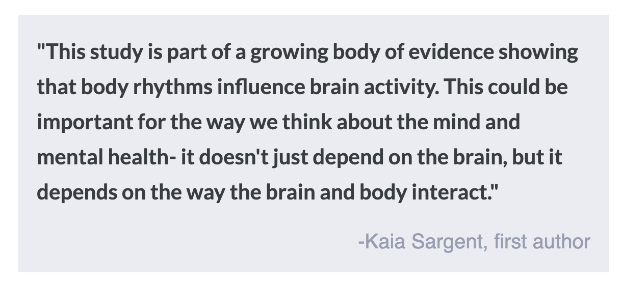
This study is the first to show that fluctuations in heart rate can modulate neural activity. The findings suggest that improvements in cardiac rhythm may enhance connectivity and communication between neurons in the brain, in turn boosting cognitive functions like emotional regulation, executive functioning, and stress management. Cardiac variables such as HRV could also be a potential therapeutic target for mental health disorders, where methods like HRV biofeedback could help improve well-being. 

How Do Scientists Study Lucid Dreaming?

Post by Laura Maile

What is lucid dreaming?

Lucid dreaming is a state of sleep where the individual becomes aware that they are dreaming. This phenomenon represents a paradoxical situation where the dreamer maintains a level of cognitive awareness while remaining asleep. The concept of lucid dreaming or “sleep awareness” has been written about across the globe for centuries, though it was not studied scientifically until the late 20th century and wasn’t accepted as a legitimate field of study until the 2000s. This phenomenon occurs in about half of individuals, but infrequently. Though lucid dreaming has become a popular field of study among sleep researchers, the small sample sizes in previous research have made the physiology and brain regions underlying this phenomenon difficult to identify.   

How do scientists study it?

In the 1970s, scientists began to study the neuroscience of lucid dreaming using electrooculography (EOG) to detect eye movements during sleep. Eye movements occur during normal dream states, but during lucid dreams, the individual can intentionally move their eyes. This allows the dreamer to communicate with the outside world while still in the dream state, giving experimenters an opportunity to observe and record information about dreaming in real-time. EOG, in combination with fMRI and electroencephalogram (EEG) to record brain activity, allows experimenters to confirm the state of lucid dreaming and collect neuroimaging data on the brain regions underlying this phenomenon. The major challenge with studying lucid dreaming is the low frequency with which it occurs. Some neuroscientists are developing methods to encourage lucid dreams to happen more often. These include exercises that train individuals to reflect on their state of mind before sleeping and during short “inverse naps” during the rapid eye movement (REM) stage of sleep. Others have used virtual reality training or direct electrical or ultrasound stimulation of specific brain regions during sleep.  

What does the future look like?

Scientists in the field of lucid dream research indicate that standard operating procedures are needed to ensure similar methods of cognitive training, experimental procedures, EOG, and EEG to enhance reproducibility and grow the field. Engaging in cross-lab collaborations and open sharing of data will allow scientists to produce more robust, powered studies. Wearable technology is now available to allow study participants to record EEG and EOG during normal sleep at home with simple, user-friendly devices, which will also greatly increase the amount of lucid dream data acquired. Citizen neuroscience also presents unique opportunities for larger-scale data collection and sharing of experiences and resources between community groups of lucid dream enthusiasts and academic researchers. 

Recent advances have allowed for two-way communication between the lucid dreamer and experimenter. Light and sound cues can be used to signal to the dreamer, while the dreamer can communicate with the experimenter using eye movements detected with EOG. Electromyogram technology can also detect subtle facial muscle movements, with the potential for uncovering dream speech. Finally, computational analysis and generative artificial intelligence give scientists new abilities to reconstruct and interpret dream states. Given the surge of interest and the recent technological advances, the future of research of lucid dreaming holds huge potential for understanding the neuroscience of dreaming in general.  

Access the original scientific publication here.

Astrocytes: The Unsung Heroes of the Brain

Post by Trisha Vaidyanathan

What are astrocytes?

Astrocytes are star-shaped cells that are found throughout the brain and contain thousands of very fine branches. Astrocytes are the largest type of a neural cell called “glia”, which is the Latin word for “glue,” because scientists originally thought they simply existed to hold the brain together. However, the field of astrocyte biology has exploded in recent years, and while many mysteries remain, it is now well-appreciated that astrocytes serve many critical functions in the brain. 

Astrocytes are an essential component of the synapse

Canonically, the neuronal synapse includes two players: the pre-synaptic neuron and the post-synaptic neuron. However, we now know that there is a third player — the astrocyte — resulting in what is called the “tripartite synapse”. Most neuronal synapses are enveloped by an astrocyte branch. By “hugging” the neuronal synapse, astrocytes can both monitor what is happening via different receptors and directly manipulate the synapse. Astrocytes can manipulate synapses in several ways, including altering ion and neurotransmitter concentrations via transporters or by releasing their own signaling molecules. Indeed, astrocytes are necessary for synapse formation, maintenance, elimination, and plasticity.

Astrocytes provide energy to the brain

Neurons require a massive amount of energy in the brain, and astrocytes take on the job of supplying that energy. Astrocytes contact blood vessels through special branches called “end-feet” where they help form the blood brain barrier, regulate blood flow, and, importantly, take in glucose from the blood. Astrocytes either store glucose as glycogen, or convert it into lactate. Lactate is then shuttled directly into hungry neurons, where it is rapidly converted into ATP, the energy currency of the cell. Astrocytes dynamically regulate glucose uptake, storage, and lactate shuttling to match the energy demands of the brain and regulate neuronal activity.

Astrocytes respond to injury and disease

A large body of evidence has demonstrated that astrocytes are able to react in the presence of disease or injury. When astrocytes become “reactive” they change their shape, gene expression, and function. Reactive astrocytes have been found in response to most types of brain insults, including traumatic brain injury, viral infections, stroke, epilepsy, neurodegenerative diseases, autoimmune disorders, cancer, and psychiatric diseases.

There is no single definition of a “reactive astrocyte” and the response varies with the type of disease/injury and severity. Interestingly, in some instances, the reactive state can be beneficial, while in other instances it can exacerbate the disease or injury. There is still much to understand about astrocyte reactivity and what drives the astrocyte to either help or hurt.

Astrocytes control circuits and behavior

Astrocytes have thousands of fine branches that allow them to communicate with many cells at once. A single astrocyte can simultaneously contact up to 100,000 synapses in the mouse brain and two million in the human brain. As such, they are uniquely suited to regulate larger neuronal circuits. The recent development of new tools has allowed scientists to identify a critical role for astrocytes in controlling many different circuits and behaviors. Astrocytes have been shown to play a pivotal role in behaviors like memory, learning, sleep, feeding, emotional regulation, motor behavior, and decision making. Precisely how astrocytes regulate these complex circuits and behaviors remains to be understood, but it’s becoming increasingly clear that astrocytes are critical to the bigger picture of the brain and behavior.

What don’t we know about astrocytes?

Relative to neurons, our understanding of astrocytes lags far behind. However, newer tools are allowing astrocyte biologists to begin exploring some basic questions about these long-ignored cells. New microscopes and genetically encoded sensors are providing insight into how astrocytes signal to the rest of the brain using calcium. New molecular tools are revealing unique features of human astrocytes that may be critical to their role in disease and injury. Lastly, new evidence suggests astrocytes may not be just a single class of cell, but instead may have specialized functions, much like different neuron subtypes.

While there is still a lot we don’t know about the fundamental functions of these cells, new advances are being made rapidly and it is becoming increasingly clear that astrocytes are essential to brain function.

References +

Chung, Won-Suk, Nicola J. Allen, and Cagla Eroglu. "Astrocytes control synapse formation, function, and elimination." Cold Spring Harbor perspectives in biology 7.9 (2015): a020370. DOI: 10.1101/cshperspect.a020370

Hasel, Philip, and Shane A. Liddelow. "Astrocytes." Current Biology 31.7 (2021): R326-R327. DOI: https://doi.org/10.1016/j.cub.2021.01.056

Kofuji, Paulo, and Alfonso Araque. "Astrocytes and behavior." Annual review of neuroscience 44 (2021): 49-67.DOI: https://doi.org/10.1146/annurev-neuro-101920-112225

Lawal, Oluwadamilola, Francesco Paolo Ulloa Severino, and Cagla Eroglu. "The role of astrocyte structural plasticity in regulating neural circuit function and behavior." Glia 70.8 (2022): 1467-1483. DOI: https://doi.org/10.1002/glia.24191

Magistretti, P., Allaman, I. Lactate in the brain: from metabolic end-product to signalling molecule. Nat Rev Neurosci 19, 235–249 (2018). DOI: https://doi.org/10.1038/nrn.2018.19

Sofroniew, Michael V., and Harry V. Vinters. "Astrocytes: biology and pathology." Acta neuropathologica 119 (2010): 7-35. DOI: https://doi.org/10.1007/s00401-009-0619-8