How the Awake and Sleeping Brain Tags Relevant Information for Memory

Post by Meredith McCarty

The takeaway

This research provides evidence for a mechanism by which relevant information is selected or tagged for memory consolidation in the hippocampus, a region of the brain critical for memory. 

What's the science?

An essential feature of brain function is selecting relevant information to be stored in long-term memory. However, the neural mechanism by which this is accomplished has yet to be fully understood. The authors hypothesized that hippocampal neurons are involved in selecting, or “tagging”, relevant information for subsequent memory storage. This week in Science, Yang and colleagues identify a potential neural mechanism explaining how relevant information is selected for memory formation. 

How did they do it?

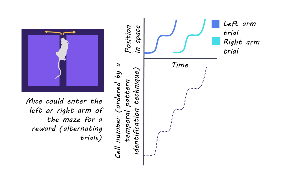
To study this mechanism, the authors had mice perform a spatial memory task. In this task, a water reward was delivered at the left or right arm of a figure 8-shaped maze (the location of this reward alternated on each trial). The mice had to navigate through this maze and choose the correct arm (left or right) to receive the water reward (the mice were water-restricted before beginning the task). Following this memory task, mice were placed in their familiar home cage and their neural data was recorded during periods of sleep. 

During the task and sleep periods, the authors recorded the spiking activity of single neurons via electrodes located in the hippocampus, measuring changes in spiking activity and identifying occurrences of sharp wave ripples (SPW-Rs). SPW-Rs are thought to be an essential feature of “offline” brain processing, during which populations of hippocampal neurons exhibit synchronized rapid firing, communicating compressed information to connected brain regions. To understand the relationship between neural activity and key decision and memory points in the task, the authors used neural decoding and analysis techniques, linking the mice’s real-time position in the maze on a given trial with the neural spiking and SPW-R recordings. 

What did they find?

First, the authors found a correlation between the mouse’s position in the maze and CA1 population spiking activity and were able to successfully decode the recent locations of the mice in the maze from the population spiking activity. When considering the activity of single neurons in isolation (which exhibit representational drift, meaning dynamic variability in activity across trials despite stable behavioral responses), the decoding accuracy deteriorated. This suggests that locations and events during the trial are represented at the population level — made up of variable activity of individual neurons — as opposed to the individual level. 

Next, when analyzing neural activity during the time after the mouse successfully navigated the maze and received a reward, the authors noted the emergence of SPW-R events on numerous trials. They were able to successfully decode task information from the content of these ripple events. These SPW-Rs were preceded by a decrease in theta activity, signifying the transition out of an active behavioral state. These findings suggest that SPW-Rs, theta power, and rewarding stimuli are related to memory replay and tagging events in the hippocampus.

When comparing awake and asleep neural activity, they found the occurrence of SPW-Rs during the awake task period to be a significant predictor of subsequent SPW-Rs during sleep. This suggests that the information tagged during awake SPW-Rs is subsequently replayed during sleep, furthering the process of memory consolidation.
What's the impact?

Based on these results, the authors propose a neurophysiological framework by which experiences are tagged for further memory consolidation in the hippocampus. The proposed mechanisms underlying this process are a decrease in theta power preceding the tagging of relevant information via SPW-Rs, and the subsequent repetitions of SPW-Rs during sleep that allow the replay of these events necessary for memory consolidation. 

This work has implications for both experimental and clinical work, to better understand how memories are formed, and how this process is disrupted in individuals with debilitating memory impairments.  

Inflammatory Effects of Trem1 are Associated with Risk of Developing Alzheimer’s Disease

Post by Shireen Parimoo

The takeaway 

Trem1 is a receptor protein found in macrophages – a type of white blood cell – that increases in older age and has pro-inflammatory effects. Deletion of Trem1 reduces inflammation by increasing mitochondrial metabolism and rescues cognitive deficits associated with aging and Alzheimer’s disease, such as memory loss. 

What's the science?

Myeloid cells like microglia and macrophages are critical for optimal brain and immune functioning. Alzheimer’s disease (AD) is characterized by pathology including the accumulation of amyloid protein and neurofibrillary tangles. Research has identified many families of genes that are associated with an increased risk of developing Alzheimer’s disease (AD). In aging mice, a receptor expressed on myeloid cells-1 (Trem1)  has pro-inflammatory effects that lead to higher rates of cognitive decline and greater amyloid accumulation. However, the exact role of Trem1 in AD is presently unclear. This week in Nature Neuroscience, Wilson and colleagues investigated the cellular mechanisms by which Trem1 regulates myeloid cell functioning and its effects on memory in typical aging and AD.

How did they do it?

The authors performed a series of experiments on several mouse models. The first included old and young wildtype mice and mice with deletion of both Trem1 alleles (Trem1-/-), which allowed them to identify the baseline effects of Trem1 deletion on myeloid cell functioning. Here, flow cytometry was used to look at Trem1 expression in myeloid cells of wildtype and mice with deleted Trem1 alleles. Next, inflammatory markers in plasma, peripheral macrophages, and the brain were quantified and compared to performance on spatial and object recognition memory tasks. The authors then used RNA sequencing to compare the differences in microglial and macrophage gene expression between the groups. Finally, as energy metabolism is important for regulating inflammatory responses, they investigated the effects of Trem1 deletion on macrophage metabolism by characterizing the expression of mitochondrial genes in the mouse models.

The second group consisted of mouse models of AD (i) without Trem1 deletion, (ii) with deletion of a single Trem1 allele, and (iii) with deletion of both Trem1 alleles. These AD mouse models enabled the authors to determine how Trem1 deletion affects the cellular responses of microglia and macrophages to amyloid burden and its effect on memory. As before, they measured Trem1 expression, inflammatory responses, memory performance, and mitochondrial metabolism. Finally, to determine whether Trem1 is implicated in human AD, the authors used human frontal cortex tissue from controls and patients with AD to quantify Trem1 levels and to examine the relationship between Trem1 and disease severity. 

What did they find?

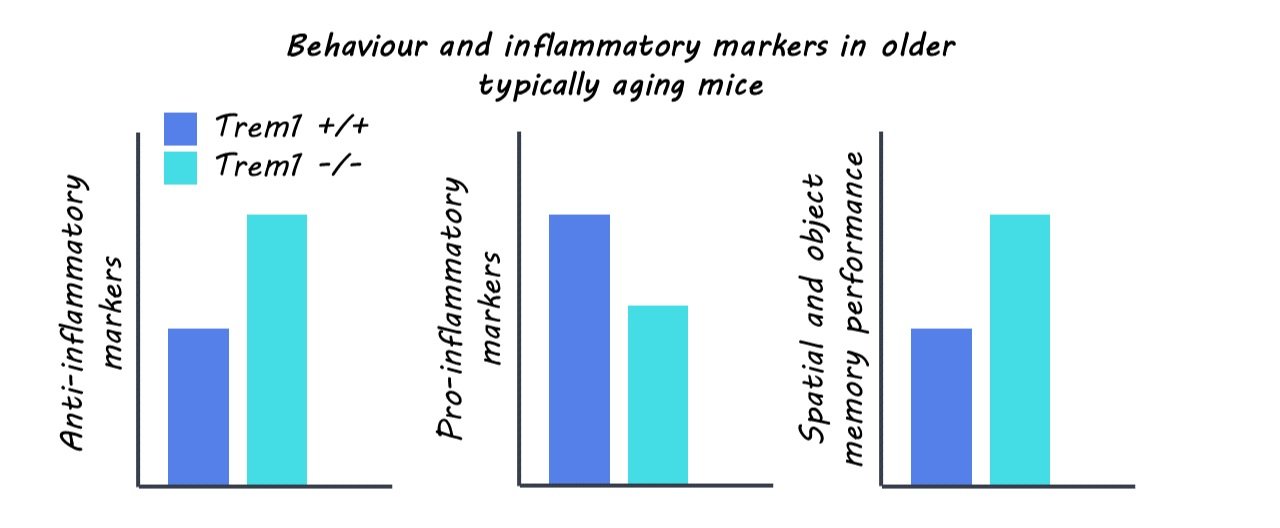
Older wildtype mice had elevated Trem1 expression when compared to younger wildtype mice, as well as higher levels of inflammatory markers in plasma. Mice with Trem1 deletion, however, showed similar levels of plasma and brain inflammatory proteins as the younger mice. Anti-inflammatory markers in macrophages were lower in older than younger mice while pro-inflammatory markers showed the opposite pattern. Within the older group, mice with the deletion of both Trem1 alleles had higher anti-inflammatory and lower pro-inflammatory markers, better spatial and object memory performance, and different gene expression profiles of proteins associated with mitochondrial metabolism and inflammatory regulation in peripheral macrophages compared to the wildtype mice. Together, these results suggest that in typical aging, Trem1 expression negatively affects memory through pro-inflammatory mechanisms while Trem1 deficiency has neuroprotective effects.

Trem1 expression was higher in post-mortem brains of humans who had AD than those who didn’t, and elevated levels of Trem1 were associated with more advanced stages of AD. In mouse models of AD, mice with and without Trem1 deficiency had similar amyloid burden as well as similar levels of Trem1 in microglia. Compared to AD mice with both Trem1 alleles deleted, the deletion of only a single Trem1 allele rescued memory performance. Trem1 deletion in AD was also associated with lower inflammatory markers in the blood, indicating that the increased presence of Trem1 in aging has adverse effects on cognition due to increased inflammatory responses in AD, irrespective of amyloid burden. 

What's the impact?

This study is the first to demonstrate the role of Trem1 signaling in the risk of developing Alzheimer’s disease. The finding that Trem1 expression increases with age and promotes inflammation via metabolic pathways provides an avenue for future research to develop treatments targeting those specific pathways. 

Access the original scientific publication here.

Different Patterns of Gene Mutation in Neurons and Glia During Aging

Post by Lani Cupo

The takeaway

Neurons and glia show different patterns of gene mutations during the aging process in humans, suggesting different processes may underlie age-related genetic mutations in different cell types. 

What's the science?

The underlying cellular and molecular mechanisms contributing to the process of typical aging (in the absence of disease) are still poorly understood. While aging-related genetic mutations in neurons have been investigated, glia, responsible for everything from providing brain structure to maintaining homeostasis, represent more than half of the cellular content of the brain and have yet to be examined for gene mutations in the aging process. This week in Cell, Ganz and colleagues present a characterization of mutations in neurons and oligodendrocytes (glia that produce myelin in the brain), finding different patterns of mutations between the two cell types.

How did they do it?

The authors extracted oligodendrocytes from the prefrontal cortex of 13 deceased individuals aged 0.4 - 83 years and accessed genome data from the neurons of 19 individuals (12 of whom overlapped with their oligodendrocyte donors). They used a method called single-cell whole-genome sequencing to identify the genetic code of these cells individually. Then, they used an algorithm to automatically identify two types of mutations in the genomes of the cells: single-cell whole-genome sequencing (sSNVs) and small insertions and deletions (indels). sSNVs occur when a single nucleotide is switched for another, and are acquired over the lifespan, rather than being inherited. Indels refer to insertions or deletions of nucleotides in the genome. With these data, the authors first estimated annual rates of accumulation of both sSNVs and indels. They also examined where in the genome the mutations occurred. Finally, they compared the patterns of mutations they observed with genomes from a database of tumor cells.

What did they find?

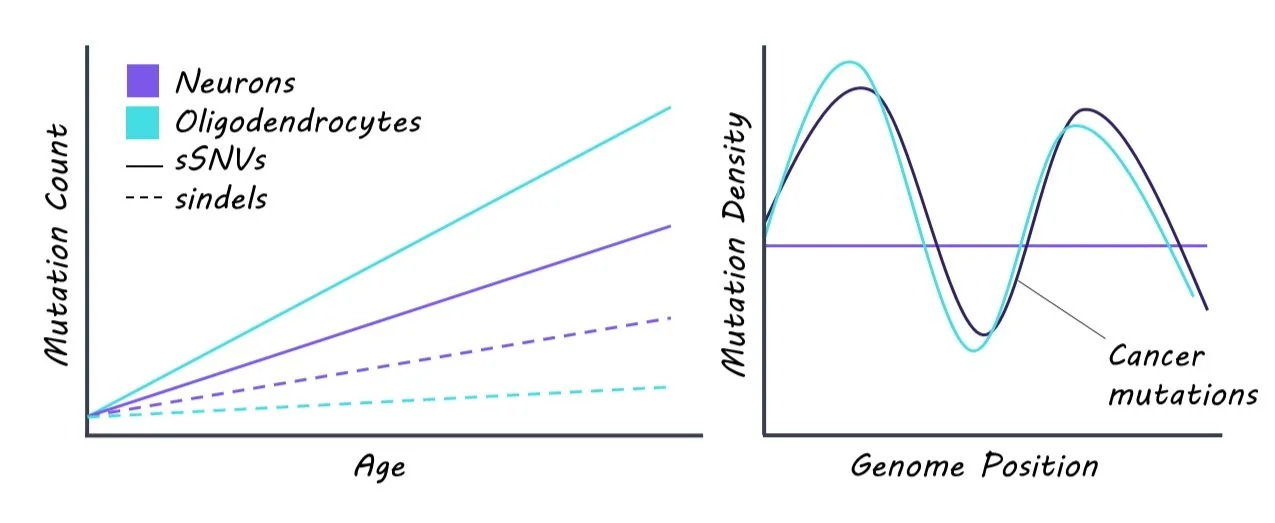
The authors found that oligodendrocytes exhibited higher rates of sSNV accumulation, but lower rates of indel accumulation compared to neurons. Deletions of nucleotide base pairs were more common than insertions in both cell types, but there were also differences in the kinds of deletions between the cell types. Oligodendrocytes mostly showed deletions of a single base pair whereas neurons showed deletions of 2-4 base pairs or 1 base pair insertion compared to the glial cells. At birth there was no significant difference between sSNVs or indels between cell types, suggesting that the changes were acquired over the lifespan and relevant to aging. The fact that profiles of mutations differed between cell types provided evidence that different mechanisms underlie aging between cell types. 

The authors also found opposite patterns in the placement of mutations between cell types. Mutations in oligodendrocytes were found to be more prevalent in intergenic regions, or parts of the genetic code that are not transcribed into proteins. In contrast, mutations in neurons were found to be more prevalent in regions in genes - regions that are transcribed into proteins. As a result of the placement, mutations in neurons were also found to have more of a functional effect on the cell. When comparing the mutation profiles of oligodendrocytes and neurons to cells from tumors, the authors found that densities of sSNVs in oligodendrocytes correlated with those in all cancer types, but neuronal mutations did not correlate with mutations in cancer cells, suggesting the mechanism contributing to mutations in glia cells, but not neurons, may be related to tumor formation.

What's the impact?

For the first time, this study compares genetic mutations in oligodendrocytes and neurons throughout the typical aging process. They found differences in the type and placement of mutations between cell types, suggesting different mechanisms contribute to the mutations. In time this may help us identify the specific mechanisms underlying what goes wrong in aging and neurodegenerative diseases.

 Access the original scientific publication here.