The Purpose of Sleep is to Restore our Brain to an Optimized State Called Criticality

Post by Trisha Vaidyanathan

The takeaway

The waking experience pushes the cerebral cortex away from “criticality”, a state of neural activity that is optimized for computation and cognition. The function of sleep is to restore the brain to criticality.

What's the science?

We spend about one third of our lives sleeping, but the purpose of sleep is debated. Broadly, we understand that sleep is “restorative”, but it is unclear how sleep contributes to brain computation and information processing. This week in Nature Neuroscience, Xu and colleagues provide new evidence to support a theory that one of the primary functions of sleep is to restore the brain to an optimized state called “criticality.” Criticality is a concept borrowed from physics that describes when a system of individual parts will be most effective at responding to an input. It makes sense that our brain should operate at criticality so that it can quickly and effectively process new information – for example, if our brain receives new visual input caused by a tiger appearing, it should quickly and effectively transmit that information to brain regions that will drive us to run away.

How did they do it?

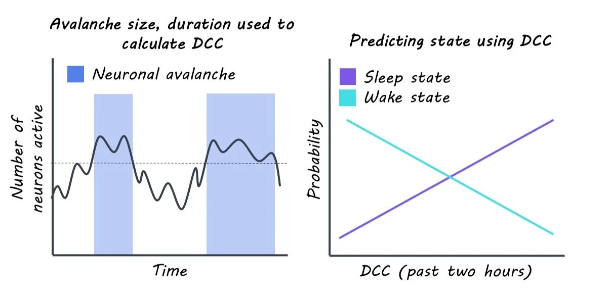
To measure how close the brain is to criticality, the authors performed continuous extracellular recordings of individual neurons in the visual cortex of rats. Criticality is characterized by neuronal avalanches, which are cascades of bursts of neuronal activity. This allowed them to create a score that measured how close the cortex was to criticality at any given time called the “deviation from criticality coefficient”, or DCC. The higher the DCC score, the further the brain is from criticality. Because rats constantly switch between wake and sleep throughout the day, the authors could assess how the DCC score fluctuated with wake and sleep.

Using the DCC score, the authors tested their theory that wakefulness pushes the brain away from criticality and sleep restores criticality. First, the authors asked how the DCC score changed during sleep and wake. Next, they asked whether the DCC score was predictive of future sleep/wake behavior and if the DCC score could be predicted by previous sleep/wake behavior. Lastly, they asked if the DCC score would change if the rats were forced to stay awake for longer periods of time.

What did they find?

First, the authors found that more time in a waking state correlated with higher deviation from criticality (i.e., higher DCC scores) and more time in sleep correlated with lower DCC scores, consistent with their theory. Interestingly, the effect was greater when the rats spent more time moving during wake and the effect was absent when the rats were awake but in the dark. This suggested that not all wake experiences are the same and that more stimulation during a waking state can result in a greater deviation from criticality.

The authors found that future sleep/wake behavior could be predicted using the DCC score. The DCC score was more predictive than other known regulators of sleep, like the time of day or prior amount of sleep. Further, the authors could predict the DCC score by using the sleep/wake behavior from the previous two hours, in support of the theory that sleep and wake drive changes in criticality.

When rats stayed awake for periods of 90 minutes, slightly longer than normal, the DCC score increased as predicted by their theory that wakefulness pushes the brain away from criticality. When the rats were allowed to sleep again, the DCC score went back down, demonstrating the restorative effect of sleep on criticality.

What's the impact?

This study addresses a big mystery in neuroscience: why do we sleep? The authors provide strong evidence that one of the primary functions of sleep at a systems level is to restore the brain to an optimal state described by the theory of criticality.

Atypical Neural Activity Facilitates Positivity After Experiencing Negative Events

Post by Rebecca Hill

The takeaway

Individuals who interpret negative circumstances in a more positive light are more easily able to weather hardships, but it is still unclear how this happens in the brain. Atypical neural activity in a brain network called the default network facilitates this positive interpretation after witnessing a negative event. 

What's the science?

The ability to be optimistic during adverse life experiences, such as a poor medical diagnosis or difficulties with interpersonal relationships, can improve physical and mental health outcomes after these events. However, the brain mechanisms that allow for these positive outlooks after negative experiences are still unknown. Previous work has suggested that the default network, which is implicated in subjective interpretation, could be involved with this positive thinking. This week in PNAS, Iyer and colleagues studied which cognitive mechanisms are used to facilitate positive interpretations of negative events.

How did they do it?

The authors had participants watch videos about cystic fibrosis while measuring their brain activity in regions throughout the default network using a functional MRI (fMRI). Subjects watched one video of patients discussing their experience with cystic fibrosis and one video of an explanation of the biological mechanisms of cystic fibrosis. This was to test whether an experience that was more open to interpretation – the video of the patient – led to more atypical neural processing than an experience that was not open to interpretation – the explanation of cystic fibrosis. After each video was played, subjects were scanned for 6 minutes during a rest period. The authors used this method to pinpoint the time that atypical thinking might be occurring to encourage positive interpretation. To measure neural activity associated with this atypical thinking, the authors calculated the activity connecting regions within the default network, and then compared this activity between participants. They used this to sort subjects into similar or dissimilar activity, as those with dissimilar activity to all other subjects experience atypical neural activity. After subjects completed their fMRI, they were asked to write descriptions of what they remembered about each video and the descriptions were then analyzed for how much positive or negative wording was used.

What did they find?

The authors found that atypical neural activity within the default network after watching the patient video was significantly related to positive descriptions from the subjects. Subjects who gave negative descriptions of the patient video instead had similar neural activity. This suggests that atypical thought processes and neural activity are necessary for interpreting negative events positively. When analyzing these responses both while subjects were watching the video and afterward, during the rest period, the authors found the most activity during the earlier half of the rest period. This suggests that positive thinking is facilitated by atypical neural activity occurring relatively quickly after experiencing a negative event. When testing for which region in the default network is most important for atypical neural activity, the authors found that the ventromedial prefrontal cortex, part of the reward system in the brain, was the only region required to facilitate a positive interpretation of negative experiences.

What's the impact?

This study is the first to describe that atypical neural activity is required to view negative experiences in a positive light. Understanding the neural mechanisms that allow for optimism during adversity is important for being able to encourage resiliency in response to negative events. Everyone goes through difficult experiences during life, so being able to identify and use effective coping strategies is essential for protecting mental and physical health.

Access the original scientific publication here

Pinpointing Target Regions for Brain Stimulation in Treatment-Resistant Depression

Post by Natalia Ladyka-Wojcik

The takeaway

This is the first large-scale randomized controlled trial to demonstrate the importance of pinpointing target regions involved in depression with magnetic resonance imaging (MRI) for treatment with transcranial magnetic stimulation (TMS). Treatment benefits can last longer than we previously understood - up to six months after TMS. 

What's the science?

Although antidepressants and psychotherapies are effective treatment methods for many people with severe major depressive disorder, some individuals have treatment-resistant depression and may benefit from therapeutic neuromodulation, such as repetitive transcranial magnetic stimulation (rTMS). rTMS for depression has been popular for the past several decades, but – critically – no data is available to support its long-term effectiveness (beyond 1-3 months). Moreover, rTMS is traditionally applied to the same site on the scalp such that individual differences in the brain circuits involved in depression may not be accounted for. A more recent approach called connectivity-guided intermittent theta burst stimulation (cgiTBS), may provide longer-lasting treatment effects than standard rTMS by personalizing the targets to each individual’s brain circuitry using MRI data. This month in Nature Medicine, Morriss and colleagues directly compared the efficacy of both therapeutic neuromodulation approaches in a multi-center, randomized controlled trial for treatment-resistant depression over 26 weeks.

How did they do it?

Participants in this study were randomly assigned to receive 20 sessions of either rTMS or cgiTBS over four to six weeks. For both groups, sessions involved placing an electromagnetic coil against the scalp to deliver magnetic pulses that can alter activity in brain circuits thought to be involved in depression. The authors pinpointed the precise location of a brain region involved in depression (the left dorsolateral prefrontal cortex) for each participant by collecting structural or functional MRI (think brain anatomy vs. brain activity) for those undergoing rTMS or cgiTBS, respectively. cgiTBS coordinates were based on the correlation between brain activity of the left dorsolateral prefrontal cortex and the right anterior insula. Crucially, a computerized tracking system, called Neuronavigation, allowed them to deliver the treatment consistently across all 20 sessions, reducing the variability in stimulation at each session. To determine if participants showed a long-lasting reduction in depression symptoms following rTMS or cgiTBS, they administered a widely used depression assessment scale at the start of the study, and later at 8, 16, and 26 weeks.

What did they find?

This study provides large-scale evidence for both rTMS and cgiTBS as effective interventions for treatment-resistant depression. Both groups showed a similar and large average decrease in symptom scores on the depression assessment scale at the 8-week follow-up compared to the start of the study and both groups maintained lower symptom scores on average at the 26-week follow-up. Participants experienced improvements in quality of life despite previously not responding to other treatment approaches for depression, with over two-thirds having reported feeling better by session 20 of stimulation. Importantly, a third of participants across both rTMS and cgiTBS groups showed a 50% drop in depression symptoms, with a fifth of participants maintaining this drop even after 26 weeks. The authors also found that participants who completed fewer than 20 stimulation sessions of either rTMS or cgiTBS showed less improvement in their depressive symptoms at the 26-week follow-up. Together, these findings demonstrate that personalizing the targeted brain sites (via either functional or structural means) for TMS using MRI in patients with treatment-resistant depression can result in long-lasting reductions of depression symptoms, even beyond what has previously been assessed in the literature. 

What's the impact?

This study is the first large-scale trial to find evidence that MRI-guided TMS (both rTMS and cgiTBS) is an effective, long-lasting approach for treatment-resistant depression. Given that around a third of all people with major depressive disorder experience treatment-resistant depression which does not respond to antidepressants and psychotherapies, the results of this study highlight the need to establish MRI-guided TMS as a standard treatment approach in these cases.  

Access the original scientific publication here.