Hippocampal Activity Can Distinguish Traumatic Memories from Sad Memories

Post by Shireen Parimoo

The takeaway

In post-traumatic stress disorder (PTSD), traumatic memories are a type of autobiographical memory that are similar to memories of sad, but non-traumatic life events. However, patterns of activation in the hippocampus (a key brain region involved in autobiographical memory) differ for traumatic and sad memories, indicating that traumatic memories symbolize a distinct cognitive state.

What's the science?

Post-traumatic stress disorder is characterized by exposure to a traumatic event that individuals subsequently re-experience through intrusive dreams and flashbacks. Individuals with PTSD show altered functioning of the hippocampus, posterior cingulate cortex (PCC), and amygdala, which are brain regions involved in memory reconstruction, re-experiencing of an event, and emotional processing, respectively. However, the same negative event (like the loss of a loved one for example) can be traumatic for one individual while being perceived as sad, but non-traumatic by another. In both cases, those same brain regions are likely to be activated, raising the question of whether traumatic and sad autobiographical memories only differ in their intensity or whether they represent distinct cognitive states. To investigate whether the neural representations underlying traumatic and sad memories are shared or distinct, a study in Nature Neuroscience by Perl and colleagues used fMRI to decode brain activity in different regions as individuals with PTSD listened to audio narratives of traumatic and sad memories from their lives.

How did they do it?

Twenty-eight adults diagnosed with PTSD were recruited for this study. During their initial visit, they were asked to recount (i) the traumatic event associated with their diagnosis (‘Traumatic’), (ii) a sad, non-traumatizing event (‘Sad’), and (iii) a positive and relaxing event (‘Calm’) from their life in as much detail as possible. The authors used these events to create audio narratives that the participants later listened to while undergoing fMRI scanning. Semantic analysis was performed to obtain a semantic representation of the content - the meaning and key ideas present within the narratives (e.g., losing a job). They used a dimensionality reduction method called t-distributed stochastic neighbor embedding to reduce the number of features in a dataset while retaining meaningful information. This then allowed them to identify and match the traumatic and sad memories on the types of semantic content, enabling them to use the sad memories as a comparison against the traumatic memories for brain activity.

The authors then examined neural activation in the hippocampus, PCC, and amygdala for each narrative category and created a neural similarity matrix. Similarly, they compared the semantic similarity within and across the narratives in each category. Next, they used inter-subject representational similarity analysis (IS-RSA) to compare brain activity to the semantic similarity of narratives across participants. This approach allowed them to see whether higher semantic similarity of the different narrative categories was associated with greater neural similarity across participants. To determine whether the severity of PTSD symptoms was associated with the neural representation of traumatic memories, they divided the participants into ‘high’ and ‘low’ symptom groups and performed the IS-RSA again. Lastly, the authors used linear discriminant analysis (a classification method) to investigate whether brain activity while listening to the narrative could be used to decode the corresponding narrative category.

What did they find?

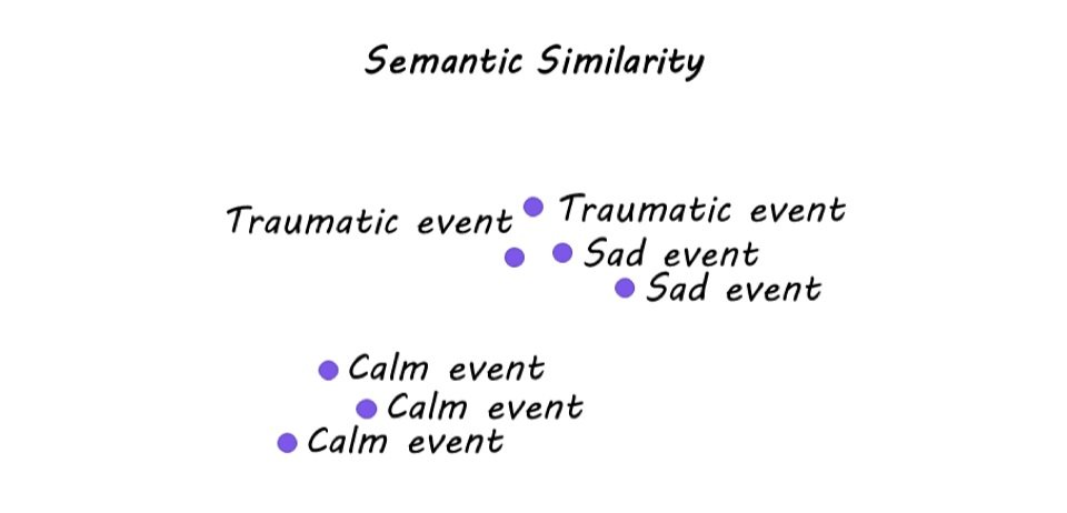
Semantic analysis revealed that Calm narratives were most similar to each other and least similar to the Sad and Traumatic narratives. On the other hand, while Traumatic narratives were highly idiosyncratic in their semantic content, they showed semantic overlap with Sad memories. In the brain, hippocampal representation of Sad and Traumatic memories varied across individuals. Specifically, greater semantic similarity of Sad memories was associated with greater similarity in hippocampal activation across participants. However, this was not the case for Traumatic memories, indicating that the hippocampus only tracks the semantic content of Sad memories. Moreover, hippocampal activity could be used to distinguish between Sad and Traumatic memories, whereas activity in the amygdala was not sensitive to the narrative category.

When neural activity was examined as a function of symptom severity, there was no difference in hippocampal or amygdala activity for Sad or Traumatic memories. In contrast, greater symptom severity was related to stronger neural representations of Traumatic – and, to a lesser extent, Sad – memories in the PCC. Together, these results demonstrate that the hippocampus differentially represents the semantic content of Sad and Traumatic autobiographical memories while the neural representations in the PCC are more sensitive to the severity of PTSD symptoms within individuals. 

What's the impact?

The authors used an innovative approach to examine brain activity while participants listened to narratives of events from their own lives. This study is the first to show that the re-experiencing of traumatic memories represents a distinct cognitive state from remembering sad or negative autobiographical memories, which has important implications for informing treatments for individuals with PTSD. 

Access the original scientific publication here.

The Hippocampus Can Predict Whether an Action Will Lead to a Reward

Post by Trisha Vaidyanathan

The takeaway

The ventral hippocampus (vHPC), via its projections to the medial prefrontal cortex, is important for encoding the context of a reward. This pathway is required to update the causal relationship between an action and a reward and adjust behavior accordingly. 

What's the science?

It has long been debated whether the hippocampus plays a role in “goal-directed actions”, something we do every day. For example, you may put money in a nearby vending machine (an action) so you can eat a candy bar (a goal). Choosing to take this action is dependent on your understanding that there is a causal relationship between putting money in a vending machine and getting a candy bar, also known as an “action-outcome contingency”. However, if candy bars randomly drop from the vending machine without your money, you may stop believing in the “action-outcome contingency” and stop putting money in the vending machine. Critically, you will only update the “action-outcome contingency” in the context of the vending machine and will likely still choose to use money at a store, or maybe even another vending machine. This week in Current Biology, Piquet and colleagues demonstrate that the ventral hippocampus (vHPC) is critical for context-specific learning that shapes behaviors in response to changing action-outcome contingencies.

How did they do it?

The authors used a behavioral test for rats to model action-outcome contingency. Rats were trained on two different action-outcome contingencies: pushing lever 1 resulted in a grain pellet, and pushing lever 2 resulted in a sugar pellet. Next, one of those action-outcome contingencies was “devalued” by giving rats access to the pellets without needing to push the lever. Finally, they tested the rats by presenting them with both levers – typically, rats are less likely to choose the lever associated with the “devalued” pellet, or the pellet they were given free access to. To test the role of the vHPC, the authors trained rats on the task after creating a lesion in the vHPC or using chemogenetics to selectively silence the vHPC neurons during specific phases of the task.

Next, the authors investigated the role of context in action-outcome contingency. Rats went through extensive pre-exposure to the behavior chamber, which is known to reduce its effectiveness as a context cue through a concept called “latent inhibition” and examined how this altered their response to the action-outcome contingency change. The authors also trained rats with or without vHPC silencing on a Pavlovian context conditioning test, in which they were trained to associate two different behavioral chambers (contexts) with two different rewards, then the authors devalued one of the rewards and tested the ability of rats to differentiate between the two chambers.

Lastly, the authors investigated whether these vHPC functions are mediated by vHPC projections to the medial prefrontal cortex by using chemogenetics to specifically silence the vHPC terminals in the medial prefrontal cortex during the action-outcome contingency task.

What did they find?

First, the authors demonstrated that rats with vHPC lesions are insensitive to changes in an action-outcome contingency. After the rats were given full access to one of the pellets, the rats with lesions failed to preferentially choose the other pellet in the “test phase”. Similar results were found when the authors chemogenetically silenced vHPC neurons during the training, but not the test phase. Together, this demonstrated that vHPC is necessary to acquire new causal information.

Next, the authors lessened the effectiveness of the behavior chamber as a context cue through pre-exposure, or “latent inhibition”, and found this caused the rats to be less sensitive to the action-outcome contingency change. This demonstrated that encoding the context of a reward is necessary to respond to an action-outcome contingency change. The authors then demonstrated that vHPC is specifically important for encoding the context of the reward. Rats with vHPC lesions or chemogenetic silencing were unable to differentiate between a “devalued” context and a “valued” context in the Pavlovian context conditioning assay.

Lastly, the authors confirmed that the vHPC exerts its effect on goal-directed actions via its projections to the medial prefrontal cortex, by demonstrating that specifically silencing the vHPC terminals in the medial prefrontal cortex had the same negative effect on behavior as the vHPC silencing.

What's the impact?

This study identifies a critical role of the vHPC-to-cortex circuit in goal-directed actions by demonstrating that the circuit is necessary to encode the context of a reward, update the causal link between action and outcome, and adapt behavior appropriately. These findings are critical to our understanding of fundamental, everyday behavior. 

Access the original scientific publication here.

Brain Volume in the Hippocampus is Influenced by Genetics

Post by Lani Cupo

The takeaway

Gray matter volume along the length of the hippocampus is influenced by genetic factors, which can be identified in living humans.

What's the science?

The volume of the hippocampus is considered to be about 80% “heritable”, meaning 80% of the variability of hippocampal volume is attributable to genetic variation. Measuring the volume of the entire hippocampus, however, neglects to consider that some regions of the hippocampus may be more susceptible to environmental influences than others. Examining the heritability of subfields, or regions within the hippocampus, may provide better estimates of whether the heritability of volume differs between regions. This week in NeuroImage, Pine and colleagues investigated the heritability of hippocampal subfields using magnetic resonance imaging (MRI) data, finding volume is influenced both by genetic factors general to the entire hippocampus, and specific to individual subfields. 

How did they do it? 

The authors used MRI from both children and adult twin pairs from three different datasets, including both dizygotic and monozygotic twins. They built biometric models to compare how highly correlated volumes were between monozygotic (single egg) and dizygotic (two eggs) twins. This method allowed them to detect differences in hippocampal subfield volume that could be attributed to sources of variance that are either genetic or environmental. For example, if there is variance in the hippocampal volumes of monozygotic twins (who begin life genetically identical) it can be attributed to an individual environmental influence (e.g. different levels of education). By examining how correlated hippocampal volumes were between monozygotic twins, compared to how shared hippocampal volumes were between dizygotic twins, the authors could estimate how much of the volume was heritable versus due to environmental influences. They compared the performance of different models to assess which one best fit their data before interpreting results. The hippocampal subfields examined were the head, body, and tail, dividing the hippocampus along its longitudinal axis (if you imagine the hippocampus as a c-shape uncurled). By including both males and females and both children and adults in this study, the authors were able to investigate how heritability by subfield differs across sexes and ages. 

What did they find?

The authors didn’t find any differences in the heritability of volume across the different subfields. This suggests there isn’t a particular subfield where volume is more heritable than others, however, it is possible that using another division of the hippocampus or treating it as a gradient, rather than distinct regions, may reveal other subtle differences. 

Nevertheless, the authors did find evidence for subregion-specific genetic components, meaning different genes might contribute to the volume of different subfields. Subfield-specific genetic contribution was found to be both sex and age-dependent. In both sexes and in both children and adults, subfield-specific genetic contributions were found for the tail of the hippocampus. In contrast, subfield-specific genetic contributions were found for the head only in children (both sexes). Finally, subfield-specific contributions were found for male children only for the body of the hippocampus. The authors reported that these sex and age differences do not follow known systematic patterns, however, it is of interest that the genetic influence on hippocampal subfield volume is not constant across ages, and other properties of the hippocampus, such as white matter, may better reflect age-related changes. 

What's the impact?

These findings suggest the head, body, and tail may not show differences in heritability of gray matter volume, however, they show subfield-specific genetic contributions that differ across subfields by age and sex. The authors also demonstrate that noninvasive imaging can be used in living humans to estimate genetically based individual differences, which can set the stage for future population studies. 

Access the original scientific publication here