­Music-Induced Emotions Affect How We Encode Memories

Post by Anastasia Sares

The takeaway

Emotion and memory are tightly linked, but it is hard to measure continuous fluctuations in emotional states reliably in the lab. This work used music to reliably induce emotions across time and examined how transitions between different emotional states affect memory.

What's the science?

We are constantly processing the continuous stream of experience that happens in life, placing similar information into different “episodes” so that we can store them efficiently in memory. Transitioning into new contexts or situations can lead to uncertainty, like walking out of a building or changing conversation partners at a party, for example. Therefore, we are generally more vigilant during these times, paying more attention to what is new and having better memory for individual items. On the other hand, within an episode, we are better at remembering relationships between different items and the order of information, such as the numbers of the rooms we pass in a hallway or the order of topics in a conversation.

Most of the above examples involve external cues to signal the boundaries between adjacent events. But we also have an inner life, and we can transition between states of mind just as easily as we walk through doors or change conversation partners. In particular, we often transition between different emotional states over time. This week in Nature Communications, McClay and colleagues used music to induce different emotional states and show how fluctuations in these emotional states affect memory formation.

How did they do it?

The authors hired trained film score composers to create emotional musical pieces (choosing from joyous, calm, sad, and anxious), with each piece having three distinct sections with different emotions. These pieces were meant to induce a range of emotions according to the circumplex model of emotion, which holds that emotions can be defined along two main dimensions: arousal and valence. Arousal refers to the energy level of an emotion: high-energy emotions include joy and anxiousness, while low-energy emotions include sadness and calm. Valence refers to the positive or negative quality of an emotion: positive valence emotions include happiness and calm, while negative valence emotions include anxiousness and sadness.

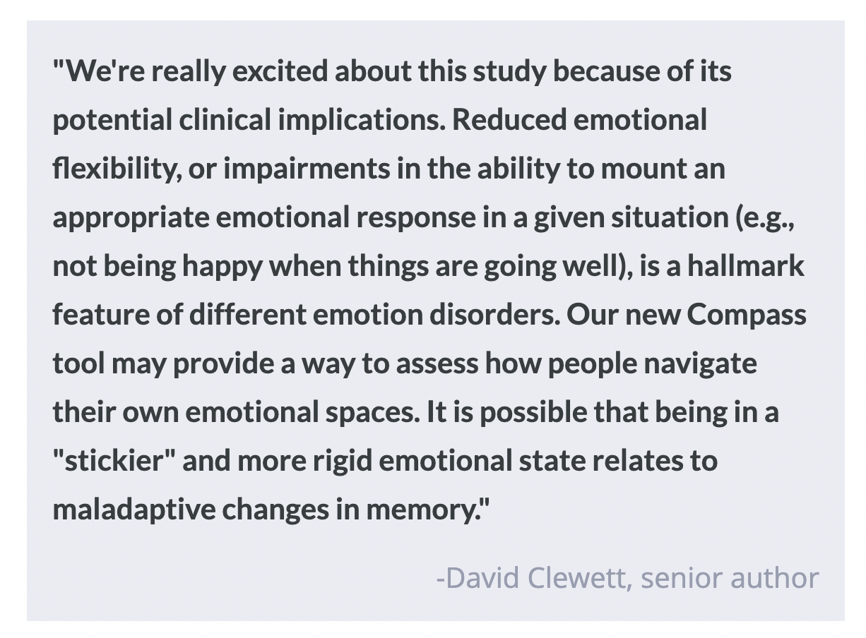
Participants first listened to the pieces in the background while trying to memorize a series of neutral images. At test time, they were presented with two objects and asked which one came first, and also how far apart the objects were in time. Finally, the participants were asked to rate the emotions they felt during the musical pieces using an “Emotional Compass”, a circle that captures a wide range of emotional valence and arousal levels. Participants rated their felt emotions in real-time, moving the mouse around on the Compass while re-listening to the musical pieces. In this way, the authors could extract measures of both the valence and the arousal levels participants experienced at each point in time and could then relate this to their memory performance. They also had a separate group of participants identify musical transitions, where the pitch or complexity changed significantly, so they could factor out the influence of these external sensory boundaries in their analyses. 

What did they find?

When two images were separated by a significant emotional transition, people experienced “time dilation” – in other words, they judged the images to be further apart in time than they actually were. Participants also had worse memory for the order of those images. Images that were shown at a boundary transition were better preserved in long-term memory overall (this was tested one day later). These effects are typical of the memory effects seen in previous studies, showing that our experience can be segmented according to our internal states just as much as our external context.

On the other hand, a large shift towards more positive emotions led to item pairs being judged as closer in time (i.e., “time compression”) and people remembered the order of those images better. High-arousal positive emotions also boosted long-term memory for accompanying items: participants could better identify which items were presented as well as when those items were encountered during the sequence. These findings indicate that positive emotions can help to fuse things together in memory, while either being in or shifting towards more negative emotional contexts may instead contribute to memory segmentation and worse memory for timing.

What's the impact?

This work shows that internal emotional states, especially emotional valence, can separate events in memory just like external changes in place and time. It also demonstrates that music is a useful tool for studying emotion in a continuous context in a realistic and reliable way.

Converting Microglia to Neurons Has Therapeutic Potential Following Stroke

Post by Laura Maile

The takeaway

In adult mammals, most neurons cannot proliferate, which means that neuronal loss following stroke and other brain injuries is irreversible. Microglia, the immune cells of the brain, maintain their capacity to divide and can be converted into neurons in mice with stroke, leading to improved neurological function. 

What's the science?

Neuronal loss is one of the major pathological effects of stroke that contributes to disability and poor health outcomes. The mammalian brain maintains limited ability for adult neurogenesis, adding to the negative effects of neuronal loss due to stroke and other brain injuries. The conversion of other cell types to neurons at the site of injury therefore presents a therapeutic opportunity that could improve functional recovery. Some researchers have had success in converting astrocytes to neurons, leading to functional improvement. In the most common cause of stroke, however, both astrocytes and neurons are depleted at the site of injury while microglia and macrophages infiltrate the injured area, making them a better target for conversion following ischemic stroke.  This week in PNAS, Irie and colleagues converted microglia and macrophages into neurons in the striatum of stroke mice using a single transcription factor and measured their functional improvements over time.

How did they do it?

The authors induced ischemic stroke using transient middle cerebral artery occlusion (tMCAO), and performed immunohistochemistry to analyze the lesioned area and the cell types located throughout the injured tissue. A week following stroke, they injected a virus carrying the NeuroD1 (ND1) transcription factor driven by a microglia-specific promoter into the injured brain area to convert infiltrating microglia and macrophages into neurons. Next, to determine whether the ND1-converted neurons became functionally integrated into the striatum, they used immunohistochemistry to stain striatal projection neurons in control and tMCAO mice and patch-clamp electrophysiology to record neuronal activity from these cells. To quantify how many microglia/macrophages were effectively transformed into neurons, they utilized a transgenic mouse line expressing cre-inducible diphtheria toxin receptor (DTR). By injecting a Cre virus with a microglia-specific promoter, they could permanently express DTR in microglia, and then count the number of DTR-expressing cells that became neurons at different time points. The authors then tested whether the conversion of microglia into neurons had functional outcomes on stroke recovery, by comparing multiple motor behaviors in injured and control mice. Finally, they ablated the ND1-converted neurons to examine whether these cells were responsible for functional improvements. 

What did they find?

They showed that following stroke, there is a significant neuronal loss in the striatum, and that while astrocytes remain at the border of the lesioned area, microglia and macrophages infiltrate into the core of the lesion. Two weeks following the injection of their virally packaged transcription factor, they showed a reduction in the microglial population and an increase in neuronal markers, which means that cells at the lesion site were reprogrammed from a microglial identity into a neuronal one. They also showed that when microglia were depleted prior to injection of ND1, they no longer observed the increase in neuronal cells at the lesion site, indicating it is likely mostly microglia that are successfully converted into neurons. Using immunohistochemistry, they demonstrated that striatal projection neurons, which are largely depleted in the lesioned area following tMCAO, show recovery following ND1 transduction. Their ND1-transduced cells became positive for a striatal neuron marker and showed functional activity that mimicked that of native striatal neurons.

They next found that one week after labeling microglia and initiating their conversion into neurons, very few labeled cells expressed neuronal markers, indicating they hadn’t yet converted to neurons at this early stage. At the eight week timepoint, however, a large number of the permanently labeled DTR cells also expressed striatal neuron markers and showed anatomically relevant connections with other neurons. This means that their strategy to convert microglia at the lesion site into neurons that would be integrated into the native circuitry was successful. Finally, they showed that following the conversion of microglia to neurons at the lesion site one week post-injury, mice showed improvements in multiple motor behaviors impacted by stroke. Damaging ND1-converted neurons blocked these improvements, suggesting that these newly converted neurons were responsible for the functional improvements observed. 

What's the impact?

This study is the first to show successful in vivo conversion of microglia into functional neurons following ischemic stroke in mice. This treatment, which led to improved neurological function in injured mice, demonstrates a promising therapeutic strategy for stroke and other injuries resulting in the loss of neurons. 

Sleeping Participants Show Evidence of High-Level Cognitive Processing

Post by Lani Cupo

The takeaway

Participants respond to external stimuli during sleep, and responses are associated with cognitive activity, suggesting sleep is not a state of complete disconnection from the external world as previously believed. 

What's the science?

Scientists have classically considered sleep a state with no reactivity to external stimuli, however, recent evidence suggests that stimuli can be processed on different cognitive levels during sleep, even to the point of learning new material. Nevertheless, few sleep studies attempt to elicit behavioral responses, as researchers consider them uniquely associated with wakefulness. This week in Nature Neuroscience, Türker and colleagues examined responsiveness to external stimuli during different sleep states, as well as lucid compared to non-lucid dreaming with an auditory decision task.

How did they do it?

The authors recruited two groups of participants: participants with narcolepsy who often lucid dream (N = 27), and healthy controls (N = 21). Participants napped in the laboratory during daytime hours while the authors spoke a combination of words and pseudowords. Participants were instructed to frown briefly three times in a row if they heard a pseudoword or smile briefly three times if they heard a real word. Facial muscle contractions were monitored with electromyography (EMG), and sleep/wake stage was assessed with a combination of electroencephalography (EEG) and electrooculography (EOG) which respectively measure electrical impulses from the brain and eye movements. After each nap, the participants reported if they dreamed, whether the dream was lucid, and whether they recalled performing the task.

In addition to the frequency of responses, response accuracy, and response rate on the task, the authors examined local brain activity associated with responses from EEG, and how well cognitive activity, measured with EEG, predicts responsiveness.

What did they find?

In terms of responsiveness, the authors found that participants could perform the task across almost any sleep state (except N3, deep sleep, in the healthy participants), meaning regardless of whether they were awake, in the lighter stages of sleep, or dreaming, they would respond to stimuli with smiles or frowns when they were presented and would not respond when no stimulus was presented. Interestingly, participants with narcolepsy responded more to stimuli across all sleep stages than healthy participants, including N3, and they reported lucid dreaming. As sleep deepened, the response rate decreased, although it increased slightly during rapid-eye movement (REM, dreaming sleep) in healthy participants. In participants with narcolepsy, the response rate increased greatly in non-lucid REM sleep and even further increased in lucid REM sleep. Examining accuracy, the authors found that all participants were more accurate than chance in responses, but healthy controls were more accurate than participants with narcolepsy, and increased depth of sleep was correlated with decreased performance. Regarding response time, as in wakefulness, during sleep participants were slower to respond to pseudowords than words, and participants were slower to respond overall while asleep than awake. Lucid dreaming was associated with significantly slower response times than non-lucid dreaming.

Using the EEG data, the authors found a signature of brain activity localized in frontal sites associated with responsiveness to stimuli across participants and sleep states. The authors computed markers of cognitive activity from all the electrophysiological data and compared these markers between responsive and nonresponsive trials, finding trials in which participants responded to the stimulus were associated with increased cognitive activity. The predictions were more accurate when tested on correct-only responses than incorrect-only responses, which provides evidence that the markers truly indicate cognitive activity, rather than merely motor activity. In contrast, lucid dreaming was associated with higher cognitive activity regardless of the response to stimuli.

What's the impact?

The results of this study demonstrate that humans maintain sensory connections to external stimuli while asleep and that they can process these external stimuli at a high cognitive level and physically respond to them. These results could precede further investigations of sleepers’ cognitive capacity and abilities, including the sleeping brain’s ability to learn new information.

Access the original scientific publication here