Learning Fear Extinction is Driven by Dopamine Neurons in the Midbrain

Post by Rebecca Hill

The takeaway

Dopamine neurons help signal fear extinction - the ability to stop fear responses to stimuli that are no longer dangerous. Neuronal pathways in the midbrain are required for dopamine signaling in fear extinction learning.

What's the science?

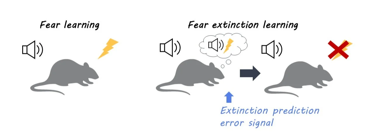
In order to adapt to signals that are no longer dangerous, animals must learn how to stop fear responses through fear extinction learning. Individuals with post-traumatic stress disorder (PTSD) also require this ability to inhibit fear responses. While previous research has shown the involvement of dopamine neurons in learning fear extinction, it is still unclear which specific neuronal pathways are involved. This week in Neuron, Salinas-Hernandez and colleagues identified these dopamine pathways in the midbrain by measuring dopamine neuron activity.

How did they do it?

In order to learn to associate signals with new and different outcomes (dangerous or not dangerous), animals must use prediction errors: where a prediction is met with a different result, an error. To capture brain activity associated with these prediction errors, the authors used fiber photometry to measure activity-dependent calcium signals from dopamine neurons while mice underwent fear extinction paradigms. This involved first playing mice a tone to habituate them to the signal. Next, the mice are conditioned to fear the tone, by pairing it with a foot shock. Last, they played the tone by itself for two days to create the extinction of the fear response. The authors measured dopamine neuron activity in the midbrain of mice both before the fear conditioning and after. They also measured the response to positive signals by rewarding mice with sugar water for nose-poking a target area. To observe whether the fear extinction response was dopamine neuron dependent, the authors optogenetically inhibited dopamine neurons in NAc. Finally, the authors used chemogenetic inhibition to test whether neurons in specific areas of the midbrain were used to signal for prediction error signals.

What did they find?

The authors found that dopamine neurons showed more activity in certain areas of the midbrain — the ventral tegmental area (VTA) and the nucleus accumbens (NAc) — during extinction than during habituation. This suggests these pathways of dopamine learning are used in fear extinction learning. Specifically, neurons in the anteromedial NAc responded with more activity than in other areas. This suggests the prediction error signal is carried to this specific area of the NAc. The authors also saw more calcium signaling, and higher activity, when mice were rewarded by sugar water, which suggests these areas also are activated by rewards. Finally, when dopamine neurons were inhibited in NAc, mice were less able to exhibit extinction learning and continued to display a fear response to just the tone. This provides further evidence that dopaminergic neurons projecting to NAc control fear extinction. Finally, when the authors inhibited dopamine neurons that projected to the VTA (a region with neurons that project to the NAc), mice could not learn fear extinction, suggesting that VTA-projecting dopamine neurons are required for prediction error generation.

What's the impact?

This study is the first to show that midbrain regions control the fear extinction learning for dopamine neurons. This work furthers our understanding of brain regions involved in fear extinction learning and could have future implications in humans. More broadly, it could point to possible treatments for disorders such as PTSD.

Access the original scientific publication here.

Medial Prefrontal Cortical Neurons Trigger REM Sleep

Post by Shireen Parimoo

The takeaway

The onset of rapid eye movement (REM) sleep – the sleep stage commonly associated with dreaming – was previously associated with activity in subcortical brain regions. Here, the authors show that REM sleep can be regulated by medial prefrontal cortex neurons exerting top-down influence on subcortical areas, thereby triggering REM sleep in mice. 

What's the science?

The rapid eye movement (REM) sleep stage is commonly associated with dreaming and is characterized by eye movements as well as an increase in the frequency of theta neural oscillations. Prior work has shown that REM sleep is triggered by subcortical structures like the hypothalamus that in turn give rise to cortical activity. However, it is not known whether cortical neurons can similarly induce REM sleep. This week in Nature Neuroscience, Hong and colleagues used optogenetic stimulation and various imaging techniques to identify the mechanisms by which medial prefrontal and subcortical neurons interact to promote REM sleep.

How did they do it?

The authors injected viral vectors into the medial prefrontal cortex (mPFC) of mice to enhance the expression of light-activated ion channels, which allowed them to stimulate or inhibit the activity of those neurons by shining a laser light. They used two mouse strains; in one strain, they optogenetically manipulated the activity of excitatory pyramidal neurons while in the other strain, they manipulated the activity of inhibitory GABAergic neurons in the mPFC. Optogenetic stimulation was performed under an open-loop setting (i.e., at random times) or under a closed-loop setting (i.e. when the animals entered the REM sleep stage). Open-loop stimulation allowed the authors to observe whether mPFC activity triggered REM sleep from the non-REM (NREM) stage, while the closed-loop stimulation provided insight into the effect of mPFC activation on various features of the REM cycle, respectively.

As the mPFC is widely connected with the rest of the brain, the authors stimulated mPFC neurons terminating in different subcortical regions to identify the subcortical regions that were predominantly activated by mPFC during REM sleep. Next, they used calcium imaging to identify the LH-projecting mPFC neurons according to the sleep stage that they were most active in, that is, during waking, during NREM, and during REM sleep. Of those neurons most active during REM sleep, they further divided them into subgroups based on whether the neurons were relatively more active during wake than during NREM (i.e., R-W-N neurons) or during NREM than during waking (i.e., R-N-W neurons).

Electrodes implanted on the skull were used to obtain recordings of oscillatory brain activity during different sleep stages. Using electroencephalography, the authors recorded changes in power across different frequency bands (e.g., theta and delta), the occurrence of phasic theta events, the duration of REM sleep episodes, and the probability of transitioning between sleep stages (e.g., from NREM to REM sleep). In addition, they performed video-oculography to record eye movement bursts in response to optogenetic manipulation.

What did they find?

Pyramidal neurons in the mPFC were more active during REM than during NREM sleep and wakefulness. Stimulating pyramidal mPFC neurons increased the transition from NREM to the REM sleep stage, longer REM cycles, and a greater frequency of phasic theta events and eye movement bursts. There was also an increase in theta power with a concomitant reduction in delta power. Inhibition of mPFC pyramidal neurons had the opposite effect, with shorter REM cycles and fewer phasic theta events, along with reductions in theta power and phasic theta events. Conversely, stimulating inhibitory interneurons in the mPFC had a similar effect as inhibiting the pyramidal neurons. These findings indicate that excitatory pyramidal neurons in the mPFC are important for promoting REM sleep.

The authors found that only those neurons projecting from mPFC to the lateral hypothalamus (LH) lateral hypothalamus were involved in REM sleep. Specifically, stimulating LH-projecting pyramidal neurons – but not inhibitory interneurons – led to longer REM sleep episodes, increased theta/sigma power along with reductions in delta power, and an increase in the frequency of phasic theta events and eye movement bursts. Inhibiting these neurons instead reduced REM sleep. Lastly, both the R-W-N and R-N-W subgroups of the LH-projecting neurons were active before the transition to REM sleep, but activity in the R-N-W neurons increased earlier than that in R-W-N neurons and had a faster decline in activity at the end of the REM stage. Moreover, the sustained activity of R-W-N neurons was related to longer REM durations and more theta phasic events, indicating that they may be important for maintaining REM sleep and induced phasic theta events. Altogether, these findings indicate that mPFC pyramidal neurons drive REM sleep through their influence on the lateral hypothalamus.

What's the impact?

This study is the first to demonstrate the mechanisms by which medial prefrontal neurons help induce and maintain REM sleep through their projections to the lateral hypothalamus. These findings provide a more comprehensive understanding of the neuronal circuits underlying REM sleep and may have important implications for treating sleep-related symptoms and disorders. 

The Grit Phenomenon: A New Discovery Or a Recycling of Old Ideas?

Post by Anastasia Sares

The takeaway

The concept of grit—passion, and perseverance for long-term goals—has taken the positive psychology world by storm. Why? It is a predictor of success that is distinct from talent, promoting the idea that consistent, hard work is just as important as raw ability. While the discussion around grit has highlighted the importance of effort and motivation in predicting success, critics argue that it may just be a new name that is redundant with already-existing concepts in psychology.

Grit becomes a buzzword

As of this writing, Angela Duckworth’s 2013 Ted talk on grit has over 30 million views, and her book on the subject has made the New York Times bestseller list. She describes her observation that students with the highest IQ did not always end up with the best grades in class. Duckworth subsequently developed questionnaires to measure what she called “grit” and showed that the scores on these questionnaires could predict success in a variety of domains— children’s spelling bee placement, whether people could make it through intense military training, how long a sales’ clerk would retain their job, and even the likelihood of divorce. The predictive power of grit even held after statistically correcting for other factors like socio-economic status, IQ, and feelings of safety.

Criticisms of grit

But wait, you might ask—is this really the first time that psychologists have thought that being a hard worker is a predictor of success, and tried to measure that relationship? Well, no, it isn’t. Other closely related personality factors include conscientiousness (one of the Big Five personality traits), self-control, work ethic, and so on. The question then becomes whether grit has anything to offer above and beyond these other personality factors. Do the questions in the grit questionnaire tap into something unique, like the idea of long-term goals specifically? And do those questions reliably access this concept?

New studies analyze large collections of questionnaires to look at the relationships between grit and other personality factors based on patterns of people’s answers. With techniques like factor analysis and structural equation modeling, they can statistically measure whether concepts are distinct or not. For example, if you know my grit score, how well can you predict my self-control score? If it’s too easy to predict one score by knowing another, they might be redundant concepts, especially if they both predict success in the same way and statistically controlling for one removes the effect of the other.

One large meta-analysis found that overall grit was very closely related to conscientiousness, but that one of its sub-scores, called “perseverance of effort,” was more independent, and also a better predictor of academic performance than the rest of the questionnaire. Other more recent work has proposed grit to be a sub-facet of self-control.

What's the impact?

Grit may have been known by different names in the past, and it may overlap with other concepts in personality psychology, but there is no question that the idea is extremely popular. This may be because it argues against a narrative of “innate talent” that one either has or doesn’t and instead promises that effort and hard work will pay off.

References +

Duckworth, A. L., Peterson, C., Matthews, M. D., & Kelly, D. R. (2007). Grit: Perseverance and passion for long-term goals. Journal of Personality and Social Psychology, 92(6), 1087–1101. https://doi.org/10.1037/0022-3514.92.6.1087

Duckworth, A. L., & Quinn, P. D. (2009). Development and Validation of the Short Grit Scale (Grit–S). Journal of Personality Assessment, 91(2), 166–174. https://doi.org/10.1080/00223890802634290

Eskreis-Winkler, L., Shulman, E. P., Beal, S. A., & Duckworth, A. L. (2014). The grit effect: Predicting retention in the military, the workplace, school and marriage. Frontiers in Psychology, 5. https://doi.org/10.3389/fpsyg.2014.00036

Meriac, J. P., Slifka, J. S., & LaBat, L. R. (2015). Work ethic and grit: An examination of empirical redundancy. Personality and Individual Differences, 86, 401–405. https://doi.org/10.1016/j.paid.2015.07.009

Credé, M., Tynan, M. C., & Harms, P. D. (2017). Much ado about grit: A meta-analytic synthesis of the grit literature. Journal of Personality and Social Psychology, 113(3), 492–511. https://doi.org/10.1037/pspp0000102

Vazsonyi, A. T., Ksinan, A. J., Ksinan Jiskrova, G., Mikuška, J., Javakhishvili, M., & Cui, G. (2019). To grit or not to grit, that is the question! Journal of Research in Personality, 78, 215–226. https://doi.org/10.1016/j.jrp.2018.12.006

Aguerre, N. V., Gómez-Ariza, C. J., & Bajo, M. T. (2022). The relative role of executive control and personality traits in grit. PLOS ONE, 17(6), e0269448. https://doi.org/10.1371/journal.pone.0269448

Van Zyl, L. E., Olckers, C., & Van Der Vaart, L. (Eds.). (2021). Multidisciplinary Perspectives on Grit: Contemporary Theories, Assessments, Applications and Critiques. Springer International Publishing. https://doi.org/10.1007/978-3-030-57389-8