Medial Prefrontal Cortical Neurons Trigger REM Sleep

Post by Shireen Parimoo

The takeaway

The onset of rapid eye movement (REM) sleep – the sleep stage commonly associated with dreaming – was previously associated with activity in subcortical brain regions. Here, the authors show that REM sleep can be regulated by medial prefrontal cortex neurons exerting top-down influence on subcortical areas, thereby triggering REM sleep in mice. 

What's the science?

The rapid eye movement (REM) sleep stage is commonly associated with dreaming and is characterized by eye movements as well as an increase in the frequency of theta neural oscillations. Prior work has shown that REM sleep is triggered by subcortical structures like the hypothalamus that in turn give rise to cortical activity. However, it is not known whether cortical neurons can similarly induce REM sleep. This week in Nature Neuroscience, Hong and colleagues used optogenetic stimulation and various imaging techniques to identify the mechanisms by which medial prefrontal and subcortical neurons interact to promote REM sleep.

How did they do it?

The authors injected viral vectors into the medial prefrontal cortex (mPFC) of mice to enhance the expression of light-activated ion channels, which allowed them to stimulate or inhibit the activity of those neurons by shining a laser light. They used two mouse strains; in one strain, they optogenetically manipulated the activity of excitatory pyramidal neurons while in the other strain, they manipulated the activity of inhibitory GABAergic neurons in the mPFC. Optogenetic stimulation was performed under an open-loop setting (i.e., at random times) or under a closed-loop setting (i.e. when the animals entered the REM sleep stage). Open-loop stimulation allowed the authors to observe whether mPFC activity triggered REM sleep from the non-REM (NREM) stage, while the closed-loop stimulation provided insight into the effect of mPFC activation on various features of the REM cycle, respectively.

As the mPFC is widely connected with the rest of the brain, the authors stimulated mPFC neurons terminating in different subcortical regions to identify the subcortical regions that were predominantly activated by mPFC during REM sleep. Next, they used calcium imaging to identify the LH-projecting mPFC neurons according to the sleep stage that they were most active in, that is, during waking, during NREM, and during REM sleep. Of those neurons most active during REM sleep, they further divided them into subgroups based on whether the neurons were relatively more active during wake than during NREM (i.e., R-W-N neurons) or during NREM than during waking (i.e., R-N-W neurons).

Electrodes implanted on the skull were used to obtain recordings of oscillatory brain activity during different sleep stages. Using electroencephalography, the authors recorded changes in power across different frequency bands (e.g., theta and delta), the occurrence of phasic theta events, the duration of REM sleep episodes, and the probability of transitioning between sleep stages (e.g., from NREM to REM sleep). In addition, they performed video-oculography to record eye movement bursts in response to optogenetic manipulation.

What did they find?

Pyramidal neurons in the mPFC were more active during REM than during NREM sleep and wakefulness. Stimulating pyramidal mPFC neurons increased the transition from NREM to the REM sleep stage, longer REM cycles, and a greater frequency of phasic theta events and eye movement bursts. There was also an increase in theta power with a concomitant reduction in delta power. Inhibition of mPFC pyramidal neurons had the opposite effect, with shorter REM cycles and fewer phasic theta events, along with reductions in theta power and phasic theta events. Conversely, stimulating inhibitory interneurons in the mPFC had a similar effect as inhibiting the pyramidal neurons. These findings indicate that excitatory pyramidal neurons in the mPFC are important for promoting REM sleep.

The authors found that only those neurons projecting from mPFC to the lateral hypothalamus (LH) lateral hypothalamus were involved in REM sleep. Specifically, stimulating LH-projecting pyramidal neurons – but not inhibitory interneurons – led to longer REM sleep episodes, increased theta/sigma power along with reductions in delta power, and an increase in the frequency of phasic theta events and eye movement bursts. Inhibiting these neurons instead reduced REM sleep. Lastly, both the R-W-N and R-N-W subgroups of the LH-projecting neurons were active before the transition to REM sleep, but activity in the R-N-W neurons increased earlier than that in R-W-N neurons and had a faster decline in activity at the end of the REM stage. Moreover, the sustained activity of R-W-N neurons was related to longer REM durations and more theta phasic events, indicating that they may be important for maintaining REM sleep and induced phasic theta events. Altogether, these findings indicate that mPFC pyramidal neurons drive REM sleep through their influence on the lateral hypothalamus.

What's the impact?

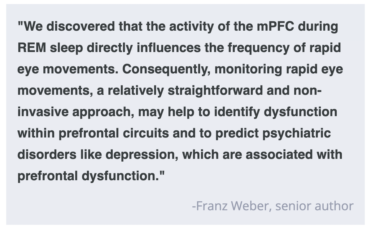
This study is the first to demonstrate the mechanisms by which medial prefrontal neurons help induce and maintain REM sleep through their projections to the lateral hypothalamus. These findings provide a more comprehensive understanding of the neuronal circuits underlying REM sleep and may have important implications for treating sleep-related symptoms and disorders. 

The Grit Phenomenon: A New Discovery Or a Recycling of Old Ideas?

Post by Anastasia Sares

The takeaway

The concept of grit—passion, and perseverance for long-term goals—has taken the positive psychology world by storm. Why? It is a predictor of success that is distinct from talent, promoting the idea that consistent, hard work is just as important as raw ability. While the discussion around grit has highlighted the importance of effort and motivation in predicting success, critics argue that it may just be a new name that is redundant with already-existing concepts in psychology.

Grit becomes a buzzword

As of this writing, Angela Duckworth’s 2013 Ted talk on grit has over 30 million views, and her book on the subject has made the New York Times bestseller list. She describes her observation that students with the highest IQ did not always end up with the best grades in class. Duckworth subsequently developed questionnaires to measure what she called “grit” and showed that the scores on these questionnaires could predict success in a variety of domains— children’s spelling bee placement, whether people could make it through intense military training, how long a sales’ clerk would retain their job, and even the likelihood of divorce. The predictive power of grit even held after statistically correcting for other factors like socio-economic status, IQ, and feelings of safety.

Criticisms of grit

But wait, you might ask—is this really the first time that psychologists have thought that being a hard worker is a predictor of success, and tried to measure that relationship? Well, no, it isn’t. Other closely related personality factors include conscientiousness (one of the Big Five personality traits), self-control, work ethic, and so on. The question then becomes whether grit has anything to offer above and beyond these other personality factors. Do the questions in the grit questionnaire tap into something unique, like the idea of long-term goals specifically? And do those questions reliably access this concept?

New studies analyze large collections of questionnaires to look at the relationships between grit and other personality factors based on patterns of people’s answers. With techniques like factor analysis and structural equation modeling, they can statistically measure whether concepts are distinct or not. For example, if you know my grit score, how well can you predict my self-control score? If it’s too easy to predict one score by knowing another, they might be redundant concepts, especially if they both predict success in the same way and statistically controlling for one removes the effect of the other.

One large meta-analysis found that overall grit was very closely related to conscientiousness, but that one of its sub-scores, called “perseverance of effort,” was more independent, and also a better predictor of academic performance than the rest of the questionnaire. Other more recent work has proposed grit to be a sub-facet of self-control.

What's the impact?

Grit may have been known by different names in the past, and it may overlap with other concepts in personality psychology, but there is no question that the idea is extremely popular. This may be because it argues against a narrative of “innate talent” that one either has or doesn’t and instead promises that effort and hard work will pay off.

References +

Duckworth, A. L., Peterson, C., Matthews, M. D., & Kelly, D. R. (2007). Grit: Perseverance and passion for long-term goals. Journal of Personality and Social Psychology, 92(6), 1087–1101. https://doi.org/10.1037/0022-3514.92.6.1087

Duckworth, A. L., & Quinn, P. D. (2009). Development and Validation of the Short Grit Scale (Grit–S). Journal of Personality Assessment, 91(2), 166–174. https://doi.org/10.1080/00223890802634290

Eskreis-Winkler, L., Shulman, E. P., Beal, S. A., & Duckworth, A. L. (2014). The grit effect: Predicting retention in the military, the workplace, school and marriage. Frontiers in Psychology, 5. https://doi.org/10.3389/fpsyg.2014.00036

Meriac, J. P., Slifka, J. S., & LaBat, L. R. (2015). Work ethic and grit: An examination of empirical redundancy. Personality and Individual Differences, 86, 401–405. https://doi.org/10.1016/j.paid.2015.07.009

Credé, M., Tynan, M. C., & Harms, P. D. (2017). Much ado about grit: A meta-analytic synthesis of the grit literature. Journal of Personality and Social Psychology, 113(3), 492–511. https://doi.org/10.1037/pspp0000102

Vazsonyi, A. T., Ksinan, A. J., Ksinan Jiskrova, G., Mikuška, J., Javakhishvili, M., & Cui, G. (2019). To grit or not to grit, that is the question! Journal of Research in Personality, 78, 215–226. https://doi.org/10.1016/j.jrp.2018.12.006

Aguerre, N. V., Gómez-Ariza, C. J., & Bajo, M. T. (2022). The relative role of executive control and personality traits in grit. PLOS ONE, 17(6), e0269448. https://doi.org/10.1371/journal.pone.0269448

Van Zyl, L. E., Olckers, C., & Van Der Vaart, L. (Eds.). (2021). Multidisciplinary Perspectives on Grit: Contemporary Theories, Assessments, Applications and Critiques. Springer International Publishing. https://doi.org/10.1007/978-3-030-57389-8

A Protein in Microglia That Influences Alzheimer’s Disease Risk

Post by Trisha Vaidyanathan

The takeaway

Two variants of the gene encoding phospholipase C-gamma-2 (PLCG2) have opposing effects on Alzheimer’s disease pathology via their opposing effects on microglia. The first variant (M28L) results in lower PLCG2 levels which reduce the microglial response to plaques and elevate disease risk, while the second (P522R) protects against Alzheimer’s disease by increasing PLCG2 activity, enhancing the ability of microglia to remove plaques and protect synaptic function.

What's the science?

Genetic studies have linked a variant of the gene PLCG2, termed PLCG2-P522R, with reduced risk for Alzheimer’s disease. PLCG2 encodes an enzyme found only in microglia and acts as a critical component of immune signaling within the brain. However, the function of PLCG2 in Alzheimer’s disease is not well understood. This week in Immunity, Tsai and colleagues investigated the “protective” P522R variant and identified a new variant that increases Alzheimer’s disease risk, called PLCG2-M28L. The authors demonstrated that both variants differently alter microglia function, leading to opposing effects on Alzheimer’s disease pathology.

How did they do it?

To investigate the function of PLCG2, the authors first generated mice that had either the “protective” P522R variant or the “detrimental” M28L variant of PLCG2 and determined the effects of this variant on PLCG2 levels. These mice were then crossed to a well-established Alzheimer’s mouse model (called 5xFAD) that is known to develop amyloid plaques, a hallmark of Alzheimer’s pathology. Throughout the study, the authors compared the mice carrying the “protective” and “detrimental” variants of PLCG2 with the typical Alzheimer’s mouse model and healthy control mice.

First, the authors used magnetic resonance imaging (MRI) and immunohistochemistry to measure the buildup of amyloid plaques. Since microglia are known to clean up amyloid plaques, the authors then investigated the proximity of microglia to plaques and measured the ability of microglia to clean up plaque proteins

Next, the authors tested the health of the neurons by measuring synaptic strength and plasticity with electrophysiology, and the mice’s cognitive ability and memory using a Y-Maze. Lastly, the authors used single nuclei RNA sequencing to identify distinct microglia subtypes and microglia functions that are altered by the PLCG2 variants.

What did they find?

The authors first determined that the “detrimental” M28L variant decreased PLCG2 levels, and is thus considered a loss-of-function mutation. In contrast, the “protective” P522R variant is known to increase PLCG2 activity and is considered a gain-of-function mutation

Compared to the typical Alzheimer’s disease mouse model, the loss-of-function M28L variant had more plaque deposits, and the plaques were associated with fewer microglia. The gain-of-function P522R variant had fewer deposits and more microglia coverage. Next, the authors found that microglia with the M28L variant took up less fluorescent amyloid, while P522R took up more, demonstrating that PLCG2 is critical for microglia to eat plaques.

Next, the authors found that mice with the loss-of-function M28L variant had impaired synaptic plasticity (long-term potentiation) and worse cognitive performance than typical Alzheimer’s mice. In contrast, the P522R variant behaved more like healthy controls, confirming that the P522R variant of PLCG2 is protective in Alzheimer’s disease and preserves brain functionality.

Lastly, single nuclei RNA sequencing revealed several subtypes of microglia, including baseline homeostatic microglia, two types of disease-associated microglia, and microglia in states of transition from baseline to disease. The disease-associated microglia expressed several genes related to immune responsiveness and are likely critical to protect against Alzheimer’s disease. Interestingly, the loss-of-function M28L variant resulted in more baseline microglia and fewer disease-associated or transitioning microglia, suggesting that PLCG2 is necessary for microglia to transition into a responsive, disease-associated state. 

What's the impact?

This study characterized two variants of PLCG2 with opposing effects on microglia and Alzheimer’s disease risk. Together, this demonstrated that PLCG2 is critical for mediating Alzheimer’s disease risk via its role in modulating the microglial response to disease. These findings may provide critical insight into PLCG2-directed therapies for Alzheimer’s disease that can enhance the protective ability of microglia to fight disease pathogenesis.  

Access the original scientific publication here.