Predicting How Adversity Changes the Brain

Post by Christopher Chen

The takeaway

Environmental stressors alter our brains. These alterations endure over time, and individual deviations from shared neural patterns associated with adversity hold the potential to predict future psychopathology, like anxiety. 

What's the science?

Brain imaging technologies like functional magnetic resonance imaging (fMRI) have enabled researchers to gather evidence that adversity, particularly during early childhood and adolescence, can lead to abnormal brain development. This may play a significant role in later psychological disorders in adulthood. For instance, brain imaging studies have demonstrated that factors like childhood trauma and poverty can impact the volume of crucial brain regions such as the hippocampus and amygdala - brain regions that are integral to emotion and cognitive function.

Specific types of adversity are thought to uniquely affect particular brain regions, however, it’s still unclear how we can predict outcomes from different types of adversity for unique individuals. Recently in Nature Neuroscience, Holz and colleagues leveraged machine learning and brain imaging data to uncover how distinct types of adversity influence the brain, how this varies across individuals, and how we can predict an individual's likelihood of developing anxiety.

How did they do it?

The researchers conducted a comparative analysis of brain images from 169 at-risk individuals and healthy controls who were part of the Mannheim Study of Children at Risk (MARS), a well-known longitudinal investigation tracking these individuals from birth into adulthood. A replication sample was derived from another imaging study known as the IMAGEN study and shared similar demographic characteristics with the MARS group. The researchers used machine learning to develop a normative model of brain development based on adversity in the MARS group. The same model was employed to generate normative brain images for the same at-risk individuals eight years later and for the replication sample from the IMAGEN study. 

The researchers then engaged in a detailed comparison of essential components within and between these three normative brain models. This included employing a measurement called the dice coefficient to assess the extent of overlap between the neural patterns associated with different types of adversity. Additionally, they harnessed individual-specific z-scores (i.e. how many standard deviations a value is from the mean) to gauge the deviation between an individual’s brain images and the normative brain model. By using linear mixed models, they could gauge how these neural deviations at the individual level predicted the manifestation of anxiety.  

What did they find?

In terms of the brain images from at-risk individuals, the researchers observed a consistent neural signature across subjects, implying that heightened adversity impacts similar brain regions. Remarkably, while brain regions like the hippocampus and amygdala, known to be affected by adversity, exhibited changes in volume, the researchers also noted persistent volume changes in non-limbic system regions like the occipital gyrus and thalamus. They further uncovered that this neural signature remained stable over time. 

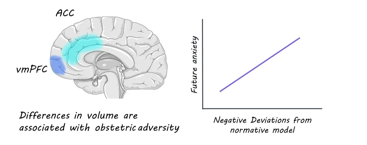
Using dice coefficients, the researchers showcased the links between specific types of adversity and corresponding changes in distinct brain regions. Adversities such as prenatal maternal smoking and obstetric challenges demonstrated lower dice coefficients, signifying their unique impact on specific brain areas. For instance, prenatal maternal smoking was closely tied to volume expansions in the hippocampus and volume contractions (i.e. reductions) in the postcentral and occipital gyrus. Meanwhile, obstetric adversity correlated with volume expansions in the ventromedial prefrontal cortex (vmPFC) and volume contractions in the anterior cingulate cortex (ACC). 

Perhaps the most intriguing revelation was the predictive capacity of the normative model. The researchers discovered that significant negative deviations (indicating volume reduction) in an individual's brain were associated with a higher predisposition to future anxiety.

What's the impact?

These findings underscore the positive correlation between adversity-induced brain changes and the likelihood of anxiety. Moreover, the research suggests that the effects of adversity on the brain could be more profound and enduring than previously believed. Although the study's scope was limited to adults and a relatively small cohort, its specificity holds promise for aiding researchers and healthcare professionals in developing more targeted and effective strategies to help individuals navigate the repercussions of adversity in their lives.

The Potential of Brain Stimulation During Sleep to Enhance Memory Function

Post by Meredith McCarty

The importance of sleep

Sleep is essential for humans and all living species. Despite being a period of apparent vulnerability during daily life, the maintenance of sleep throughout evolution suggests that sleep is fundamental for neural and bodily function. Sleep deprivation can lead to numerous deficits including altered attention, memory, and learning (Krause et al., 2017).  

While humans spend about a third of their lifetimes asleep, the duration and nature of our sleep differ from that of our nearest primate relatives. Humans spend less time asleep than other primates, and relatively more time is spent in rapid eye movement relative to non-REM sleep (Nunn & Samson, 2018). There are many interesting theories as to the evolutionary origin of such changes in sleep quality and duration (see Nunn et al., 2016 for review), and increasing evidence for the essential role of sleep in memory consolidation. 

Memory consolidation during sleep

Memory consolidation describes the process by which information learned from the environment is transferred from temporary short-term memory into more distributed and permanent long-term memory. There is growing evidence that slow-wave sleep (SWS), a period of non-REM sleep marked by low-frequency, high-amplitude brain waves, is pivotal for memory consolidation (Klinzing et al., 2019).  

During SWS, cortical and subcortical regions, namely the hippocampus, thalamus, and neocortex, exhibit distinct patterns of neural oscillations (Ngo et al., 2020). These dynamics are described as thalamo-cortical spindles, hippocampal ripples, and cortical slow oscillations (Latchoumane et al., 2017). Hippocampal ripples, or brief periods of synchronized oscillatory activity, are thought to facilitate communication between the hippocampus and cortical and subcortical regions (Todorova & Zugaro, 2020; Brodt et al., 2023). 

The neural mechanism by which these dynamics may enable memory consolidation is through phase-locking of brain activity between different brain areas, enabling the transmission and nesting of neural signals between brain regions. Animal research has shown that when hippocampal ripples are disrupted, memory consolidation is impaired (Ego-Stengel & Wilson, 2010).

Can memory be enhanced during sleep?

Studies investigating what happens when we disrupt SWS in human and non-human animals have shown that disruption of oscillatory dynamics during SWS can lead to deficits in memory tasks.  But what about the possibility of enhancing memory through sleep?

In a recent study, Geva-Sagiv and colleagues performed closed-loop brain stimulation during sleep in human patients implanted with intracranial electrodes (Geva-Sagiv et al., 2023). The participants performed a cognitive memory task, by which memory accuracy was compared following natural sleep and sleep during which closed-loop stimulation was precisely applied during active phases of SWS. They found enhanced sleep spindles and synchronized spiking between interconnected brain regions following stimulation SWS. Additionally, stimulation during SWS correlated with improved memory accuracy in the behavioral task. These data suggest that the synchronized brain activity during SWS can be increased via external stimulation, correlating with enhanced memory consolidation.  

What’s next?

Sleep-related memory enhancement has enormous implications for clinical application. Sleep deprivation is incredibly detrimental to human health, for both the brain and the body. Insight from sleep research can enable improved treatment for the effects of sleep deprivation and insomnia, as well as the many disorders where sleep disruption occurs. Further research will help to progress our understanding of sleep-related memory enhancement, and how it can be used to make an impact in the future.

References +

Krause et al., 2017. The sleep-deprived human brain. Nature Reviews Neuroscience.

Nunn & Samson, 2018. Sleep in a comparative context: Investigating how human sleep differs from sleep in other primates. American Journal of Physical Anthropology.

Nunn et al., 2016. Shining evolutionary light on human sleep and sleep disorders. Evolution, Medicine, and Public Health.

Klinzing et al., 2019. Mechanisms of system memory consolidation during sleep. Nature Neuroscience.

Ngo et al., 2020. Sleep spindles mediate hippocampal-neocortical coupling during long-duration ripples. eLife.

Latchoumane et al., 2017. Thalamic Spindles Promote Memory Formation during Sleep through Triple Phase-Locking of Cortical, Thalamic, and Hippocampal Rhythms.

Todorova & Zugaro, 2020. Hippocampal ripples as a mode of communication with cortical and subcortical areas.

Brodt et al., 2023. Sleep-A brain-state serving systems memory consolidation. Neuron.

Geva-Sagiv et al., 2020. Augmenting hippocampal-prefrontal neuronal synchrony during sleep enhances memory consolidation in humans. Nature Neuroscience.

The Role of Serotonin in Postnatal Plasticity in the Brain

Post by Trisha Vaidyanathan

The takeaway

There is a brief window after birth in which the brain corrects mistakes formed during embryonic development. This window of rewiring is dependent on serotonin and is negatively affected by preterm birth.

What's the science?

Proper wiring of neuronal circuits is critical for brain function and the brain has a remarkable ability to adapt to errors made during development in a process called neuronal plasticity. However, little is known about the timing of this plasticity or what neuronal signals regulate the process. This week in Proceedings of the National Academy of Sciences (PNAS), Sinclair-Wilson and colleagues investigated how the brain corrects for an embryonic developmental error that prevents sensory neurons in the thalamus from reaching their correct targets in the cortex.

How did they do it?

The authors used a powerful genetic mouse model (Ebf1cKO mice) that disrupts the ability of thalamic sensory neurons from finding their appropriate cortical target — a process that would typically occur during embryonic development. As a result of this mutation, somatosensory thalamic neurons invade the visual cortex instead, while visual thalamic neurons never reach their cortical target. Interestingly, it has previously been shown that these embryonic errors in Ebf1cKO mice are corrected postnatally.

To examine appropriate wiring of sensory circuits, the authors used retrograde labeling to quantify how many visual cortex inputs came from the appropriate visual area of the thalamus, rather than the incorrect somatosensory region of the thalamus. In a subset of their experiments, the authors also used in situ hybridization to visualize molecular markers of cortical regions in individual neurons, revealing the sharpness of boundaries around the visual and somatosensory cortex.

First, the authors performed retrograde labeling at successive postnatal developmental ages and identified the specific time window in which the embryonic errors of the Ebf1cKO mice were corrected. Next, the authors induced preterm labor in pregnant Ebf1cKO mice with the drug mifepristone to ask whether preterm birth affected the window of plasticity and ability to correct wiring deficits. Lastly, the authors gave mice pharmacological drugs to either increase or decrease serotonin levels to investigate the hypothesis that serotonin – which prematurely decreases after preterm birth – regulates postnatal plasticity in Ebf1cKO mice.

What did they find?

First, the authors observed that Ebf1cKO embryonic deficits were corrected early in postnatal development, by postnatal day 2. Although sensory thalamic regions in Ebf1cKO mice underwent significant cell death, ultimately the surviving axons were able to find their correct cortical targets and the sensory cortical areas were structurally and functionally intact. This revealed a brief window for postnatal plasticity shortly after birth.

Second, the authors found that inducing preterm birth in Ebf1cKO mice impaired postnatal plasticity. Preterm Ebf1cKO mouse pups still had visual cortex inputs that originated in somatosensory regions of the thalamus, while at the corresponding postnatal day, full-term Ebf1cKO mice had correctly rewired. This demonstrated that preterm birth negatively affects postnatal plasticity and could impact healthy postnatal development

Lastly, the authors investigated the hypothesis that serotonin, which prematurely decreases in preterm offspring, is necessary for postnatal plasticity. The authors found that daily administration from postnatal day 1 to 3 of a serotonin synthesis inhibitor (parachlorophenylalanine; decreases serotonin levels) impaired plasticity in full-term Ebf1cKO mice, resulting in sensory wiring and cortical boundaries that resembled the preterm Ebf1cKO mice. When the authors administered a selective serotonin reuptake inhibitor (SSRI, fluoxetine) that increases serotonin levels, they were able to rescue the plasticity in preterm Ebf1cKO mice, allowing the sensory thalamic neurons to reach their correct cortical targets and form sharp cortical area boundaries. This revealed that serotonin is a key component of the signaling that underlies postnatal development and the negative impacts of preterm birth.

What's the impact?

This study identified a brief window of plasticity that allows for the correction of errors in embryonic development, and that this window is negatively affected by preterm birth in a serotonin-dependent fashion. Together, this work provides critical insight into the effect of birth timing on brain development and sheds light on potential therapeutic tools that could be used to rescue developmental defects.

Access the original scientific publication here