How Do Sleep Oscillations Promote Long-Term Memory Storage?

Post by Trisha Vaidyanathan

The takeaway

In 1924, a study found that participants remembered a list of nonsense syllables better if they slept – rather than stayed awake – after learning. Since then, many studies have demonstrated how sleep promotes memory. A prevailing theory is that long-term memory is formed during sleep where short-term memories in the hippocampus are transferred to the cortex for long-term storage. This memory storage process is thought to rely on the precise timing of three distinct neural oscillations in the brain. 

What is long-term memory storage?

Memory consolidation, also known as long-term memory storage, is the process by which newly formed “short-term” memories are transformed into long-term, stable memories. Short-term memories are largely encoded in the hippocampus, where the neural representation of a memory is prone to fading. However, these hippocampal representations can be transferred to the cortex for long-term storage during memory consolidation, where there is unlimited capacity for new memories throughout our lifetime.

Memory consolidation starts with “hippocampal replay”

A new memory is composed of several features, including sound, vision, taste, and even emotion. The hippocampus integrates all these features into one unique neural representation or a precise pattern of neuronal activity. As such, new memories are initially completely dependent on the hippocampus. During sleep, the hippocampal neuronal representation of a memory continually “replays” and the sequence of neuronal activity repeats over and over again. Hippocampal replay events mostly occur during a specific stage of sleep called non-rapid eye movement (NREM) sleep and represent the starting point by which these memories are transferred to the cortex for long-term storage.

Three key oscillations coordinate to promote long-term memory storage

How exactly is information transferred from the hippocampus to the cortex during sleep? The prevailing theory is that this transfer occurs because of the precise timing of 3 different types of neuronal oscillations. A neuronal oscillation is generated when a population of neurons continually alternates between synchronous activity and synchronous silence. The precise timing of these oscillations drives communication between brain areas because of spike timing dependent plasticity, or the phenomenon in which two co-active neurons will strengthen their connection. 

The first key oscillation is the sharp wave ripple, a high-frequency oscillation (150-250Hz) generated in the hippocampus during NREM sleep. The sharp wave ripple is critical for memory consolidation since hippocampal replay occurs during the burst of activity that is generated in the active phase of a sharp wave ripple. 

The second key oscillation is the sleep spindle. Sleep spindles are slower oscillations (12-15Hz) that originate in the thalamus during NREM sleep and spread to the cortex and hippocampus. In the hippocampus, sharp wave ripples tend to nest into the troughs – or the active phases – of sleep spindles. This spindle-ripple coupling forms the first bridge by which neuronal activity is transferred outside of the hippocampus.

The last key oscillation is the slow oscillation, a low-frequency oscillation (<1Hz), generated within the cortex in NREM sleep. The active phase of the slow oscillation also called the UP state, can drive the thalamus to generate sleep spindles (which, as mentioned above, are associated with hippocampal sharp wave ripples). This slow-oscillation-spindle-ripple coupling is thought to be the foundation of memory consolidation from the hippocampus to cortex.

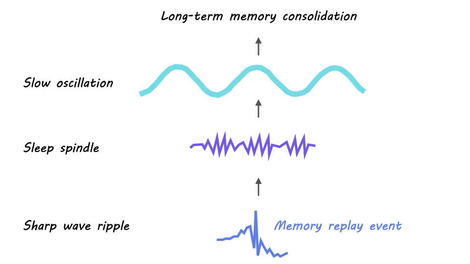
In sum, a prevailing model of memory consolidation is that the cortex opens a window for memory consolidation during sleep when a cortical slow oscillation drives the thalamus to generate a sleep spindle, which in turn synchronizes a hippocampal sharp wave ripple containing a replay event. The resulting synchronous activity – the replay event nested in the sharp wave ripple, nested in the sleep spindle, nested in the slow oscillation – across the cortex, thalamus, and hippocampus is believed to drive memory consolidation through neuronal spike timing dependent plasticity. 

How does this affect our memories?

The theory that long-term memory storage relies on the transfer of memory from the hippocampus to the cortex suggests that our memories could be susceptible to alteration during this process. In fact, there is evidence that suggests the cortex can integrate new memories into pre-existing stored information. This may underlie our ability to extract general principles from a series of individual memories. Additionally, other factors like emotional state may bias how memories are stored. However, further research is needed to better understand these transformations of memory and how they occur.

References +

Goode, T. D., Tanaka, K. Z., Sahay, A. & McHugh, T. J. An integrated index: engrams, place cells, and hippocampal memory. Neuron 107, 805–820 (2020). https://doi.org/10.1016/j.neuron.2020.07.011

Guskjolen, A., Cembrowski, M. S. Engram neurons: Encoding, consolidation, retrieval, and forgetting of memory. Mol. Psychiatry (2023) https://doi.org/10.1038/s41380-023-02137-5

Jenkins, J. G., Dallenbach, K. M. Obliviscence During Sleep and Waking. The American Journal of Psychology, 35, 605–612 (1924). https://doi.org/10.2307/1414040

Joo, H.R., Frank, L.M. The hippocampal sharp wave–ripple in memory retrieval for immediate use and consolidation. Nat Rev Neurosci 19, 744–757 (2018). https://doi.org/10.1038/s41583-018-0077-1

Klinzing, J.G., Niethard, N. & Born, J. Mechanisms of systems memory consolidation during sleep. Nat Neurosci 22, 1598–1610 (2019). https://doi.org/10.1038/s41593-019-0467-3

Skaggs, W. E., McNaughton, B. L. Replay of neuronal firing sequences in rat hippocampus during sleep following spatial experience. Science 271, 1870–1873 (1996). https://doi.org/10.1126/science.271.5257.1870

A Key Micronutrient Identified in Breast Milk Promotes Synaptic Growth

Post by Lani Cupo

The takeaway

Myo-inositol (MYO) is a micronutrient identified in human milk from around the world which helps establish synaptic connections between neurons early in life.

What's the science?

Diet can have a big impact on brain development early in life and preservation later in life, however, it is unclear what micronutrients are important in contributing to connectivity between neurons. This week in PNAS, Paquette and colleagues analyzed samples of human breast milk from around the world to identify a micronutrient MYO that is present during periods when neuronal connections are formed. They then validate the ability of MYO to promote synapse formation in cultured neurons and mouse models.

How did they do it?

First, the authors collected samples of breast milk from women from Mexico City (N = 10), Shanghai (N = 10), and Cincinnati (N = 10) at 5 time points throughout the first year postpartum. Focusing on inositol, a sugar that is elevated in human milk compared to cow’s milk, the authors quantified the amount of MYO, a form of inositol not bound in other molecules, across time. Second, using human induced pluripotent stem cells the authors cultivated human glutamatergic neurons to examine the impact of MYO on synaptic sites.

Next, the authors cultivated hippocampus neurons from rats in vitro (in a Petri dish) and exposed them to differing dosages of MYO to examine the impact on the synapses between neurons. Then, the authors conducted an analysis in mice by supplementing them with MYO from birth to 35 days old and examining synaptic sites in the visual cortex. Finally, they examined the impact of MYO in mature tissue - aging neurons from the mouse hippocampus - before exposing them to the sugar.

What did they find?

First, the authors found that MYO levels are highest in the first weeks of lactation, a time period associated with dramatic increases in synaptic density in babies. Second, introducing MYO to cultured neurons increased post-synaptic staining intensity, an indicator of increased sites for synaptic connections.

Next, the authors found that MYO increased synapse formation in a dose-dependent manner with higher dosages leading to an increased impact. These results provided further evidence of MYO’s role in promoting synapse formation for both excitatory and inhibitory neurons. Then, the authors found enlarged synaptic sites in the visual cortex of mice exposed to supplements of MYO after giving birth. This further emphasizes the role of MYO across species and brain regions. Finally, the authors found evidence of increased synapses in mature tissue as well, suggesting a potential preservative effect of MYO in adults.

What's the impact?

The authors highlight the importance of the micronutrient MYO in synapse formation and neuronal connectivity, both in development and mature tissue. Their results provide avenues to improve pediatric nutrition products, and may, in time, promote synapse protection in aging.

Access the original scientific publication here

How Do Pesticides Affect the Brain?

Post by Anastasia Sares

The takeaway

Pesticides kill unwanted weeds, pests, and fungi, often with molecules affecting the nervous system, but their mechanisms of activation can cause havoc in “non-target” species, including the humans that use them. Over the years, some pesticides have been banned for their toxic effects and new ones have been developed. Now we’re moving from recognizing acute poisoning situations to being able to assess long-term detriments, especially to cognitive function in humans.

How has pesticide use evolved?

Humans have used pesticides for a long time to increase crop yields or kill unwanted guests in their homes and bodies. We’ve tried everything from chrysanthemums to nicotine to arsenic to DDT. However, we walk a fine line with pesticide use: on the one hand, it can increase food security and decrease disease (for example, killing malaria-carrying mosquitoes), both of which preserve human life. On the other hand, the effects of pesticides may decrease biodiversity and have toxic effects on the human body.

When developing a pesticide, it would be ideal to come up with a chemical that disrupts only the pest’s biological processes and is otherwise “harmless.” Unfortunately, as we are all a part of the same tree of life and share many biological processes with other organisms, it is hard to find chemicals that match this ideal. Pesticides like DDT have been introduced and commercialized, only to be later banned (at least in some countries) when toxic effects were observed. But some pesticides act in more subtle ways or require accumulated doses, and the link to age-related diseases like Parkinson’s and Alzheimer’s has only emerged after longer periods of observation. Scientists have been hard at work trying to understand the more subtle mechanisms of pesticide neurotoxicity.

Oxidative stress is a common factor

Though different pesticides have different primary mechanisms for killing their target pest, many of them also cause oxidative stress in cells. Oxidative stress is a process that disrupts the normal functioning of mitochondria, the energy generators of the cell.

Normally, mitochondria convert molecules derived from our food and oxygen from our lungs into water and carbon dioxide through a series of controlled chemical reactions. The energy generated from these reactions is used to “charge up” molecules called ATP (adenosine triphosphate), and these ATP molecules travel throughout the cell and act as little batteries to provide energy for other chemical reactions that need it. Sometimes the electron transport chain fails and instead of producing carbon dioxide and water, it produces a rogue molecule with a negatively charged oxygen called a reactive oxygen species, or ROS. These ROS can leave the mitochondria and do damage to other parts of the cell, like membranes, proteins, or even DNA, which can lead to cell death (see this video at 5:15).

In a healthy cell, the activity of ROS is kept to a minimum, but pesticides can make the production of ROS more likely, causing the cell to lag in energy production and accumulate damage to DNA and proteins. This is the state of oxidative stress, and neurons are particularly sensitive to it.

What are some other mechanisms of neurotoxicity?

Pesticides can cause a myriad of other effects besides oxidative stress, including the accumulation of dementia-related proteins such as amyloid beta and tau, toxic buildups of signaling molecules like acetylcholine or glutamate, inflammation and immune cell activation, DNA damage and suppression (methylation), altered neuron structure and growth, and abnormal activation of growth factors, to name a few.

Some mechanisms of neurotoxicity are quite complex, and have only recently been brought to light: for example, glyphosate (a weed-killer) interrupts a biological process not present in human cells, which made it seem safe to use. However, it turns out that glyphosate can affect our gut bacteria which produce tryptophan, a precursor for the neurotransmitter serotonin. This change in the serotonin production chain can lead to anxious or depressive symptoms. Researchers will continue looking for complex reactions like these to better estimate the true effects of pesticide use. In addition, new research may need to focus on the synergistic effects of multiple pesticides instead of looking at one pesticide at a time.

What's the impact?

Regulation of pesticides is absolutely necessary to maintain the right balance between pest/disease control and human health, and continued research on the effects of pesticides is needed to inform those regulatory decisions.

References +

  1. Aloizou, A.-M., Siokas, V., Vogiatzi, C., Peristeri, E., Docea, A. O., Petrakis, D., Provatas, A., Folia, V., Chalkia, C., Vinceti, M., Wilks, M., Izotov, B. N., Tsatsakis, A., Bogdanos, D. P., & Dardiotis, E. (2020). Pesticides, cognitive functions and dementia: A review. Toxicology Letters, 326, 31–51. https://doi.org/10.1016/j.toxlet.2020.03.005
  2. Costa, L., G. (2008). Neurotoxicity of pesticides: A brief review. Frontiers in Bioscience, 13(13), 1240. https://doi.org/10.2741/2758
  3. Richardson, J. R., Fitsanakis, V., Westerink, R. H. S., & Kanthasamy, A. G. (2019). Neurotoxicity of pesticides. Acta Neuropathologica, 138(3), 343–362. https://doi.org/10.1007/s00401-019-02033-9
  4. Franco, R., Li, S., Rodriguez-Rocha, H., Burns, M., & Panayiotidis, M. I. (2010). Molecular mechanisms of pesticide-induced neurotoxicity: Relevance to Parkinson’s disease. Chemico-Biological Interactions, 188(2), 289–300. https://doi.org/10.1016/j.cbi.2010.06.003
  5. Rueda-Ruzafa, L., Cruz, F., Roman, P., & Cardona, D. (2019). Gut microbiota and neurological effects of glyphosate. NeuroToxicology, 75, 1–8. https://doi.org/10.1016/j.neuro.2019.08.006