How Does Episodic-Like Memory Formation Develop?

Post by Baldomero B. Ramirez Cantu

The takeaway

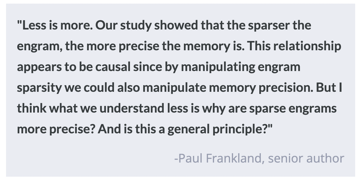
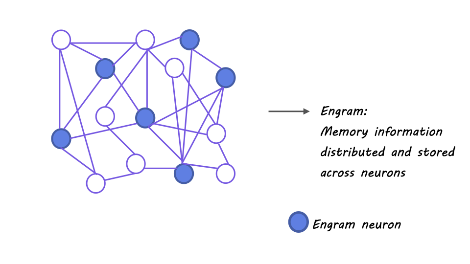
The development of the hippocampus from birth involves multiple stages of functional maturation, which are responsible for increases in the precision of episodic and episodic-like memory as we age. Engram sparsity (i.e. how scattered or distributed it is across brain regions) and the maturation of a specific subset of inhibitory interneurons are key features of this development.

What's the science?

Episodic memory refers to an individual's personal recollection of experiences, events, and situations, which is not innate but rather developed through brain maturation. However, the specific neurobiological mechanisms underlying the transition from gist-like memories to more precise, detailed memories remain unclear. This week in Science, Ramsaran and colleagues provide insights into the underlying neurobiological basis of the development of episodic-like memory. 

How did they do it?

The authors used a variety of techniques in order to investigate the development of hippocampal engrams (i.e. physical memory traces in the brain) and episodic-like memory in mice. They primarily relied on behavioral assays of memory, optogenetic and chemogenetic manipulations, and immediate early gene expression markers. First, the authors trained mice in contextual fear conditioning tasks at different ages in order to test their ability to recall a particular context as a function of their age. They optogenetically silenced the activity of the CA1 region of the hippocampus while the mice performed this task in order to first check whether the task was hippocampus dependent in juvenile and adult mice.

Next, they manipulated CA1 activity by expressing either excitatory or inhibitory designer receptors exclusively activated by designer drugs (DREADDs) and observed the effect that these manipulations had on engram size and sparsity. The authors then tested the role of CA1 neurons in memory recall in mice aged 20, 24, and 60 days by expressing both excitatory and inhibitory opsins in the same subset of neurons. They expressed an inhibitory DREADD into parvalbumin expressing (PV) inhibitory interneurons to test the role of these neurons in engram density and sparsity. Furthermore, the authors perturbed the expression of perineuronal nets (PNNs) — crucial for the appropriate development and function of parvalbumin inhibitory interneurons — using a viral vector approach.

What did they find?

Inhibiting a subset of CA1 neurons with inhibitory DREADDs prior to memory acquisition decreased engram size and led juvenile mice to display adult-like memory precision. Conversely, activating these neurons with excitatory DREADDs increased engram size and caused adult mice to exhibit juvenile-like memory. Silencing of CA1 neurons using the bidirectional optogenetic strategy described above hindered fear recall in 24 and 60 day old mice, but not in 20 day old mice. This suggests that information is stored in a much broader subset of neurons in the 20 day old mice and therefore that the sparser the engram is, the more precise the memory is. Therefore inhibiting only a subset of these neurons is not sufficient to fully impair memory recall. Chemogenetic inhibition of PV neurons during learning caused the expression of juvenile-like memory allocation and imprecision. These findings support the hypothesis that immature PV neuron function may underlie juvenile-like memory phenotypes. Perturbing and destabilizing CA1 PNN resulted in imprecision of contextual memory, while accelerating the maturation of CA1 PNN led to adult-like engram formation and precise contextual memory acquisition.

What's the impact?

This study expands our understanding of how memory develops and is stored in the brain and can inform the development of strategies for enhancing memory function and treating memory-related disorders. In addition, this research provides important insights into the complex processes of neural development and maturation. 

Targeting the Gene that Produces Tau to Fight Alzheimer’s Disease

Post by Christopher Chen

The takeaway

MAPT is a gene in the body that codes for tau, one of the key pathological markers of Alzheimer’s disease (AD). By using a new therapeutic designed to disrupt MAPT, researchers found that treated patients saw more than a 50% reduction in tau levels.

What's the science?

Affecting over 50 million people worldwide, AD is a neurodegenerative disease resulting in severe cognitive decline and decreases in quality of life. A critical pathological marker in AD is tau, a protein in neurons that at certain levels will prevent a neuron’s ability to “talk” with other neurons, a process called neurotransmission. Animal models that express AD-like phenotypes show marked increases in tau accumulation as well as decreased overall brain function. Conversely, researchers have found that lowering tau production in animal models leads to enhancements in memory and overall brain functioning.

In humans, the gene MAPT codes for tau production. Thus, strategies aimed at reducing MAPT activity may lead to reductions in tau levels and improvement in symptoms of AD. Synthetic molecules called antisense-nucleotide oligonucleotides (ASO) can hijack a gene’s ability to make new proteins (like tau), thus making them good potential therapeutic agents to use in the fight against AD. In a recent article in Nature Medicine, researchers conducted a clinical trial where they injected patients with AD with an ASO called MAPTRx to see if doing so would lead to a reduction in tau levels. 

How did they do it?

The randomized, double-blind, placebo-controlled clinical trial was conducted over a 36-week period (13-week treatment period + 23-week post-treatment period) across multiple research centers and focused on 46 total patients (34 experimental, 12 placebo) with mild AD. In short, patients were given a spinal injection of either MAPTRx or placebo at fixed intervals throughout a 13-week period with tau levels in the cerebrospinal fluid (CSF) taken at specific timepoints during the 36-week period.  

One of the trial’s primary goals was the overall safety of MAPTRx so researchers employed a diverse set of techniques to measure physiological and cognitive health throughout the trial. Specifically, researchers recorded any adverse events patients experienced, gave neurological and physical exams, took bloodwork, and measured readouts from MRI and electrocardiograms (EEG).

There were also therapeutic goals that focused on how well patients metabolized the drug and whether it lowered overall tau levels. To understand MAPTRx’s pharmacokinetics (PK), or how the drug was metabolized in the body, researchers used a variety of specialized measurement techniques to quantify whether increasing dosages of MAPTRx resulted in higher baseline levels in the body. As for quantifying the amount of tau, researchers used similar measurement techniques to compare the concentration of tau in the CSF from experimental and control cohorts.  

What did they find?

Considering the trial was early phase, researchers were primarily focused on the safety of MAPTRx. First, injection of MAPTRx did not lead to any adverse events deemed severe or serious, with all symptoms being recorded as either mild (88%) or moderate (12%). The most common symptom was a post-lumbar puncture headache, which was a mild headache following the spinal injection. As for the drug’s metabolic profile, MAPTRx concentrations were found in dose-dependent concentrations, showing that the body did not eliminate it immediately and suggesting that the drug remains in the body long enough to exert its therapeutic effects.

In terms of lowering overall tau concentration, MAPTRx-treated patients expressed on average ~50% reduction in CSF tau concentration compared to controls. However, no significant therapeutic effects of these reductions were seen on cognitive assessments in post-treatment analysis. It should be noted that this study was not powered to show a cognitive benefit; such a trial would require larger numbers of participants. The authors are exploring this in a phase 2 trial that is ongoing. The value of the exploratory cognitive measures is in how they will inform future clinical trials more focused on the effects of MAPTRx on AD-related health outcomes.

What's the impact?

This clinical trial was the first of its kind using ASO-mediated treatment of AD. It showed that the drug was relatively well-tolerated in the body and that such treatment did not lead to any severe side effects in patients. Perhaps most significantly, it showed target engagement: treated patients saw on average a 50% reduction in tau levels in the body. While the drug’s overall ability to slow the cognitive decline associated with AD is still unclear, researchers acknowledged that more comprehensive trials are needed to determine the full therapeutic potential of MAPTRx.

Access the original scientific publication here.

Decoding “Zoom Fatigue”

Post by Anastasia Sares

The takeaway

During the COVID pandemic, the term “Zoom fatigue” was coined as people participated in more virtual meetings than ever before. Now, since virtual work is here to stay, scientists are trying to dissect this phenomenon to better understand what it is about virtual conferences that is so draining. It turns out there are many factors that may be at play, and not all of them have to do with the technology itself.

What’s different about video-conferencing?

Through conceptual analysis, it is possible to collect theories from different areas of psychological research about how virtual communication can cause fatigue. One important area of difference is eye contact: this is a crucial element of human nonverbal communication, and it works very differently in virtual meetings. In in-person meetings, we use eye contact sparingly, taking turns looking at different speakers, whereas, during an on-screen meeting, everyone can look at everyone else during the entire meeting—the experience of being stared at by 8-10 people for an hour straight can change our body’s arousal and stress responses. At the same time, full and mutual eye contact is impossible, because for one person to experience full eye contact, the other person has to look into their camera. Being in different spaces and unable to focus on the same things in the environment or experience the three-dimensional movement of another person further hampers nonverbal communication.

The lag in video calls is another important factor that can hamper behavioral and brain synchrony. People are very sensitive to conversational timing, and while spontaneous coordination can arise in person, it can often be disrupted in virtual environments. In-person, eye contact can also synchronize brain waves between individuals, but this effect is attenuated in virtual situations (as covered in a previous BrainPost).

Call quality can also increase the amount of effort needed for communication. Noisy audio can increase listening effort, which in turn reduces the neural resources left over for memory. In addition, poor video quality can increase visual fatigue. People on virtual calls may also feel the need to exaggerate their voice or expressive gestures in order to be understood, leading to heightened social monitoring and more fatigue.

Figure from Döring et al.

But wait, what if it’s not the technology?

As research into Zoom fatigue continues, it will be important to account for factors that are less related to the technology itself, and more related to the situations people might use it in. For example, when scheduling virtual meetings, the number and length of meetings might differ, as well as the number of breaks afforded, and the reason for needing a virtual meeting in the first place (home obligations, financial issues precluding travel, a global pandemic) might create a situation of stress before the meeting even starts. In addition, an individual’s technological ability and personality, including their opinion of virtual meetings, may impact their experience.

What's the impact?

As researchers continue to explore the underlying factors of "Zoom fatigue," a better understanding of the multifaceted reasons behind the exhaustion experienced in virtual meetings can be achieved. This knowledge can then be used to develop improved strategies and tools for virtual communication, ultimately enhancing the well-being and productivity of remote workers. With virtual work becoming an integral part of modern society, addressing the issue of Zoom fatigue is vital for fostering healthier, more effective communication in the digital era.

References +

  1. Döring, N., Moor, K. D., Fiedler, M., Schoenenberg, K., & Raake, A. (2022). Videoconference Fatigue: A Conceptual Analysis. International Journal of Environmental Research and Public Health, 19(4), 2061. https://doi.org/10.3390/ijerph19042061
  2. Troje, N. F. (2023). Zoom disrupts eye contact behaviour: Problems and solutions. Trends in Cognitive Sciences, 27(5), 417–419. https://doi.org/10.1016/j.tics.2023.02.004
  3. Bailenson, J. N. (2021). Nonverbal overload: A theoretical argument for the causes of Zoom fatigue. Technology, Mind, and Behavior, 2(1). https://doi.org/10.1037/tmb0000030
  4. Zamm, A., Debener, S., & Sebanz, N. (2023). The spontaneous emergence of rhythmic coordination in turn taking. Scientific Reports, 2023(13), 3259. https://doi.org/10.1038/s41598-022-18480-6
  5. Schwartz, L., Levy, J., Endevelt-Shapira, Y., Djalovski, A., Hayut, O., Dumas, G., & Feldman, R. (2022). Technologically-assisted communication attenuates inter-brain synchrony. NeuroImage, 264, 119677. https://doi.org/10.1016/j.neuroimage.2022.119677