The Impact of Corticosteroid Treatments on Hippocampal Function

Post by Baldomero B. Ramirez Cantu

The takeaway

This study provides evidence that corticosteroid treatments can disrupt the circadian regulation of the hippocampus, leading to impairments in hippocampal function and plasticity.

What's the science?

Circadian rhythms are natural 24-hour cycles that regulate various physiological, biological, and behavioral processes in organisms. Corticosteroid treatments are commonly used to manage inflammatory and immunologic disorders, but they can produce side effects such as mental health issues and memory deficits. Despite their widespread use, the underlying mechanisms behind these side effects remain poorly understood. This week in PNAS, Birnie, Claydon and colleagues identify a molecular basis for the memory deficits in patients treated with corticosteroids and provide insights into the influence of corticosteroid treatment on hippocampal function.

How did they do it?

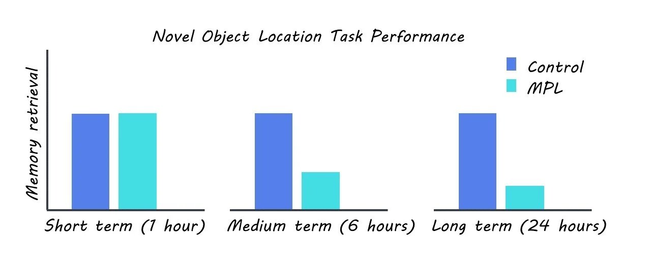
The authors used a rat model to mimic corticosteroid treatment by administration of the commonly prescribed corticosteroid methylprednisolone (MPL). The rats were housed in a 12-hour light-dark and were administered MPL orally. The authors screened for behavioral, genetic, and neurophysiological changes in the MPL group relative to a control group. They monitored changes in locomotion and body temperature between the two groups in order to screen for any potential impacts of the treatment on these behaviors. They also performed a novel object location task to assess changes in short, intermediate, and long-term memory. RNA sequencing was used to measure changes in gene expression in the two groups. Protein levels were assayed using Western blotting. Intracellular ex-vivo recordings were used to explore changes in synaptic plasticity as a result of corticosteroid treatment.

What did they find?

The authors observed that corticosterone treatment caused disruptions in synaptic physiology and gene expression in the hippocampus. Specifically, the authors found that corticosterone treatment impaired long-term potentiation (a measure of synaptic plasticity that is associated with long-term memory), altered the expression of genes involved in circadian regulation and synaptic plasticity, and disrupted the amplitude and frequency of miniature excitatory postsynaptic currents (mEPSCs) in the hippocampus. The authors also showed that corticosterone treatment inhibited NMDAR-dependent plasticity, which is a key mechanism underlying learning and memory in the hippocampus.

In addition, their behavioral task found that rats treated with corticosterone exhibited impaired intermediate and long-term memory, but not short-term memory. The authors further investigated the molecular basis for these effects by analyzing gene expression in the hippocampus. They found that corticosterone treatment dysregulated genes that are known to be crucial for memory processing in the hippocampus and circadian regulation, including CAMKII and CLOCK. Crucially, the authors found that the time of day was a potent modulator of hippocampal activity and that this temporal regulation is disrupted by corticosteroid treatment.

What's the impact?

This study sheds light on the underlying mechanisms of the cognitive side effects of long-term corticosteroid treatments, which are very commonly used to treat many inflammatory and immunologic disorders. Furthermore, these findings could lead to the development of new therapeutic approaches for patients who experience cognitive side effects of corticosteroid treatments. 

Access the original scientific publication here.

Using Virtual Reality to Probe Human Spatial Learning and Decision Making

Post by Anastasia Sares

The takeaway

Using a virtual reality environment and fMRI, researchers can study human navigation and how we can learn what is rewarding in our environment. This article shows how the orbitofrontal cortex, a region located in the frontal lobe just above the eyes, interacts with our hippocampus (the brain’s map-making region) in situations where knowledge about our surroundings can help when we face new decisions.

What's the science?

One of the difficulties in trying to understand how humans learn about objects in their environment is that objects close to each other in space also tend to be close to each other in time. That is, as we explore spaces, we tend to encounter close-by objects sooner and far-away objects later. However, since everyone navigates their environment in slightly different ways, they experience their own unique sequence of events while reconstructing the same space in their head.

This week in Nature Neuroscience, Garvert and colleagues designed a virtual reality experiment to pull apart the time and space aspects of our navigational abilities and to show what happens when one of those dimensions (here: space) can inform new decisions.

How did they do it?

The authors conducted a three-day experiment where participants learned their way around a virtual arena with monsters in it; the knowledge they acquired in the arena could later be used to maximize rewards.

On day 1, participants started by exploring the arena, which had some landmarks around the edge, and hidden monsters scattered about (they would not appear until the person “walked” to just the right position). The goal on the first day was just to memorize the locations of the monsters. After exploring, participants were tested by walking to the locations of the monsters they were tasked to find.

On day 2, participants had some additional practice and were put into the functional magnetic resonance imaging (fMRI) scanner. While their brains were scanned, they viewed images of the monsters and were encouraged to think about their location in the arena. Sometimes, two monsters would appear, and they would have to choose the one whose location was closest to the monster presented previously. This allowed the researchers to investigate how the monsters were represented in the brain.

On day 3, participants practiced locating the monsters again and entered the scanner for a second time. This time, they were presented with pairs of monsters and told to select the one with a higher reward, knowing that monsters that lived in close proximity would be worth a similar number of points. Points were presented after they made their choice on each trial. In reality, monsters close to a certain point in the arena were worth more points, and those further away were worth less. At the end of the choice task, participants were presented with two monsters whose reward value had never been shown in the choice task and asked to predict the reward value. Participants could successfully do this, demonstrating that they used knowledge about the monster locations to predict unseen rewards. Finally, participants completed the viewing task again, just like they had on day 2, this time thinking about both the spatial location and reward value of each monster.

What did they find?

Participants had a variety of exploration strategies in the virtual environment, and thus each person visited the monsters in a unique order.

However, in the choice task, the spatial relationships and not the order in which they saw the monsters could be used to maximize rewards. The authors compared different models to see which best explained the participants’ choices: one that relied only on predictions based on spatial relationships, one that relied on predictions from that person’s exploration order, and one that combined both. It was the “both” model that ended up making the best predictions. People use a combination of time (the order of their experiences) and space (the map representations they have built) to represent the relationship between two objects. Some people leaned more towards spatial relationships and were better at updating them, while others relied more on the order in which they had experienced things.

This was also true in the brain. The team focused on the hippocampus, a region that is known to help us make cognitive maps and navigate our environment. The authors were able to use activity in this region to predict how much people used their knowledge about the relative distances between monsters to infer the values of monsters whose values they had not experienced, yet, on day 3. So, this region is likely involved in helping people generalize about properties of their environment such as whether it’s rewarding or not. However, it was not the only brain region involved: activity in the orbitofrontal cortex was related to the participant’s ability to update and refine their strategy, focusing on spatial aspects to get the optimal reward. This is a neat confirmation of a similar animal-based result in a recent brainpost.

What's the impact?

Learning to navigate to rewards is a crucial skill for any animal’s survival—we must have maps to rewards like food sources or safe places to sleep. The interaction of the hippocampus and the orbitofrontal cortex seems to be key to this skill, now being shown in both animals and in humans.

Finding Better Ways to Target Epilepsy Zones in the Brain

Post by Christopher Chen

The takeaway

Surgically removing the epileptogenic zone (EZ) – the brain region where epileptic seizures are generated – can allow patients with drug-resistant epilepsy (DRE) to live seizure-free, but it is often challenging to locate the EZ’s precise location. By mapping the propagation of interictal spikes - brief spontaneous neural discharge between seizures - researchers discovered key components of successful surgical resections that may help improve health outcomes in patients with DRE.

What's the science?

In patients with drug-resistant epilepsy (DRE), complete surgical removal of the epileptogenic zone (EZ) – the brain region where seizure activity is generated – can allow patients to live a life free from seizures. Currently, clinicians locate the EZ using a biomarker called the seizure onset zone (SOZ) which is identified by using a technique called intracranial electroencephalography (iEEG). However, iEEG has several technical limitations that may lead to defining a SOZ that does not encompass the entire EZ, thus leading to incomplete EZ resection and poor clinical outcomes.

To offset these limitations, clinical researchers have devised a new technique combining electric source imaging (ESI) with iEEG to provide more precise identification of the EZ. ESI allows for the mapping of propagating electric signals following a seizure (i.e., interictal spikes) and clearer delineation of brain function following a seizure. In a recent article in Brain, researchers used this strategy on patients with DRE to identify physical and network signatures that correlate with both good and poor surgical outcomes. 

How did they do it?

To characterize brain activity in patients with both good and poor surgical outcomes, researchers retroactively analyzed brain imaging and network activity data from roughly 40 children with DRE at a single hospital site in the United States over a six-year period. Working with a team of clinicians, researchers analyzed patient data to generate 3D images of each patient’s brain along with functional maps of brain activity.

Researchers then needed to characterize the physiological and network characteristics of patient brains. They used ESI to help reconstruct three key spatiotemporal zones of interictal spike propagation: spike onset, early spread, and late spread. In other words, they measured and identified the physical location of the spike signal in the brain over the first 10% of its spread (onset), the next 10% of its spread (early spread), and the final 80% of its spread (late spread). Researchers also measured the level of functional connectivity between these three zones as well as their degree of physical overlap with the surgically-resected region. These data points were then compiled and analyzed in order to compare profiles of patients who had good and poor outcomes from surgical resection.  

What did they find?

There were several notable findings pertaining to the patient profiles from good and poor surgical outcomes. In terms of the type of electric events, there were no major differences in the type of spike activity (isolated vs. propagating) and only slight differences in the descriptive characteristics of the spikes themselves (e.g. spike velocity) between patient groups. However, there was a key difference in the actual direction of the spike spread. Specifically, spike spread was more organized and hierarchical in patients with good outcomes (onset -> early spread -> late spread). Researchers also found that patients with good outcomes had surgical resections physically closer to the onset, early and late spread zones. 

Furthermore, when researchers delved deeper into the patient profiles with good outcomes, they found that their surgical resections were closest to one zone in particular, the spike onset zone and that surgical resection of the spike onset zone was an accurate predictor of surgical outcome. Interestingly, researchers also found that resection of the clinically-defined SOZ was not an effective predictor of good surgical outcomes. Overall, these findings point to the idea that patient profiles with good surgical outcomes were highlighted by more directionally-organized information flow and resection that was close to the spike onset zone.

What's the impact?

This research highlights network connectivity signatures and spike propagation analysis as critical strategies in identifying the EZ in patients with DRE. Researchers were able to link resection of the spike onset zone to good surgical outcomes suggesting that locating the onset zone in patients with DRE may be another critical step in enhancing the probability of a good outcome in surgical resection. While the sample size was relatively small and concentrated in a single hospital site – their findings should help inform therapeutic interventions clinicians can employ to help patients with DRE live seizure-free.