The Impact of Air Pollution on the Brain

Post by Christopher Chen 

The takeaway

Exposure to air pollution has been linked to negative effects on cardiovascular and respiratory health, but its impact on brain function remains unclear. Scientists discovered that short-term exposure to air pollution in controlled conditions negatively affected fMRI measurements of brain connectivity, suggesting that air pollution may be harming our brains. 

What's the science?

The harmful effects of traffic-related air pollution are well-documented, with numerous studies showing how exposure to air pollution harms cardiovascular and respiratory health. Further, researchers have linked air pollution to negative outcomes on brain health, with some reports suggesting that air pollution particles transmitted to the brain via the olfactory bulb may enhance neuroinflammation. However, the effects of traffic-related air pollution on the brain remain relatively unexplored, at least under controlled conditions. A recent article in Environmental Health investigated the effects of exposure to traffic-related air pollution on the brain and cognitive function. 

How did they do it?

Twenty-five healthy adults were selected and screened for inclusion in this study. They were then chosen at random to be initially exposed to either: diluted diesel exhaust (DE) or filtered air (FA) under double-blind conditions at the Air Pollution Exposure Lab at the University of British Columbia. Immediately before exposure, researchers measured brain activity using fMRI. Following initial fMRI measurement the two-hour exposure began with the participants lightly cycling on a stationary bike for fifteen minutes in order to generate a representative level of activity. Immediately following the two-hour exposure, brain activity was again measured using fMRI. Two weeks later, participants first exposed to DE were exposed to FA while those first exposed to FA were exposed to DE. 

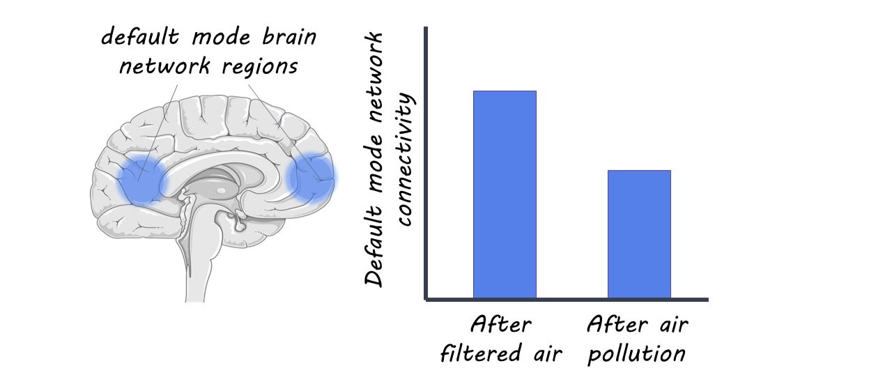
Following the conclusion of the experimental portion of the study, investigators analyzed fMRI data for changes in the blood oxygen level-dependent (BOLD) signal following DE and FA exposure. Specifically, they focused on brain regions linked to the default mode network, a well-studied functional brain network involved in higher-level thinking skills and memory that includes regions that are active together during wakeful rest. 

What did they find?

The fMRI data revealed several key differences in brain activity following exposure to DE and FA. The fMRI data also showed that under control conditions (exposure to FA), subjects saw a significant increase post-exposure in brain activity in the right middle temporal gyrus and occipital fusiform gyrus, brain regions known to be linked to the default mode network. This increase was not present after DE exposure. Furthermore, when comparing widespread changes in brain connectivity, researchers showed that brain connectivity in FA conditions was significantly enhanced compared to DE conditions. In other words, there was decreased brain connectivity after DE exposure compared to after FA exposure, suggesting that the brain isn’t functioning as well after exposure to diesel air. 

What's the impact?

Investigators found that traffic-related air pollution decreased brain connectivity compared to exposure to clean air. Overall, these findings suggest that there are negative effects of short-term exposure to air pollution on the brain. Future studies are needed to examine pollution exposures over longer durations to better understand the long-term impact.

Neurons in the Brainstem Modulate Pain Sensation

Post by Baldomero B. Ramirez Cantu

The takeaway

This study provides evidence that neurons located in the medulla oblongata are involved in modulating pain sensation. These neurons exert their inhibitory effects through a tract that connects the cortex and spinal cord, regulating the perception of pain.

What's the science?

The detection of stimuli that could be perceived as painful typically begins with nociceptive neurons in the peripheral nervous system, which transmit signals to higher brain centers to produce an appropriate sensory response. However, the perception of pain is not fixed and can be modulated to suit a specific context. For example, individuals may exhibit a higher pain tolerance while pursuing a goal, or a lower pain tolerance while a particular region of the body is undergoing repair following an injury. The medulla is thought to play a role in top-down pain modulation. This week in Nature Neuroscience, Gu and colleagues elucidate the mechanisms by which neurons in the ventrolateral medulla (VLM) play a role in modulating pain sensation via the locus coeruleus-spinal cord (LC-SC) pathway.

How did they do it?

The authors used a variety of techniques to probe the role of the medulla oblongata in pain sensation and regulation in adult mice. First, the authors used the neural activity marker c-Fos to confirm the activation of neurons in the ventrolateral medulla (VLM) in response to painful stimulation with capsaicin, the active component of chili peppers. They then used multiple-label immunohistochemistry staining and viral vector tracing to further characterize the identity and connectivity of the pain-responding neuronal population found in the VLM. Next, the authors used viral vectors to express a fluorescent calcium indicator, GCaMP6, in VLM neurons, which allowed them to observe neuronal activity in-vivo using fiber photometry. Finally, the authors used chemogenetics (DREADDs) and optogenetics to manipulate neural activity in these circuits.

What did they find?

The authors observed an increase in c-Fos expression in the caudal VLM following capsaicin stimulation. Double-label immunohistochemistry revealed that neurons labeled for tyrosine hydroxylase, a crucial enzyme in the synthesis of neurotransmitters such as dopamine, were also labeled for c-Fos. Further analysis confirmed their molecular identity as noradrenergic and dopaminergic neurons. Anterograde viral tracers injected in the spinal cord showed no projections to the noradrenergic neurons of the cVLM, supporting the role of the cVLM in supraspinal processing of painful stimuli. In-vivo fiber photometry showed that cVLM-TH neurons responded to various noxious stimuli including capsaicin, noxious heat, and noxious mechanical pinch, indicating their preference for noxious stimuli.

Modulating the activity of cVLM-TH neurons modified mice’s behavioral response to noxious stimuli. Specifically, chemogenetic activation of cVLM-TH neurons led to a suppression of responses to heat-detection tests. Conversely, chemogenetic suppression of the activity of cVLM-TH neurons resulted in a reduction of the latency of withdrawal responses in heat-detection tests, suggesting that these neurons normally provide inhibition to this nociceptive response. In other words, inactivating cVLM-TH neurons caused the mice to withdraw from a heating plate earlier, while activating them delayed their withdrawal. These findings were recapitulated using optogenetic manipulation of the neurons. 

Viral tracing revealed that cVLM-TH neurons project strongly to the locus coeruleus (LC), a major source of norepinephrine release, and a brain region long implicated in exerting analgesic effects via its projections to the spinal cord. To further understand the connectivity between cVLM-TH and LC neurons, the authors employed a combination of viral tracing, photometry, and electrophysiological techniques. Activation of cVLM-TH neurons using several modalities revealed responses in LC-SC neurons that project to the spinal cord. These findings suggest that cVLM-TH neurons modulate nociceptive signals via their connections with LC-SC neurons in the spinal cord. The authors then conducted an experiment to investigate the role of norepinephrine in the cVLM-TH mediated analgesic effects. They blocked norepinephrine transmission while chemogenetically activating the cVLM and observed that there was an increase in heat sensitivity. These activation and subsequent inactivation manipulations provide evidence for the involvement of norepinephrine released by LC neurons in the analgesic effects mediated by cVLM-TH.

What's the impact?

This study contributes to the understanding of the neural basis of pain and could inform the development of new analgesic treatments. Overall, this study has the potential to have a significant impact on the field of pain research. 

Decoding the Neural Signature of Reward

Post by Leanna Kalinowski

The takeaway

Researchers have established a whole-brain machine-learning model that can predict the brain’s response to different levels of reward.

What's the science?

Our actions towards positive and negative outcomes are often dependent on the brain’s ability to process rewarding and punishing stimuli. Dysregulations in the brain’s reward processing system are therefore a hallmark sign of many neuropsychiatric disorders, including substance use disorders. Previous researchers have developed mathematical models that can predict how the brain responds to rewarding and punishing stimuli; however, these models often rely on the activity from single brain regions, making it difficult to generalize their findings. This week in NeuroImage, Speer and colleagues ran a series of reward tasks and developed a whole-brain machine learning model to predict the brain’s response to reward.

How did they do it?

To establish the machine learning model (referred to as the Brain Reward Signature or BRS), the researchers administered the Monetary-Incentive-Delay task to 40 participants. Each trial began with a cue phase, where participants are shown a cue that signals the monetary reward or punishment associated with the trial (potential reward of 5€, potential loss of 5€, or no monetary outcome). Following a brief delay, they were asked to press a button when a target square appeared for a limited amount of time. Depending on the trial cue and their accuracy in pressing the button, they were then informed as to whether they lost or gained money. Participants underwent 108 trials of this task, with brain activity being simultaneously measured using functional magnetic resonance imaging (fMRI).

To test the accuracy of their BRS model, the researchers then applied it to results from a publicly available dataset that used a slightly different version of the Monetary-Incentive-Delay task. In this task, everything but the cues were the same as before; instead of being shown one of three cues, the participants were shown one of five cues (potential reward of 5€, potential reward of 1€, potential loss of 5€, potential loss of 1€, or no monetary outcome).

To test whether their BRS model is specific to the neural signature of reward and not other emotional responses (i.e., disgust), the researchers then applied it to a newly developed Disgust-Delay Task. In this task, participants were asked to press a button when a target rectangle appeared and then were given feedback on whether they hit or missed the target. Then, if they hit the target, they were shown a neutral image; if not, they were shown a disgusting image. Participants completed 72 trials of this test, again with brain activity being measured using fMRI.

What did they find?

The researchers first found that their BRS model could predict the neural signature of monetary rewards versus losses in the Monetary-Incentive-Delay task with high accuracy. Brain regions critical to the BRS were often brain regions previously associated with monetary or reward-related tasks in previous studies (as identified by the NeuroSynth database). When testing the accuracy of their model on a dataset that used an expanded version of the task, the researchers found that it not only could predict monetary rewards versus losses but also could predict the magnitude of rewards and losses. When testing whether their model was specific to the neural signature of reward, the researchers found that their model could predict unsuccessful versus successful trials from the Disgust-Delay task (i.e., a non-monetary reward), but it could not predict neural differences in viewing a neutral versus disgusting image (i.e., a non-reward emotional response).

What's the impact?

This BRS model can successfully and accurately predict the magnitude of rewards and losses across different samples and tasks. Further, this model is specific to reward and not generalizable to other emotional responses (e.g., disgust). As this model is trained on full-brain responses, it is much more generalizable and reproducible than previous models that were trained on specific brain regions.

Access the original scientific publication here.