Inflammation in the Brain Drives Neurodegeneration in Tauopathy

Post by Elisa Guma

The takeaway

Neurodegeneration and tauopathy, but not amyloid deposition, are associated with increased immune markers in the brain of humans and mouse models. Importantly, reducing inflammation in mouse models is associated with a decrease in disease progression. 

What's the science?

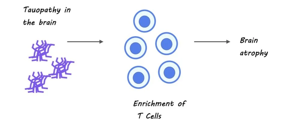
Alzheimer’s disease is characterized by the deposition of amyloid-B plaques and intracellular tau neurofibrillary tangles in the brain, together with brain atrophy. Interestingly, regional patterns of brain atrophy mirror regional patterns of tau accumulation, but not amyloid deposition in the brain. While the pathology of Alzheimer’s disease remains to be fully elucidated, evidence suggests that the immune system may play an important role in disease pathology. This week in Nature, Chen and colleagues investigate the relationship between the immune system and neurodegeneration in two different mouse models of Alzheimer’s disease, one with amyloid-B deposition and the other with tauopathy, to better understand the contribution of the immune system to each of these hallmark features of the disease.

How did they do it?

The authors compared the immune system function in the brains of two transgenic mouse models, one with amyloid-B-deposition and the other with tauopathy, both created by crossing transgenic mice with human-APOE-knock-in mice. The authors performed single-cell RNA sequencing of immune cells from the meningeal and parenchymal lining surrounding the brains of male mice. They also performed immunohistochemical analyses of the parenchyma to characterize further the presence of T cells, microglia, and antibodies in both mouse models. To compare the findings in the mouse models to human Alzheimer’s disease, they performed the same immunohistochemistry experiments on brain samples of patients with Alzheimer’s disease at different levels of disease severity.

Next, the authors wanted to understand the specific role of several immune modulators in the immune response to tauopathy. They tested each of these by administering a neutralizing antibody to the mice. The first one they tested was IFN-gamma, a cytokine that can augment the immune response. The second one was T cells, and the third was the programmed cell death protein 1 (PDCD1), an immune checkpoint for T cells. They then evaluated the immune profile, accumulation of phosphorylated tau in the brain, and behavior. 

What did they find?

First, the authors found that only mice with tau pathology showed brain atrophy at 9.5 months of age, with regional patterns mirroring human disease. The authors found that 9.5-month-old tau mice had an increased presence of adaptive immune cells, including T cells, dendritic cells, and macrophages in their parenchyma and meninges compared to amyloid mice. Immunohistochemistry of the parenchyma confirmed that tau mice had elevated levels of T cells, enriched for INF-gamma transcripts, and microglia compared to amyloid mice. Importantly, similar elevations in T cell number were observed in the brain of humans with Alzheimer’s disease, particularly in regions with more tauopathy.

Next, the authors found that anti-IFN-gamma treatment resulted in attenuated brain atrophy in tau mice. Similarly, the T cell depletion treatment resulted in decreased brain atrophy, and a reduction in the overall number of microglia, suggesting that T cells in the brain of tau mice can indeed augment the number of microglia. Furthermore, T cell depletion improved performance on several memory tasks (short-term, hippocampal- and amygdala-dependent), and resulted in a decrease of phosphorylated tau (the conformation that allows it to accumulate into fibrils), resembling that of earlier disease stages. Blockade of PCDC1 also led to a decrease in tau-mediated neurodegeneration and p-tau staining. These data suggest that a reduction in immune mediators in the brain can attenuate some of the key features associated with disease progression in Alzheimer’s disease.

What's the impact?

This study suggests that tauopathy and neurodegeneration are linked to an immune system signature of activated microglia and T cells and that a reduction in the presence of these immune markers can delay disease progression. These mechanistic insights may aid in identifying therapeutic targets for preventing or slowing down neurodegeneration in Alzheimer’s disease. While these findings are compelling, the experiments were mostly performed in male mice - the need to replicate these findings in female mice is of paramount importance.

Increased Heart-Rate Leads to Increased Anxiety-Like Behavior in Mice

Post by Lani Cupo

The takeaway

The authors find evidence that emotional states emerge not only top-down, with the brain influencing the body, but also in a bottom-up fashion, with changes to the body (increased heart-rate) increasing anxiety-like behavior.

What's the science?

In acting, there are two techniques to embody a character and scene: a popular inside-out approach where the actor uses a variety of approaches to feel an emotion internally and then expresses that internal state, and an outside-in approach where actors mold the voice and body to capture the emotion and allow it to influence their internal feelings. However, to what degree the physiological state, such as heart and breathing rate, can contribute to the development of an emotional state (like anxiety) is still debated scientifically. This week in Nature, Hsueh and colleagues found that experimentally controlling the heart rate of mice increased anxiety-like behavior, identifying the brain structures involved in the effect.

How did they do it?

First, the authors used cutting-edge genetic engineering to develop a mouse whose heart-rate they could control with a laser mounted on a vest and directed towards the chest (towards the heart, but over the skin). By pulsing the light, the authors could stimulate a heart-rate up to 900 beats per minute, although they could not slow the heart-rate below baseline rates. Mimicking patterns of increased heart-rate observed during stressful contexts, the authors examined the behavior of these “paced” mice compared to controls in two different anxiety tests. They also included an operant test, which examined reward-seeking in a stressful context—during mild foot shocks of variable frequencies.

Next, the authors used an ex-vivo assessment (CLARITY and cell-staining for neural activation) to examine what brain regions might be involved mechanistically in the observed effect. To confirm the role of the identified brain regions in cardiac pacing, they recorded activity from neurons in this region in live mice while increasing the heart rate. Finally, the authors investigated whether inhibiting activity in this brain region inhibited the anxiety-like behaviors observed during increased heart-rate.

What did they find?

The authors observed increased anxiety-like behavior in paced mice compared to controls on both the open-field test and the elevated-plus maze. Importantly, there were no baseline differences between mobility or anxiety levels, suggesting the differences were due to the increased heart-rate rather than the experimental manipulations. In the operant test, there were also no baseline differences in reward-seeking behavior of the experimental mice, however when mild foot shocks were delivered with the reward in 10% of trials, the experimental mice had suppressed reward-seeking. This indicates an apprehensive behavior, where the risk of a foot shock decreases the mouse’s reward-seeking behavior.

Next, the authors identified the posterior insular cortex (pIC) as a region of interest - a brain region known to play a role in interoception. These results were further supported by the authors’ findings that neurons in the pIC were more active when the heart-rate was increased.

Finally, the authors found that inhibiting activation in the pIC reversed the effects of the increased heart-rate in reward-seeking. That is, mice with increased heart-rates but also inhibited pICs no longer differed from controls in their reward-seeking behavior, even with the risk of a foot shock. The authors also tested whether inhibition of another brain region (medial prefrontal cortex) or pIC inhibition without increased heart-rate decreased anxiety-like behavior, and found that they did not. This provides very strong evidence that the pIC is crucially involved in the connection between increased heart-rate and an anxious state.

What's the impact?

This study presents strong evidence that increased heart-rate can evoke anxious states, and that the pIC is integral in this relationship. The methods used in this research add new techniques to the neuroscientist’s tool box, and these findings can help to pave the way for effective interventions for those suffering from panic and anxiety disorders. 

Access the original scientific publication here

The Effect of Bilingualism on Subcortical Brain Structures

Post by Megan McCullough

The takeaway

Speaking two languages is a cognitively demanding task that has a measurable impact on the volumes of subcortical brain structures. However, this relationship can be non-linear and reaches a plateau after a certain level of bilingual experience has been reached.

What's the science?

Bilingualism, the ability to fluently speak two languages, is a complex cognitive skill. Previous research has demonstrated that engaging in cognitively demanding tasks can increase grey matter volumes in the brain, but this increase is nonlinear, plateauing or even decreasing after a certain point. The expansion-renormalization model of experience-dependent neuroplasticity explains these findings. However, so far it is unclear whether these nonlinear changes apply also to bilingualism. In a recent study published in Scientific Reports, Korenar, and colleagues investigated the impact of bilingual experiences on subcortical brain regions involved in cognitive control and language selection, specifically the basal ganglia and thalamus.

How did they do it?

Participants in this study were native Slavic language speakers who had a good command of English. Bilingualism is not a one-size-fits-all phenomenon, so the study did not consider participants as bilingual just because they spoke two languages. Instead, each participant was given a composite score to reflect the richness of their individual bilingual experience. The authors effectively treated bilingualism as a continuum of experiences, which allowed them to study the relationship between bilingual experience and changes in subcortical structure volume. Magnetic resonance imaging was used to measure the volume of the caudate nucleus, globus pallidus, putamen, nucleus accumbens, and thalamus. These structures are important for controlling behavior and managing two languages in the brain. The authors then used non-linear statistical modeling, specifically generalized additive mixed models (GAMMs), to investigate whether bilingual experiences had an impact on subcortical volumes that were consistent with the model of experience-dependent neuroplasticity. By using GAMMs, the authors were able to model the possible non-linear effects of bilingual experience on subcortical volumes.

What did they find?

The authors of the study have discovered that bilingualism has a measurable impact on the volume of subcortical structures such as the basal ganglia and thalamus. Interestingly, this relationship is non-linear and dependent on the bilingual composite score, which measures the level of bilingual experience. Specifically, there is a positive association between the bilingual composite score and the volumes of the caudate nucleus and the nucleus accumbens. However, this relationship reaches a plateau in individuals with extensive bilingual experience, indicating a renormalization process. The Dynamic Restructuring Model provides a possible explanation for these findings. According to this model, acquiring and using an additional language leads to an expansion of brain structures as new neural pathways are formed. However, this expansion is followed by a contraction as less efficient pathways are pruned, leaving only the most efficient connections. This suggests that becoming more proficient in handling two languages allows the brain to selectively keep only the most efficient neural pathways for this task. The study's data suggest that the bilingual composite score can be used as a predictor of subcortical structure volumes important for using two languages. Moreover, the study shows that bilingualism impacts subcortical structure volumes in a manner similar to other cognitively demanding tasks previously studied, like playing a musical instrument.

What's the impact?

Overall, this study is the first to investigate the non-linear relationship between bilingual experiences, volumes of subcortical brain regions, and a continuous measure of bilingualism. The findings suggest that acquiring and speaking a new language can prompt the brain to grow and shrink, as a function of efficiency, similar to other cognitively demanding tasks.