Aggression-Specific Mirror Neurons Exist in the Hypothalamus

Post by Lincoln Tracy

The takeaway

A subset of neurons essential for aggression in the ventromedial hypothalamus of the mouse is activated when observing aggression in other animals, providing an example of mirror neurons for this specific social behavior. 

What's the science?

Mirror neurons are a subtype of neurons activated both during seeing and undertaking a behavior. Consequently, they have been suggested to be an important component of the cognitive substrates underlying successful social interactions. Activating neurons in the ventrolateral section of the ventromedial hypothalamus – the ‘attack’ center of the brain – elicits aggression in mice, but it’s unknown whether these neurons are also activated when observing aggression between other animals. This week in Cell, Yang and colleagues used calcium imaging and multiple types of genetically modified mice to determine whether neurons in the ventrolateral section of the ventromedial hypothalamus could perceive aggressive encounters between other mice.

How did they do it?

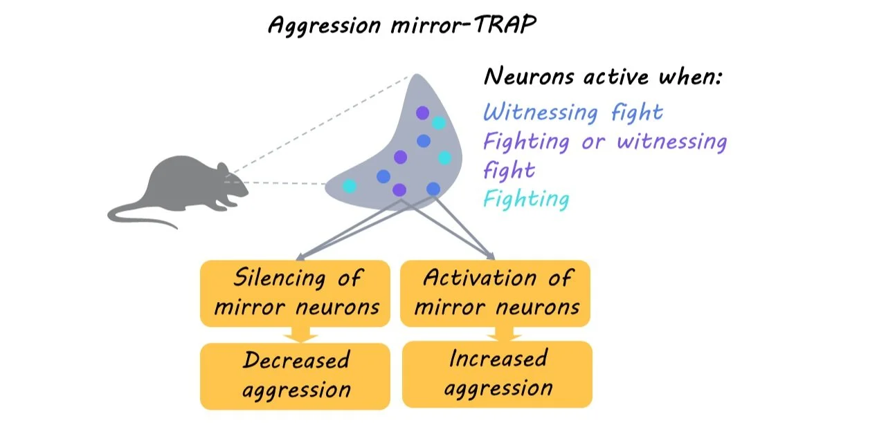
First, the authors used fiber photometry to determine whether aggressor neurons in the ventromedial hypothalamus of male mice were activated in response to social interactions with a male intruder in their cage. Second, they tested whether observing, but not participating in, aggression between two other mice activated the neurons in a similar fashion by separating the mice with clear, perforated plastic partition. Third, they tested which specific aspects of observing aggression were required for neuronal activation by altering the mice’s ability to smell and see the other animals. Fourth, they used socially naïve and experienced males to determine whether aggression mirroring requires the mouse to have previously participated in aggressive behaviors. Fifth, they used a TRAP2 mouse model to specifically test whether neurons activated in observers were also activated when the mouse participated in aggressive behaviors. Finally, they tested if the aggression-mirroring neurons were functionally relevant for fighting by using an aggression mirror-TRAP (aggression mirror neurons tagged with TRAP) to inhibit these neurons in a territorial aggression paradigm. 

What did they find?

First, the authors found aggressor neurons in the ventromedial hypothalamus of male mice were activated when a mouse interacted with and attacked a male intruder that had been placed in its cage. Second, just as when the mouse participated in aggressive behaviors, observing other mice be aggressive resulted in similar activation of ventromedial hypothalamic neurons. Third, they found that visual input is necessary for the activation of aggression-mirroring hypothalamic neurons, as repeating the experiment under infrared light did not activate aggressor neurons. Fourth, they found prior experience was not needed, as ventromedial hypothalamic activation was similar for both socially naïve and experienced mice when observing aggressive behaviors between other mice. Fifth, they found that there was an overlap of ventromedial hypothalamic neurons that were activated in observers and participants of aggression. Finally, they found the aggression mirroring neurons were essential for territorial fighting, as forced inhibition of these neurons reduced aggressive, but not non-aggressive social, behaviors. In additional studies, they also found that activating aggression mirroring neurons was sufficient to elicit aggression.

What's the impact?

These findings provide a genetic platform that can help us gain molecular and cellular insights into how individual neurons represent social behavior like aggression with respect to both mirroring and performing actions.  

Treating the Gut in Parkinson’s Disease

Post by Christopher Chen

The takeaway

A common symptom in people with Parkinson’s Disease (PD) is a compromised, pro-inflammatory gut bacteria profile that may lead to neuroinflammation and neurodegeneration associated with PD. Clinicians found that a short-term regimen of prebiotic fiber helped restore the gut’s anti-inflammatory environment in patients with PD and may even attenuate clinical symptoms. 

What's the science?

Though classified as a neurodegenerative disease, Parkinson’s Disease (PD) also manifests itself in the gut microbiota. Specifically, patients with PD express increased levels of pro-inflammatory bacteria (family Enterobacteriaceae) and decreased levels of anti-inflammatory bacteria (family Lachnospiraceae). This imbalance may lead to intestinal “leakiness” which allows for the infiltration of harmful substances like lipopolysaccharide (LPS) which may increase neurodegeneration. However, research has shown that anti-inflammatory bacteria can restore gut balance by producing molecules called short-chain fatty acids (SCFA). Scientists know that the body’s breakdown of substances called prebiotic fibers recruits the helpful bacteria that generate SCFAs. Recently in Nature Communications, Hall and colleagues investigate whether treating PD patients with prebiotic fiber can increase SCFA production, decrease pro-inflammatory bacteria, and potentially alleviate clinical symptoms of PD.    

How did they do it?

The researchers first determined the effects of different prebiotic fibers on SCFA production. To do so, they treated the stool of age-matched healthy controls and patients with PD with different types of prebiotic fiber and measured how much each fiber increased SCFA. Based on this data, the scientists created an edible bar containing an optimized mixture of prebiotic fibers (30% resistant starch, 30% resistant maltodextrin, 30% stabilized rice bran, and 10% agave branched inulin).

Over the course of ten days, the bar was given to two groups of PD patients, the first group being newly diagnosed and non-medicated PD patients and the second being medicated PD patients. Following the ten-day regimen, researchers assessed participant stool to assess microbiota composition and also assessed participants on a range of symptoms linked to gastrointestinal and neurological function, including UPDRS, a neurological assessment specifically linked to PD symptomology. These results were then compared to data from the same assessments taken prior to the prebiotic fiber intervention.  

What did they find?

Following treatment, both participant groups exhibited changes at the microbiome and cellular level as well as in behavioral assessments. In terms of microbiome composition, patient stool showed a decrease in the overall abundance of pro-inflammatory bacteria and an increase in the overall abundance of anti-inflammatory, SCFA-producing bacteria. Researchers also took blood samples from patients, which showed an increase in SCFA following the treatment. Additionally, a metabolic pathway linked to acetyl-CoA fermentation known to be upregulated in PD was downregulated following prebiotic fiber intervention. It should be noted, however, that while the overall amount of SCFA-producing bacteria increased, several species of SCFA-producing bacteria were downregulated following the treatment. In terms of neurological effects, no neuroinflammation markers showed decreases in patients, though levels of a neurodegeneration marker called NfL were reduced.   

Finally, an exploratory analysis was conducted to assess the effects of the prebiotic fiber regimen on clinical and behavioral outcomes. Most notably, both patient groups had minimal GI discomfort and scored significantly lower on the UPDRS following treatment, suggesting an improvement in PD symptomology.   

What's the impact?

Taken together, evidence from this study suggests that diets high in prebiotic fiber may be conducive to restoring anti-inflammatory, SCFA-producing bacteria in patients with PD. Furthermore, the resulting increase in SCFA-producing bacteria may be linked to functionally significant decreases in PD symptomology. Though this study was conducted on a relatively small patient group (twenty total participants) and the long-term effects of prebiotic fiber remain unclear, it offers a compelling glimpse into the therapeutic potential of non-pharmacological, microbiome-centric treatments for neurological disorders such as PD. 

Access the original scientific publication here.

How Do Psychedelics Promote Neuroplasticity?

Post by Leanna Kalinowski

The takeaway

Activating serotonin 2A receptors within the cell membrane is necessary for promoting the neuroplasticity-inducing and antidepressant-like effects of psychedelics.

What's the science?

Several neuropsychiatric diseases, including depression, are marked by a decrease in dendritic spine density in the cortex. Due to the ability of psychedelics to promote neuroplasticity (e.g., the regrowth of these dendritic spines) in the brain, psychedelics have emerged as a promising treatment for neuropsychiatric diseases. While it is known that neuroplasticity can be promoted by activating serotonin 2A receptors (5-HT2ARs) in the brain, the mechanisms by which this occurs following psychedelic administration are still poorly understood. This week in Science, Vargas and colleagues uncovered the mechanisms by which psychedelic-induced activation of 5-HT2ARs promotes neuroplasticity and antidepressive-like behaviors in mice.

How did they do it?

In the first experiment, the researchers determined the primary location of serotonin 2A receptors (5-HT2ARs) within neurons. To do this, they first tagged in vitro (i.e., in a petri dish) human embryonic kidney cells (a control) and cortical neurons with a fluorescent marker, and then tagged b2 adrenergic receptors (control receptors) and 5-HT2ARs with a different fluorescent marker. The location of each receptor type was then mapped relative to the cell membrane of each cell type.

In the second experiment, the researchers tested whether it was necessary for psychedelics to cross the cell membrane in order to promote neuroplasticity. To do this, they treated in vitro rat embryonic cortical neurons in a petri dish with either (1) psychedelics that are capable of crossing cell membranes (i.e., DMT & psilocin) or (2) versions of these psychedelics that were chemically modified into substances that are incapable of crossing cell membranes (i.e., TMT & psilocybin). Half of these substance administrations were done in the presence of electroporation, which creates temporary openings in the cell membrane so membrane-impermeable substances can cross, while the other half were not. Following substance administration, the researchers measured dendritogenesis, which is a form of neuroplasticity defined as the formation of new dendrites.

In the third experiment, the researchers tested whether importing serotonin into neurons promotes neuroplasticity in mice. To do this, they first engineered mouse neurons to express serotonin transporter (SERT), which acts as a gate to allow serotonin into the cell. Half of the mice received an intra-mPFC injection of the virus that causes its neurons to express SERT, while the other half of the mice received a control injection into the mPFC. Then, after three weeks, both groups of mice were given an intraperitoneal injection of para-chloroamphetamine (PCA), which is a drug that causes the release of serotonin. 24 hours following this injection, markers of neuroplasticity (i.e., dendritic spine density) were assessed.

The fourth and final experiment was like the third experiment, except this time, the researchers tested whether importing serotonin into neurons promotes antidepressant-like behaviors in mice. Three weeks after the procedure to create SERT-positive neurons in the mPFC, mice were placed in a container of water and given a baseline forced swim test to test depressive-like behaviors without the presence of serotonin. In this test, mice who stop trying to swim to escape the container after a shorter period of time are considered to be exhibiting more depressive-like behaviors. Two days after the baseline forced swim test, mice were injected with PCA to facilitate the release of serotonin, after which they were once again administered a forced swim test.

What did they find?

From the first experiment, the researchers found that within kidney cells (controls), both receptor types (b2 adrenergic & 5-HT2ARs) were localized along the cell membrane. However, within cortical neurons, b2 adrenergic cells (controls) were localized on the cell membrane, while 5-HT2ARs were localized within the cell membrane. This is unique from most other G-protein-coupled receptors that are generally located along the cell membrane.

From the second experiment, the researchers found that regardless of whether electroporation was applied, membrane-permeable psychedelics (i.e., DMT and psilocin) always promoted neuroplasticity within cortical neurons. On the other hand, membrane-impermeable psychedelics (i.e., TMT and psilocybin) were only able to promote neuroplasticity when temporary openings in the cell membrane were present due to electroporation. Together, these findings suggest that psychedelics can only promote neuroplasticity when they are able to cross neuronal cell membranes.

From the final two experiments, the researchers first found that SERT-expressing mice that were administered PCA displayed higher markers of neuroplasticity (i.e., dendritic spine density) compared to controls. In addition, they found that these mice also displayed a reduction of immobility in the forced swim test, which is indicative of anti-depressive-like behaviors. Together, these results suggest that importing serotonin into neurons promotes neuroplasticity and antidepressant-like effects in mice.

What's the impact?

Results from this study have the potential to transform how scientists think about psychedelics and other drugs that target the serotonin system. Now that we know that (1) 5-HT2ARs are located within the cell membrane, (2) serotonin generally cannot cross the cell membrane to bind to these 5-HT2ARs, and (3) many psychedelics can cross the cell membrane to bind to these 5-HT2ARs, scientists are equipped to develop future treatments for neuropsychiatric disorders in which 5-HT2ARs are implicated. Future studies should evaluate the potential of other drug classes to bind to intracellular targets and produce therapeutic effects. 

Access the original scientific publication here.