CBD’s Anti-Seizure Mechanism of Action

Post by Baldomero B. Ramirez Cantu

The takeaway

Hippocampal hyperactivity is a hallmark of a variety of neurological diseases. Cannabidiol (CBD) is capable of modulating the excitatory-inhibitory balance of hippocampal neurons and its effects are mediated by interactions with the GPR55 receptor.

What's the science?

CBD is known to reduce seizure activity in animal models and patients with treatment-resistant epilepsies. However, the underlying mechanisms that enable CBD to reduce seizure activity and severity remain unknown. This week in Neuron, Rosenberg and colleagues investigate the role of the GPR55 receptor and its endogenous ligand LPI in CBD’s anti-seizure action.

How did they do it?

The authors tested if the GPR55 receptor is the site of action for CBD's anti-seizure effects by using mice with functional or genetically-removed GPR55 receptors. They conducted experiments on hippocampal neurons in-vitro to study how GPR55’s natural ligand, LPI, affects neuronal excitability and how CBD interacts with LPI’s effects. Finally, to investigate changes in GPR55 receptor expression after seizures in control or CBD pre-treatment conditions, the authors used qPCR and immunohistochemistry. Seizures were induced in the mice using the drug pentylenetetrazole or lithium-pilocarpine.

What did they find?

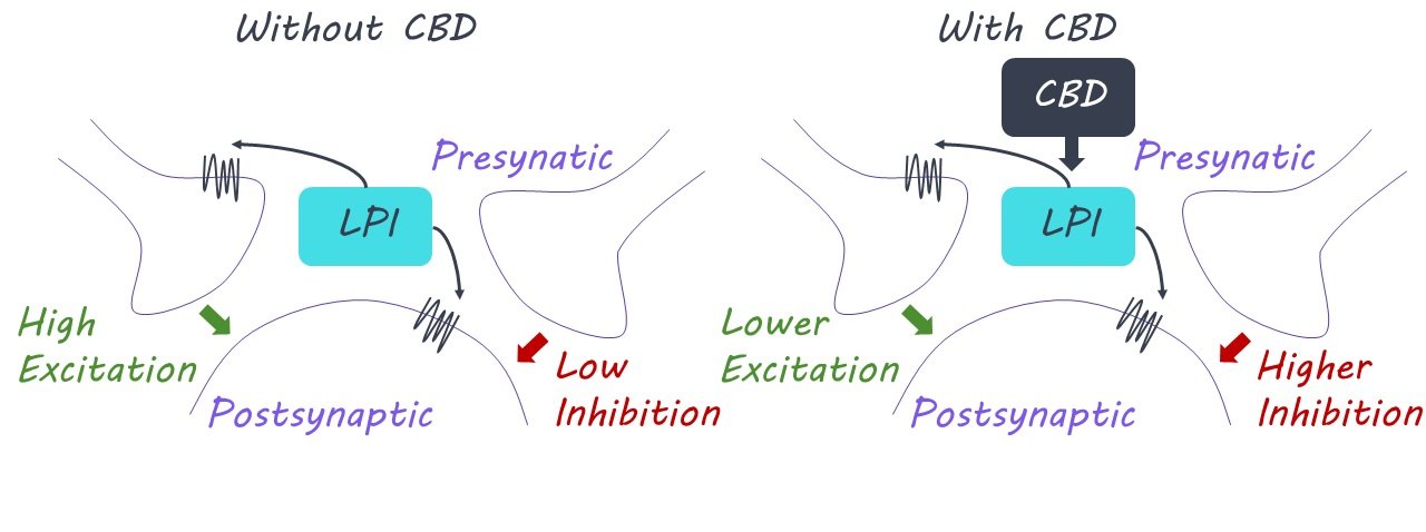
The authors found that seizures were only reduced by CBD if mice expressed GPR55 receptors - there was no improvement if these receptors were removed. This finding suggested a critical role of the GPR55 receptor in enabling CBD to attenuate seizures. They conducted neural recordings from the hippocampus and found that the application of LPI increased neuronal excitability in neurons from normal mice, but not in those from genetically modified mice lacking GPR55 receptors, indicating that LPI drives GPR55-dependent increases in neuronal excitation. LPI was found to disrupt neuronal activity in several ways, such as interfering with the impact of crucial inhibitory interneurons that are essential for maintaining an appropriate excitatory-inhibitory balance in the hippocampus. Importantly, hippocampal neural recordings showed that the presence of CBD counteracts the increased excitability produced by LPI. This provides evidence for the GPR55 receptor as a functional target of CBD’s anti-seizure action. Mice that received CBD pre-treatment showed significantly lower increases in GPR55 immunoreactivity and receptor mRNA expression after induced seizures compared to the control group.

What's the impact?

This is the first study to identify the two-pronged molecular signaling, affecting both excitation and inhibition, underlying CBD’s anti-seizure action. These findings could serve as the foundation for the development of new, targeted anti-epileptic therapies

Mitochondria Set the Tempo for Neuronal Development

Post by Elisa Guma

The takeaway

The development of neurons and mitochondria in the cerebral cortex takes considerably longer in the human brain than in other species, such as mice. Accelerating mitochondrial metabolism seems to accelerate human neuronal maturation, indicating that they are important regulators of the pace at which the brain develops.

What's the science?

There are striking species differences in the amount of time required for brains to develop, wherein the human brain develops over the course of months to years, while the mouse brain develops over the course of weeks. It is unclear what is responsible for these differences, however, given the important role that mitochondria play in driving cell maturation, they may also play a key role in modulating the differences in developmental timelines of cortical neurons. This week in Science, Iwata and colleagues investigate the role that mitochondria have in determining the developmental timelines of cortical neurons across the human and mouse brain.

How did they do it?

To investigate the relationship between mitochondrial metabolism and neuronal maturation, the authors used cultures of human and mouse cortical pyramidal neurons derived from pluripotent stem cells, as well as embryonic mouse brain neurons. For the pluripotent stem cells, the authors devised a method that stages neurons based on their birthdate to allow them to compare neurons in the same stage of development. To birthdate the neurons, the authors tagged the NeuronalDifferentiation1 gene – a gene that is active when the neuron enters a specific maturational stage – with a green fluorescent protein, allowing them to identify and separate it from the other neurons.

Within neurons of the same maturational stage, the authors also tagged mitochondria with an emerald-green fluorescent protein allowing them to visualize the morphology and location of these organelles within the neuron. To monitor mitochondria in developing cortical neurons in the mouse brain, the authors labeled mouse cortical neurons with a fluorescent protein using in utero electroporation in mid-late gestation, or they transplanted fluorescently labeled human neurons in the mouse brain. This allowed them to use light and electron microscopy to examine patterns of mitochondrial development and identify the age at which they reached maximal levels of growth and size.

Following a characterization of mitochondrial development, the authors examined the metabolic activity of mitochondria in both mouse and human cortical neurons at similar times after birth, focusing on mitochondrial oxidative phosphorylation and electron transport chain capacity (two indicators of metabolic function). Finally, to test whether enhanced mitochondrial activity would also accelerate neuronal development, the authors accelerated mitochondrial metabolism by inhibiting specific enzymes that are important in glucose metabolism in human cortical neurons. 

What did they find?

In mouse pluripotent stem cells and newly born neurons (from the embryonic brain), the authors found that mitochondria were initially small and sparse but grew in quantity over the 3-week maturational window of these neurons. In contrast, pluripotent stem cells derived from human cortical neurons, as well as their mitochondria, showed a much slower pattern of maturation, taking several months. These data suggest that mitochondrial morphology and development follow a species-specific timeline that is highly correlated with neuronal maturation.

Consistent with morphological development, mitochondrial metabolism was higher in the mouse neurons than in the human neurons in the early stages of development and continued to increase at a faster rate across development. In human cortical neurons, they also observed lower levels of oxidative stress compared to the mouse neurons, consistent with lower activity of mitochondrial metabolism.

When the authors increased mitochondrial activity in human neurons, they observed an increase in oxidative phosphorylation with no significant alterations to mitochondrial morphology. However, they did observe an increase in the speed at which neurons were maturing both in terms of function (based on synaptic currents and membrane potentials) as well as morphology (larger neuronal size and increased dendritic length and complexity). This indicates the crucial role of these organelles in regulating timelines of neuronal development.

What's the impact?

This study provides evidence for the role of mitochondrial metabolic activity in regulating the species-specific developmental timeline of cortical neurons. When mitochondrial metabolism was enhanced, neurons showed accelerated morphological and functional maturation. This may in part explain why the human brain develops across much longer time courses than other species, such as the mouse. Future work is needed to understand the downstream effects of mitochondrial metabolism on brain function, plasticity, and neurodevelopmental disorders. 

Infants Have an Innate Perception of Musical Patterns

Post by Leanna Kalinowski

The takeaway

Infants can perceive recurring patterns in music (called “meter”), regardless of whether the patterns are marked by obvious tones, in a similar manner to adults.

What's the science?

Across different cultures, music has several universal features that often cause humans to bob their heads or tap their feet in time with it. Within a given song, there is a small set of recurring patterns and accents, called “meter”, that allows humans to synchronize their perception of music and move along to it together. Sometimes, meter is made obvious by including tones that indicate its pulse, but other times, meter is much more implicit and marked by periods of silence. While we know that the adult brain can perceive both obvious and more implicit meter, less is known about how early this perceptual ability develops in infants. Recently in Developmental Science, Lenc and colleagues recorded brain activity in infants that were exposed to rhythms that are known to induce the perception of meter in adults.

How did they do it?

The researchers recruited 20 infants between five and six months old to participate in this study, during which they were exposed to two rhythms: a “strongly-periodic” rhythm and a “weakly-periodic” rhythm. The two rhythms typically produce the same perceived meter in adults, but the tones in the strongly-periodic rhythm matched the pulse of the typically perceived meter, while the tones in the weakly-periodic rhythm did not. The infants were played each rhythm twice -- once with a high-pitched tone, and once with a low-pitched tone – for a total of four sound clips. Brain activity while listening to each sound clip was measured using electroencephalography (EEG).

What did they find?

The researchers found that listening to both rhythms induced brain activity in infants that matched the frequency of the meter in a similar pattern to what is seen in adults. This brain activity was present not only for the rhythm with beats that matched the meter (i.e., strongly periodic) but also for the rhythm that did not have beats that matched the meter (i.e., weakly periodic). They also found that meter-related brain activity of both rhythm types was enhanced when the rhythm was produced by bass sounds (i.e., low-pitched tones) as compared to high-pitched tones. Together, these results suggest that high-level neural processes that facilitate music perception are already present soon after birth.

What's the impact?

Results from this study show that infants are able to perceive musical patterns shortly after birth – even when these patterns are not marked by obvious tones – well before infants even develop the motor ability to bob along to the music. These findings may help pave the way for developing age-appropriate interventions for developmental disorders that rely on auditory stimulation and rhythm perception.  

Access the original scientific publication here.