Both Cardiovascular and Behavioral Threat Responses Contribute to Defensive States

Post by Lincoln Tracy

The takeaway

A new framework developed in mice involving microstates, macrostates, specific behaviors, and heart rate dynamics has proven beneficial in learning more about the complex neural states and associated systemic functions in response to an external threat.

What's the science?

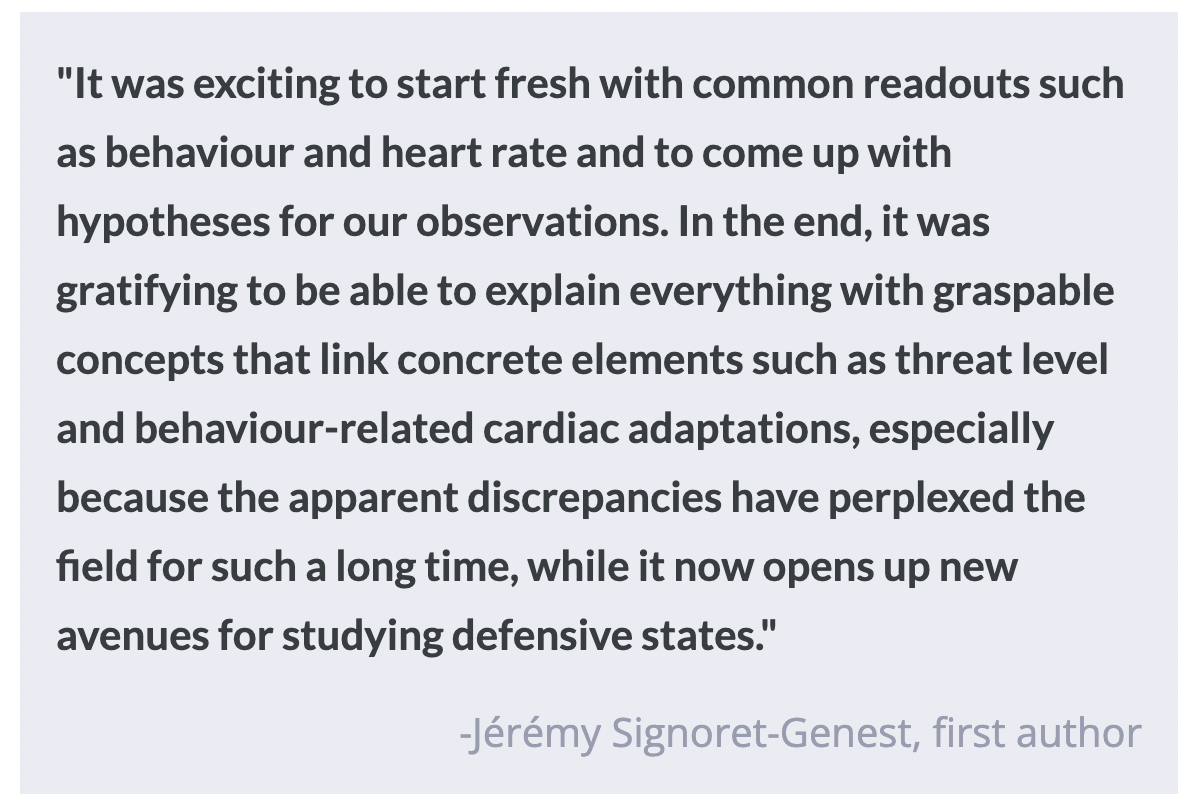
The typical defensive reaction to an external threat, a key part of fear or anxiety, involves multiple behavioral and physiological responses that are controlled by our neural circuitry. There have been attempts to propose a unified and species-preserved concept describing defensive responses, or ‘states’, but such attempts have largely focused on behavioral mechanisms and ignored autonomic responses. This week in Nature Neuroscience, Signoret-Genest and colleagues provide a novel framework for characterizing integrated cardiovascular and behavioral defensive states using behavioral, heart rate, and thermal imaging data from freely moving mice across several experimental paradigms. They show how this allows linking of specific defensive states to key brain circuitry such as the periaqueductal gray (PAG).

How did they do it?

The authors implanted electrocardiogram electrodes in mice, which allowed them to record changes in heart rate while the mice were exposed to a series of environments that varied in threat intensity and acuteness, each eliciting a different emotional state. The environments included low-threat situations (i.e., their home cage) to high-threat, fear and anxiety-evoking experiments such as a conditioned fear (or ‘flight’) paradigm, the open field test, and the elevated plus maze. The authors were interested in comparing the association between immobility, or ‘freezing’ behaviors, and decreases in heart rate when the mice were exposed to different threat environments. Finally, they used optogenetics to manipulate the activity of glutamatergic neurons as well as subtypes of this population (vesicular glutamate transporter 2 [Vglut2] and Chx10) in the PAG in an attempt to identify the specific neural circuits involved in the control of these defensive states.

What did they find?

First, the authors identified threat ‘microstates’, involving immobility and bradycardia (a decrease in heart rate) during the conditioned flight paradigm. They noticed that the immobility-associated bradycardia increased as the conditioning session went on, and hypothesized there were other underlying processes interfering with the heart rate changes at both a global level and in the defensive microstates. They called the dynamic changes operating at an extended timescale ‘macrostates’. This suggests that rather than simply following changes in behavioral activity (rearing or immobility), threat-induced changes in heart rate reflect integrated defensive microstates involving both behavioral and autonomic components. The authors also found the changes in heart rate were strongly influenced by the pre-existing state of the animal. Furthermore, they identified that the integrated defensive response was context-dependent, with higher contextual threat levels resulting in more constrained heart rate changes. Finally, they found stimulating Vglut2+ neurons evoked intensity-depended behavioral and cardiovascular responses (low intensity stimulation led to immobility and bradycardia, while high intensity stimulation led to a mixture of flight responses and immobility accompanied by bradycardia) but stimulating Chx10+ neurons led to robust immobility and bradycardia. This suggests Chx10+ neurons in the midbrain periaqueductal gray mediate a particular defensive microstate associated with both immobility and bradycardia.

What's the impact?

The findings from this study act as a starting point for a more complete understanding of the neuronal mechanisms underlying emotions such as fear and anxiety. The novel framework teases the possibility of returning to a translational research pathway (from mice to humans) and the potential ability to explore maladapted fear and anxiety responses across species.

“Sleep Fingerprints” Can Identify Signatures of Psychiatric Disease

Post by Anastasia Sares

The takeaway

Researchers have developed a way to concisely quantify the activity of tens of thousands of waveforms occurring in a person’s brain during sleep, uncovering stable profiles of different people. This will help us understand individual differences in sleep, from the normal to the pathological: as a first step, it has revealed new insights into the brains of people with schizophrenia.

What's the science?

Our brains don’t turn off when we go to sleep. They remain hard at work, forming memories, dreaming, and recuperating from the day. We know this, in part, because we can observe them through a technique called polysomnography, where we record the electrical activity of the sleeping brain. The signal we get from polysomnography is composed of a symphony of overlapping brain frequencies, as well as short bursts of oscillatory activity called transient oscillations. Among transient oscillations, sleep scientists are especially interested in sleep spindles, which are involved in memory consolidation and are altered in disorders such as Alzheimer’s, autism, and schizophrenia. However, the current methods for defining sleep spindles are based on methods from the early 1900s and are therefore biased toward the waveforms people could see easily by eye in paper tape traces of brain activity. 

This month in the journal Sleep, Stokes and colleagues developed a new approach, which, rather than just looking at traditional spindles, identifies tens of thousands of spindle-like waveforms at all times and frequencies during sleep. They summarized these waveforms in a sleep “fingerprint” that can be used to identify signatures of neurological health and disease.

How did they do it?

The authors analyzed data from both healthy, neurotypical people and people with schizophrenia who had had recordings taken of their brain waves in the lab while they slept. They transformed the signal from each person into a time-frequency plot, with high frequencies (fast oscillations) near the top and low frequencies (slow oscillations) near the bottom, and time going from left to right. They then applied an image processing technique called the watershed algorithm to identify regions with spindle-like brain waveforms. Finally, they created visual summaries showing how these tens of thousands of waveforms continuously evolve across the night. This is more effective than dividing activity into the traditional “sleep stages” because it retains more information.

What did they find?

The team generated these visualizations for each participant, showing how brain activity changed as a person went deeper and deeper into sleep, and also how brain activity related to the slowest sleep oscillations, like tiny waves superimposed on a rising and falling tide. Surprisingly, they found a variety of activity patterns in healthy, neurotypical participants, yet each individual had the same pattern from night to night. Therefore, these representations acted like a fingerprint: different across individuals but consistent over time. They then demonstrated the clinical usefulness of this method by comparing the control and schizophrenia cohorts, which revealed two new classes of spindle-like events that traditional methods had been unable to capture.

What's the impact?

The sleep fingerprints developed by this team offer a highly informative way to summarize a large amount of data: electrical signals gathered over the course of an entire night. What’s more, these fingerprints are different between people but stable within the same person—meaning we may be able to use them to inform clinical approaches for psychiatric conditions, sleep disorders, and more. The researchers have released an open-source toolbox to allow other scientists to use this powerful technique.

Using Magnetic Fields to Treat Alzheimer’s Disease

Post by Christopher Chen

The takeaway

Repetitive transcranial magnetic stimulation (rTMS) can be used as a noninvasive therapy to alleviate some symptoms of Alzheimer’s disease (AD). Applying rTMS to a brain region called the precuneus of patients with AD may slow the disease’s progression and even enhance brain activity in the precuneus itself.

What's the science?

In Alzheimer’s disease (AD), a specific network in the brain called the default mode network (a group of brain regions that are functionally connected) undergoes pathological changes that underlie AD symptoms. The precuneus is a brain region found in the posterior cortex and is one of the primary brain regions included in the default mode network. Research shows that the precuneus is one of the earliest regions within the brain to display amyloid plaques and neurofibrillary tangles, well-known markers of AD. Unsurprisingly, research also links these pathologies to compromised precuneus function, resulting in overall dysfunction in the default mode network. Thus, restoring precuneus activity and connectivity to the default mode network may provide therapeutic benefits to patients with AD. rTMS, which provides indirect magnetic stimulation to specific parts of the brain, has been shown to restore cognitive function in patients with mild forms of AD when applied over a short (two-week) period. Recently in Brain, Koch and colleagues explored whether long-term application of rTMS to the precuneus carries therapeutic value to patients with AD.

How did they do it?

The study consisted of fifty patients with mild to moderate forms of AD. All patients had been prescribed an independent pharmacological treatment for AD. Before the study, the patients were given a battery of assessments designed to measure cognitive function by a team of clinicians and researchers. Following these assessments, patients were divided into the experimental group which would receive rTMS to the precuneus, and the control group which would receive a procedure that resembled rTMS but was not (sham control).

In the first two weeks of the experiment, all patients received extensive experimental or sham rTMS treatment five times a week. The final twenty-two weeks was the maintenance period where patients received experimental or sham treatment once a week. This maintenance period also included a mid-study assessment of cognitive function at twelve weeks. Following the six-month period, patients underwent a final round of assessments to measure cognitive function. Single-pulse TMS combined with EEG was also used to assess precuneus activity and oscillatory activity.

What did they find?

Researchers compared scores from all clinical and behavioral assessments as well as functional readouts from brain imaging assessments in the experimental and sham groups. While both groups showed a generalized decrease in performance on the cognitive tests over time, patients in the experimental group showed smaller decreases on every cognitive assessment both at the mid-study (twelve-week) point and end-study (twenty-four week) point. The brain imaging assessments – which measured precuneus signaling activity using a noninvasive electroencephalogram (EEG) – revealed significant differences in precuneus activity between experimental and sham groups. In fact, the experimental group showed an increase in precuneus activity following the study’s conclusion.

What's the impact?

While short-term rTMS has been used to alleviate AD symptomology, this study is the first to examine its effects over the long term. Additionally, rTMS treatment in AD patients has been largely focused on the prefrontal cortex, not the precuneus, a region of the brain known to exhibit some of the earliest signs of AD pathology. Based on the beneficial changes precuneus-specific rTMS treatment had on patients with AD, this study shows that the precuneus may be a compelling therapeutic target for AD treatments.  

Access the original scientific publication here.