A Brain Region Critical in Creating Cognitive Maps

Post by Anastasia Sares

The takeaway

The lateral orbito-frontal cortex (lOFC), is a brain region in the frontal lobe, just above the eyes, that helps us interact with the world by mapping associations between different events. While previously it has been given the role of “deploying” these maps (deciding when to use them), this new study suggests that the lOFC might be involved in creating those association maps in the first place. 

What's the science?

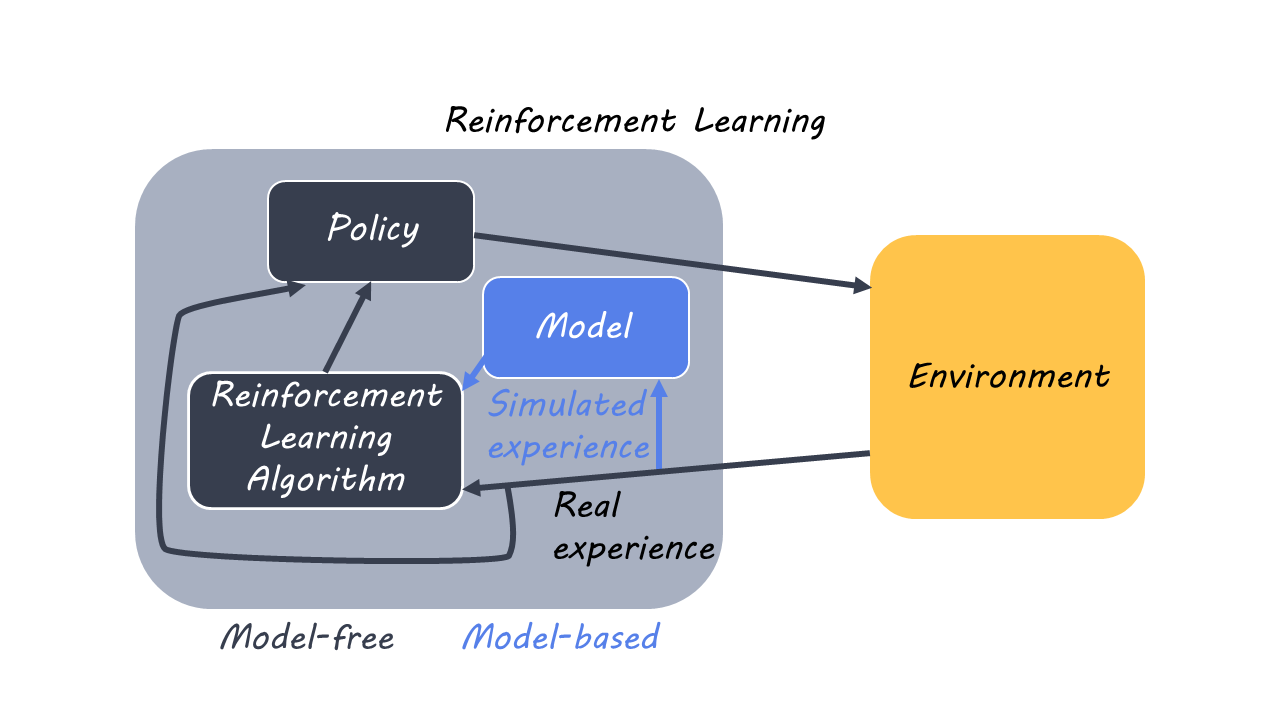
Reinforcement learning is how we develop knowledge through our interactions with the environment. When we perform actions, we get feedback about the results of those actions, and if they result in a reward, we are more likely to repeat those actions again later. Reinforcement learning can be divided into model-free learning, where we learn directly from the consequences of our actions, and model-based learning, where we create a map of associations (in other words, a model) that can help us make decisions based on context.

This week in Nature Neuroscience, Costa and colleagues tested what happens when the lOFC is taken out of commission.

How did they do it?

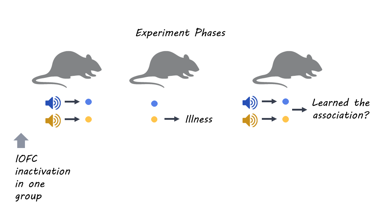
The authors first trained rats to associate sound stimuli with getting food pellets, with one sound predicting banana-flavored pellets and another sound predicting bacon-flavored pellets. The rats learned that sounds were associated with a food reward, but at this phase, there was no reason for them to learn the distinction between the two sounds.

In the second phase, both types of pellets were again given to the rats, but after they ate one kind of pellet (let’s say the banana one), they would be administered lithium chloride, which made them feel nauseous. Here, the rats learned a connection between one type of pellet and nausea, but the sounds were not involved.

In the final phase, the rats were again presented with the sounds, and the authors measured how long the animal searched for a food pellet after hearing the sound. The rats had never learned a direct association between the sounds and nausea, so they would have to create this connection themselves based on information from the first two phases. Specifically, they would need to rely on an internally-created “model” or associative map linking the relevant sound to the bad pellet (even though the type of sound had not mattered before).

Two groups of rats participated in this experiment. The first was a control group, and the second group had been bio-engineered (by applying a custom viral agent) so that the lOFC could be temporarily “turned off” by administering a drug just before the learning session in the first phase. If the lOFC was involved in creating association maps, then turning it off for in the first learning phase should make the rats unable to learn the association between sound cues and nausea. However, if the lOFC is only involved in “deploying” these association maps, then turning it off in phase 1 should have no effect on their ability to use them later.

What did they find?

When the control rats heard the specific sound that predicted the “bad” pellets (the ones that had made them nauseous), they did not go to the food bowl as often as when they heard the other sound. This means they had clearly learned the distinction between different sounds, the foods they signaled, and the predicted result of eating those foods, and were able to put this information together in the final test to guide behavior. In contrast, rats whose lOFC was deactivated in phase 1 reduced their trips to the food bowl for both sounds at test time. These rats thus had the ability to form a rudimentary map to guide behavior, however, this map lacked the specific information that would allow them to choose the “good” pellets and avoid the “bad” ones.

The authors also put the rats through an object recognition task, which required them to distinguish between new and old objects. In this case, the rats with the deactivated lOFC performed similarly to the control rats, indicating that the lOFC is not involved in basic learning.

Finally, the authors created some mathematical models to try and reproduce the results. They found that the best explanation of the impaired animals’ behavior was an imprecise mapping from sounds to pellet flavors during the first learning phase. 

What's the impact?

Model-based reinforcement learning is a crucial function of the brain, and abnormalities in this system can lead to maladaptive behavior. For example, problems with association maps are present in mental illnesses such as schizophrenia, substance abuse, and obsessive-compulsive disorder. Therefore, understanding more about how this area of the brain works may help us better diagnose and treat these conditions.

Access the original scientific publication here.

How Habitual Checking of Social Media Changes the Adolescent Brain

Post by Christopher Chen

The takeaway

Social media use has become nearly universal among American teenagers but its possible effects on adolescent brain development remain unclear. A new study indicates that habitual checking of social media may be disrupting the normal development of brain circuits linked to reward processing and cognitive control in the young adult brain.

What's the science?

The brain undergoes drastic changes during adolescence, particularly in regions associated with motivation, reward processing, and cognitive control. Furthermore, the maturation of these regions allows for developmentally normative neural and behavioral responses to social feedback. With its use of immediate feedback in the form of “likes” or notifications as well as its widespread use in adolescent populations, exploring how habitual social media use affects social feedback-based networks in the adolescent brain is more relevant than ever. In a recent article in JAMA Pediatrics, Maza et al. investigate differences in brain activity levels in regions associated with social feedback in adolescents who habitually check social media. 

How did they do it?

The experiment looked at approximately 200 students aged 12-13 from three middle schools in rural North Carolina. First, experimenters had the students self-report how often they checked three social media sites (Facebook, Instagram, and Snapchat). Based on this data, the participants were divided into three groups based on their rate of checking social media: habitual, moderate, and non habitual. Experimenters then used functional brain imaging (fMRI) to measure brain activity of participants during a Social Incentive Delay task, a cognitive task designed to measure anticipation of social feedback. Following initial measurements, the students took part in the same experiment each year for the next two years.

Following the completion of the study, experimenters compiled and combined data from both the Social Incentive Delay task and fMRI imaging to measure activation levels of specific brain regions from each participant during the cognitive task. They then used these individual datasets to make a general linear regression model measuring the change in brain activity levels in all three groups over time.  

What did they find?

From their generalized linear regression models, experimenters found that brain activation patterns were significantly different in habitual and non habitual checkers of social media. Interestingly, these patterns were most distinct in brain regions linked to social feedback: the insular and prefrontal cortex, ventral striatum, and amygdala. Habitual social media checkers showed a decreased sensitivity to social anticipation at 12 years of age.

In habitual checkers of social media, linear regression models revealed an increase in brain activity during social anticipation across all four brain regions over time. In non habitual and moderate checkers of social media, linear regression models revealed the opposite: brain activity decreased in all four regions. These divergent results in brain activity changes in habitual and non habitual checkers of social media suggest high social media usage impacts developmental trajectories of neural circuits linked to social feedback and cognitive control.

What's the impact?

The negative functional consequences – if any – of these increases in brain activity in habitual checkers of social media are unclear. Whether the rate of social media usage directly causes or is simply correlated to these neurological changes also remains to be seen. However, this study is the first to show distinct differences in brain development in adolescents who habitually check social media, suggesting that social media is indeed changing the young adult brain.     

Access the original scientific publication here.

Social Health and Brain Reserve as Protective Factors for Dementia

Post by Megan McCullough

The takeaway

Good social health in older adults was associated with slower cognitive decline over time and higher cognition levels at baseline. High brain reserve, a measure of the brain’s resilience to damage, was also associated with slower cognitive decline suggesting that social health and brain reserve may be indicators of the propensity of an individual to develop dementia.

What's the science?

Although there are no current pharmacological treatments for dementia prevention, numerous social risk factors have been identified, indicating that dementia prevention may be possible. Previous research has shown that social support, social engagement, and other social health behaviors may be linked to a reduced risk of dementia development. Additionally, the brain reserve model of dementia suggests that individuals with more neurons or increased synaptic density are less at risk of cognitive deterioration. Although the social health model and brain reserve model both suggest protective factors against the effects of dementia, the interaction between these two models has not been investigated. This week in Annals of Neurology, Marseglia and colleagues aimed to investigate the interaction of social health and brain reserve on cognitive changes in a cohort of dementia-free older adults.

How did they do it?

Participants included 368 Swedish adults over the age of 60 who did not have dementia. These participants were followed for 12 years to assess the interactions between social health, brain reserve, and cognition over time. At the baseline assessments, a social health score was generated for each participant; this score was created from a questionnaire about social connections and social support. The authors also used magnetic resonance imaging (MRI) data to assess volumes of grey matter, white matter, and cerebrospinal fluid in each individual. These volumes were totaled and used as a measure of brain reserve. Cognitive function tests were administered throughout the study to assess different facets of cognition including episodic and semantic memory. Statistical analysis including one-way ANOVA, linear mixed-effect models, and stratified analyses were then run to explore the relationships between social health and brain reserve in the context of cognitive function.

What did they find?

The authors found that moderate to good social health and high brain reserve was associated with high cognitive performance and slower rates of cognitive decline over time. The statistical analyses that examined the interplay between these two factors showed that good social health was associated with higher cognition levels only in individuals who exhibited moderate to large brain reserves. This suggests that there are multiple protective factors that interact in slowing or preventing the development of dementia.

What's the impact?

This study found that a healthy social life and high levels of brain reserve are protective factors against age-related cognitive decline. Specifically, good social health can protect against cognitive decline in individuals with high levels of brain reserve. This research shows the relevance of promoting healthy social lives for individuals at risk of developing dementia. 

Access the original scientific publication here