How Aversive Memories Are Stored and Retrieved

Post by Leanna Kalinowski

The takeaway

Researchers have found that neuronal ensembles in one brain region recruit presynaptic neurons in other regions during the retrieval of aversive memories. 

What's the science?

Each of our memories is stored in neuronal ensembles – sparse groups of co-active neurons – across multiple different brain regions. Previous research has shown that the allocation of memories to these brain regions is not a random process; it is dependent on the level of CREB protein expressed within individual neurons. However, it is unclear how these cross-region connections are organized at the time of learning to ensure that the neuronal ensembles from different brain regions are coordinated when the memory is retrieved. This week in Neuron, Lavi and colleagues tested whether allocating aversive memories to neurons in one brain region impacts memory allocation in interconnected brain regions in mice.

How did they do it?

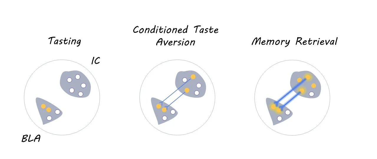
First, the researchers injected mice with a combination of three viruses into the basolateral amygdala (BLA; experiment 1) or insular cortex (IC; experiment 2), which are two interconnected brain regions that are crucial for conditioned taste aversion memories. The first virus carried the CREB gene, which biases the allocation of memories to a random subset of neurons within the injected brain region. The second virus was the rabies virus coupled with mCherry (a fluorescent marker), which allows researchers to visualize the location of presynaptic neurons that project to the injected region via retrograde tracing. The third virus encoded TVA, which is a protein that is essential for the rabies virus to travel to presynaptic neurons.

Four days after the viral injections, the mice underwent a conditioned taste aversion task, which teaches mice to associate a particular taste with an aversive stimulus (i.e., illness). During the habituation phase of this task, which lasted three days, mice received a daily water ration for 30 minutes per day from two tubes. Then, on the following day, the plain water was replaced with sugar water, and the mice were subsequently treated with an injection of lithium chloride. This injection induced feelings of illness, thereby leading mice to develop a learned aversion to sugar water. Aversion to sugar water was then tested three days later, after which their brains were collected and analyzed for neural activity using c-fos immunohistochemistry

To test whether these effects extend to other senses beyond taste (i.e., hearing), the researchers then injected the same three viruses into the BLA of a new cohort of mice and subsequently subjected them to an auditory fear conditioning task. In this task, mice were trained to associate an auditory stimulus (14kHz tones) with a foot shock. Aversion to the auditory stimulus was then tested three days later, after which their brains were collected and analyzed in the same manner as above.

What did they find?

Overall, the researchers found that the allocation of aversive memories in one brain region leads to the retrograde activation of presynaptic neurons (i.e., the target neuron sends a signal back to the presynaptic neuron) in other brain regions during memory allocation and retrieval. Specifically, using CREB to allocate taste aversion memories to the BLA leads to the recruitment of presynaptic neurons in the IC. This pattern was also observed in the opposite direction (i.e., allocating taste aversion memories to the IC leads to the recruitment of neurons in the BLA), suggesting that a bidirectional relationship between these two brain regions is necessary for coordinating taste aversion memory retrieval. Similar coordination was observed between the BLA and auditory cortex during auditory fear conditioning, demonstrating that these effects extend to other senses beyond taste aversion

What's the impact?

This study uncovered the mechanisms that underlie cross-regional memory allocation and retrieval, particularly for aversive memories. These results may pave the way in better understanding how cross-regional memory coordination is disrupted in neurological and psychiatric diseases. 

Access the original scientific publication here.

The Link Between Inflammation and Cognitive Impairment in Severe Mental Illness

 Post by Lani Cupo

The takeaway

A large, multivariate investigation of inflammation and cognition in severe mental illness reveals patterns of immune activation associated with poor verbal learning and processing speed in individuals across diagnoses.

What's the science?

The link between inflammation and mental illness has been investigated, however, the associations between immune activation and cognitive ability in severe mental illnesses, like schizophrenia and bipolar disorder, have often glossed over individual differences in both domains. To help clarify the relationship, variability amongst both healthy individuals and those with severe mental illness must be examined. This week in Molecular Psychiatry, Saether and colleagues attempt to capture individual variance between cognitive ability and inflammation in health and severe mental illness with a multivariate analysis technique, canonical correlation analysis (CCA).

How did they do it?

The authors had access to a large pool of participants (770 healthy controls [HC], 343 with schizophrenia, and 289 with bipolar disorder). Diagnoses were determined by clinical assessments conducted by trained psychologists and medical doctors. Clinical psychologists and trained researchers administered cognitive assessments, recording variables including fine-motor speed, psychomotor processing speed, mental processing speed, verbal learning, attention, verbal memory, semantic fluency, working memory, and cognitive control. To quantify inflammation, the authors included 22 markers of neuroinflamation from a blood sample. CCA is a technique that relates two independent set of variables (each called a variate) to one another, generating new linear combinations of variables (canonical variate pairs) reflecting patterns of covariation between variates. The significance of each resulting variate pair is assessed with permutation testing, allowing the authors to determine whether each pattern of covariance is likely to arise due to chance. The participant demographics can be mapped onto the correlations to examine subgroups within the patterns. The authors organized these “loading scores” (how individual participant scores mapped onto the canonical variates) into clusters using a clustering procedure (hierarchical clustering) to establish subgroups. They confirmed the stability of the identified clusters with a resampling technique.

What did they find?

From the CCA, the authors derived two significant canonical variate pairs representing relationships of covariance among the variables. Of these, the first canonical variate pair was determined to be more reproducible in a set of un-tested samples and, therefore, further examined. This canonical variate pair captured a correlation between poor verbal learning and psychomotor processing speed with an increased presence of 4 markers of innate immune activation. Mapping diagnoses onto the correlation revealed the potential for subgroups.

The clustering analysis revealed two subgroups; Cluster 1 included worse scores on cognitive domains and increased inflammatory markers, compared to Cluster 2. The majority of HCs were in Cluster 2, while the majority of participants with severe mental illness were in Cluster 1. Of the participants with severe mental illness, those in Cluster 1 exhibited worse functioning and symptomatology and higher pharmaceutical usage than those in Cluster 2. Overall, Cluster 1 represented a group that captured most of the participants with severe mental illness and exhibited decreased cognitive ability and increased inflammation. However, group assignments did transcend diagnosis.

What's the impact?

This study is one of the first to show how the relationship between cognitive dysfunction and inflammation transcends diagnosis. Regardless of diagnosis or impairment, there is a relationship between increased innate inflammation and cognitive dysfunction, emphasizing the importance of examining heterogeneity between individuals. In a subset of individuals with severe mental illness, this may provide an important link for future investigation and treatment approaches.

Access the original scientific publication here

A New and Effective Tool for Improving Memory in Older Adults

Post by Megan McCullough

The takeaway

A smartphone application called HippoCamera was shown to be an effective tool for improving the recall of episodic memories and increasing different memory representations in the hippocampus in older adults. 

What's the science?

There is a relationship between age and the ability to re-experience the past and recall specific memories about life events. Previous research has shown that as humans get older, there is a decline in memory recollection that may correlate with a decrease in the differentiation of hippocampal activity. Despite numerous studies that have confirmed these findings, there are limited behavioral interventions that have been shown to successfully target episodic memories. This week in PNAS,  Martin and colleagues aimed to test the effectiveness of a smartphone application on the ability of older adults to improve episodic memory recall.

How did they do it?

The authors tested their self-developed smartphone app on older adults, as age has been correlated with a decrease in the ability to recall details about specific everday life events that they wish to remember. HippoCamera allows users to create memory cues of specific events that they personally value and hope to remember. These cues consist of a short verbal description created by the user as well as a short video recording of the event. The users then can rate the significance of the event in the app. Cues were randomly assigned to either a replay condition, where the user could view it multiple times, or a control condition, where the cue was never replayed. This study consisted of two experiments, either a 2-week intervention, or a 10-week one. In both experiments, the authors conducted two memory tests to test the effectiveness of the smartphone app on memory recall. The authors also performed functional magnetic resonance imaging scans (fMRI) one weeks after the end of the intervention (both experiments) to measure brain activity related to memory of events evaluated in the memory tests.

What did they find?

The authors found that HippoCamera was an effective and user-friendly memory aid intervention for older adults. The authors conducted memory tests of personal life events that the participants recorded with the app both one week after the end of HippoCamera intervention and around 3 months after the use of the HippoCamera. The data support the conclusion that the use of HippoCamera enhances the recall of personal, episodic memories. After the 10-week intervention, participants were able to recall even more details about the event versus after the 2-week intervention. As for the results of the fMRI scans, they showed that use of HippoCamera altered how episodic information was represented in the hippocampus. The app promoted differentiation of memory-related activity patterns in the brain which correlated with the increase in recollection of event details.

What's the impact?

This study tested a non-invasive, user-friendly memory intervention that can be utilized to improve memory for life events in older adults. HippoCamera was shown to improve episodic recollection and increase differentiated activity in the hippocampus. Effective and safe tools such as HippoCamera can potentially improve memory for meaningful life events and therefore the quality of life of individuals of aging adults.

Access the original scientific publication here