Understanding Ketamine-Induced Dissociation

Post by Leanna Kalinowski

The takeaway

Ketamine-induced dissociation is driven by a “switch” in neuronal activity in the neocortex, where previously inactive neurons become active and previously active neurons become inactive.

What's the science?

Dissociation is an altered state of consciousness that is marked by feelings of disconnect from one’s thoughts, memories, feelings, or internal sense of self while experiencing vivid, internally generated experiences. It can naturally occur during periods of extreme stress or trauma, or it can emerge following treatment with psychedelics or ketamine. There is increasing interest in uncovering the neural mechanisms that underlie dissociation following ketamine treatment, which has been coined a “dissociative anesthetic”. However, the neural underpinnings of ketamine-induced dissociation are currently unknown. This week in Nature Neuroscience, Cichon and colleagues uncovered the neural activity that underlies ketamine-induced dissociative-like behaviors in mice.

How did they do it?

The researchers used in vivo two-photon microscopy, which is an imaging technique that allows for neural activity to be visualized in live mice. First, mice were bred to express GCaMP6f in their excitatory neurons, which is a fluorescent marker of calcium activity that causes neurons to light up when they are active. Next, the mice underwent surgical implantation of a transparent window into their skull, which allows for unobstructed visualization of the brain region of interest. Then, the brain was imaged by mounting the mice into a head stabilizer and recording images through the transparent window using a two-photon microscope.

They were interested in imaging the primary somatosensory cortex (S1), which is responsible for processing somatosensory (e.g., touch, pain) perception and is highly implicated in dissociation. Neuronal activity in the S1 was first measured at baseline. Following a single injection of ketamine, neuronal activity in the S1 was measured again, and differences in each neuron’s activity level were calculated. Mice also underwent a battery of behavioral tests to measure dissociative-like behaviors: (1) the tail suspension test, where dissociation was marked by a reduction of escape behaviors and the presence of a vertical head twitching motion, (2) the marble burying test, where dissociation was marked by fewer marbles buried, (3) the adhesive removal test, where dissociation was marked by an increased time to remove a piece of adhesive from their snout, and (4) the failed forelimb withdrawal test, where dissociation was marked by a failure to withdraw their paw in response to an air puff. 

What did they find?

The researchers found that the neurons that were highly active at baseline became less active following ketamine administration, while a subset of neurons that had low activity at baseline became more active following ketamine administration. These neuronal changes were accompanied by dissociative-like behaviors in mice. This effect was mirrored in additional brain regions that were later measured -- including the primary motor cortex (M1), secondary motor cortex (M2), and retrosplenial cortex -- suggesting that this ketamine-induced switch of neuronal activity is uniform across excitatory neurons in the neocortex.

What's the impact?

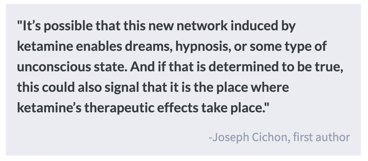
This study found that ketamine-induced dissociation is driven by a switch in activity between active and inactive neurons. Results from this study may help us better understand the neurological underpinnings of dissociation not only following ketamine exposure but also in psychiatric disorders where dissociation is a symptom (e.g., schizophrenia). 

From Repressed Memory to Dissociative Amnesia

 Post by Anastasia Sares

The takeaway

In the 1990s, there was heated scientific debate about whether people could recover “repressed” memories, or whether therapists were instead inducing false memories in their patients. One might assume this debate has been resolved, but it has cropped up under differing forms into the late 2010s, especially when it comes to deciding whether to admit such memories as testimonies in court. These kinds of recovered memories can exist in specific situations, but unreliable memories are also possible.

From repression to dissociation

The term “repressed memory” has generally fallen out of favor; however, the fifth edition of the Diagnostic and Statistical Manual for Mental Disorders (DSM 5) uses the term “dissociative amnesia” and defines a variety of dissociative disorders. This reflects the consensus of the scientific community, that traumatic memories—particularly, those that are very intense and experienced early in life—can be forgotten for a period of time, at least consciously (for example, of a woman who had a traumatic experience in an elevator and now refuses to take an elevator, though she does not know why). Studies that follow up with abuse victims whose childhood trauma history is recorded (medical record, etc.) find that some do indeed forget about their abuse and later remember it, and their recovered memories are just as reliable as those that were remembered continuously from the time of trauma.

Testing false memory in the lab

Now, this is not to deny the fact that memories can be unreliable. For example, when participants are presented with a list of related words, like “bed” “tired” “yawn” and then when asked to recall the words, they may include “sleep,” which was related to the other words on the list but not actually presented. When people are asked leading questions about whether or not they have experienced something, they may agree they have experienced it, even if they did not. Some recent evidence has shown that certain types of therapies (like Eye Movement Desensitization and Reprocessing also known as EMDR) can make people more prone to these sorts of errors. In addition, people with specific mental illnesses, such as dementia, schizophrenia, or Korsakoff syndrome may confabulate, or invent memories.

What's the impact?

As with many debates, there is at least a grain of truth on both sides. Both false and genuinely recovered memories can exist. Dissociative amnesia related to trauma or abuse is most likely to happen to people who experienced intense trauma at a young age. A person’s memory of an event, regardless of its status as “repressed,” would be bolstered by external evidence of that event in court. In the end, each case must be treated with its own unique sensitivity and scrutiny.

References +

  1. Brand, B. L., Dalenberg, C. J., Frewen, P. A., Loewenstein, R. J., Schielke, H. J., Brams, J. S., & Spiegel, D. (2018). Trauma-Related Dissociation Is No Fantasy: Addressing the Errors of Omission and Commission in Merckelbach and Patihis (2018). Psychological Injury and Law, 11(4), 377–393. https://doi.org/10.1007/s12207-018-9336-8
  2. Ghetti, S., Edelstein, R. S., Goodman, G. S., Cordòn, I. M., Quas, J. A., Alexander, K. W., Redlich, A. D., & Jones, D. P. H. (2006). What can subjective forgetting tell us about memory for childhood trauma? Memory & Cognition, 34(5), 1011–1025. https://doi.org/10.3758/BF03193248
  3. Houben, S. T. L., Otgaar, H., Roelofs, J., & Merckelbach, H. (2018). Lateral Eye Movements Increase False Memory Rates. Clinical Psychological Science, 6(4), 610–616. https://doi.org/10.1177/2167702618757658
  4. Lorente-Rovira, E., Santos-Gómez, J. L., Moro, M., Villagrán, J. M., & Mckenna, P. J. (2010). Confabulation in schizophrenia: A neuropsychological study. Journal of the International Neuropsychological Society, 16(6), 1018–1026. https://doi.org/10.1017/S1355617710000718
  5. Otgaar, H., Howe, M. L., Patihis, L., Merckelbach, H., Lynn, S. J., Lilienfeld, S. O., & Loftus, E. F. (2019). The Return of the Repressed: The Persistent and Problematic Claims of Long-Forgotten Trauma. Perspectives on Psychological Science, 14(6), 1072–1095. https://doi.org/10.1177/1745691619862306
  6. Williams, L. M. (1995). Recovered memories of abuse in women with documented child sexual victimization histories. Journal of Traumatic Stress, 8(4), 649–673. https://doi.org/10.1002/jts.2490080408

How Do Grid Cells Emerge from Neural Circuits?

Post by Andrew Vo

The takeaway

Artificial neural networks can be used to model and study the complex structure and function of brain circuits. Compared to traditional hand-designed models, trained networks better fit neural responses and generalize to other environments.

What's the science?

Grid cells are found in the entorhinal cortex of animals and humans, and their hexagonal firing patterns form spatial maps of the environment important for navigation. To better understand the biological and computational mechanisms underlying these grid-like representations, artificial recurrent neural networks (RNNs) have been used. Existing models are typically hand-tuned with parameters based on potentially biased assumptions. Consequently, it remains unclear if grid-like representations observed in hand-tuned models arise naturally and if they generalize to other environments. This week in Neuron, Sorscher et al. demonstrate how trained RNNs can innately give rise to hexagonal grid cells with greater accuracy than hand-designed models.

How did they do it?

The authors built an RNN that modeled entorhinal neuron activity from simulated mice exploring an environment. Critically, they did not assume beforehand that grid-like representations would emerge in the output. Instead, they trained their network to path integrate—when a network uses cues from an animal’s movement, such as head or body velocity, to compute and remember the animal’s spatial location—and explored whether grid cells would naturally appear from their RNN. They also tested whether their trained model could predict the entorhinal neuron firing patterns in actual electrophysiological recordings in mice.

What did they find?

Comparable to previous models, the authors found that their trained RNN achieved path integration and developed similar grid-like representations. Importantly, this observed output resulted from the network learning through training rather than being hand-tuned with optimal parameters in the first place. They also found that making small changes to the training procedure, such as allowing nonnegative firing rates and center-surround input structure, resulted in the spontaneous emergence of hexagonal grid-like representations and improved generalizability of their network to new environments beyond training. Finally, they found that their trained model was able to account for the firing patterns of actual entorhinal neurons with greater accuracy than traditional hand-tuned models.

What's the impact?

The present study showed that a simple RNN trained to path integrate resulted in the natural emergence of hexagonal grid cells. This model performs in an unbiased manner, without beforehand assumptions that confound traditional hand-tuned models. Such an approach to designing artificial neural networks allows us to test and provides insight into our conceptual understanding of complex neural circuits.

Access the original scientific publication here.