Long-Term Memory Formation Occurs Differently During Wakefulness and Sleep

Post by Lincoln Tracy

The takeaway

Long-term memories can be formed while we are awake, not just while we are asleep. However, the quality of memory consolidation varies depending on whether we are awake or asleep.

What's the science?

Sleep has long been heralded as a key promotor of consolidating newly encoded information into actual memories. Recent research suggests long-term memory consolidation can also occur while we are awake, but the mechanisms of wakeful memory consolidation are not well understood. This week in PNAS, Sawangjit and colleagues explored how long-term memory formation following a novel object recognition (NOR) task occurs in rats that had slept or remained awake after the initial encoding period.

How did they do it?

Rats were placed in an open field area and allowed to explore two identical objects for 10 minutes to familiarize themselves with the room, the area, and the objects (encoding phase). Immediately after, the rats had a two-hour post-encoding (or consolidation) phase where they were either allowed to sleep or kept awake. One week later, rats were returned to the area for the retrieval phase, where one of the objects was novel (retrieval or recall phase). The authors compared the time the rat devoted to exploring the novel object in comparison to the old object they had seen the week before. Rearing behavior (i.e., the rat standing on its hind legs) was also analyzed to assess how environmental context contributed to memory recall.

First, the authors tested the effects of sleep and wakefulness on long-term memory formation and recall. Second, they investigated whether the hippocampus makes an essential contribution to long-term memory formation during both sleep and wakefulness. To do this, they injected muscimol, a GABA-A receptor agonist, into the dorsal hippocampus 30 minutes after the rats fell asleep (and at a comparable time for the rats who remained awake) after the initial encoding session. Memory recall was tested one week later. Third, they tested NOR memory recall in a different context to what the rats were originally exposed to. That is, the recall component took place in a different arena, in a different room, with a different experimenter observing the rats. Finally, they analyzed electroencephalography (EEG) recordings of rats that slept after the NOR task to identify the specific mechanisms contributing to sleep-dependent memory consolidation. Like the previous behavioral tasks, they also tested the effect of muscimol on these potential mechanisms.

What did they find?

First, the authors found that although the rats that were kept awake displayed some long-term memory recall after a week, rats that slept after the encoding phase spent more time exploring the new object and less time exploring the familiar object (i.e., enhanced NOR performance). This suggests wakeful long-term memory consolidation is less effective than sleep consolidation. In addition, the rats that slept displayed fewer rearing behaviors in the retrieval phase compared to the encoding phase, while the rats that stayed awake displayed the same level of rearing across both sessions. This indicates the rats who were kept awake had no recollection of spatial context. Second, the authors found muscimol administration during sleep decreased NOR performance and maintained a higher level of rearing behavior in retrieval testing. In contrast, rats that were kept awake and received muscimol displayed enhanced NOR performance and did not affect rearing behavior during retrieval testing. Taken together, this suggests hippocampal inactivation influences memory consolidation during sleep and wakefulness in opposite directions, as well as selectively preventing rearing behaviors during sleep. 

Third, the rats who slept after encoding showed no improvements in NOR performance when tested in a different context, while rats that were kept awake displayed robust NOR performance during retrieval testing. This implies object memory after wakeful consolidation is less well integrated with the context in which the information is learned, meaning it can be retrieved more easily in a different context. This implication is corroborated by the changes in rearing behavior. Rats that slept displayed increased rearing behavior during retrieval testing, while rats that were kept awake displayed similar levels. Finally, the authors found sleep spindles and slow oscillation-spindle events were associated with enhanced NOR performance.

What's the impact?

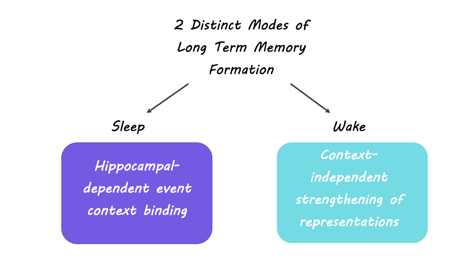
This study found there are two distinct ways that long-term memory formation can occur, albeit with differences in quality and quantity of memory encoding. During sleep, memory consolidation relies on hippocampal involvement and may involve linkages between event and context information. In contrast, wakeful consolidation is disrupted by hippocampal activation and implies that the context in which information was learned is less important.

How the Brain Integrates Our External and Internal Worlds

Post by Lani Cupo

The takeaway

Humans are capable not only of intelligible communication with others, but also private internal thought, both of which are represented separately in the brain. The interaction between the two is facilitated by convergence of these systems in two hub regions of the brain.

What's the science?

As humans, we perceive the external world through senses, and act upon it with various muscle groups, allowing communication with others. We are also capable of internal thought, allowing us to consider abstract concepts and plan for the future. The brain regions underlying these processes have been fairly well characterized as two segregated systems. There is the dorsal system, which is dedicated to analyzing the external world in terms of space and time, allowing us to perceive and act upon it, while the ventral system is dedicated to meaning, allowing our internal world to comprehend more than the immediate environment. It was previously unknown if and how these systems interact, however this week in NeuroImage, Weiller and colleagues identified hub regions in the brain where the dorsal and ventral systems interact, connecting the “dual-loop” of the dorsal and ventral systems and regulating syntax across cognitive domains, both internal and external.

How did they do it?

The authors used diffusion magnetic resonance imaging (dMRI) data, a technique which allows researchers to estimate reconstructions of white matter tracts in the brain by analyzing the characteristics of water molecules’ diffusion. First the authors modelled the fiber tracts with information about their directional trajectories, known as “streamlines”, as well as where each streamline terminates (where the white matter tract ends). They specifically examined regions of interest corresponding with the dorsal and ventral language systems of the brain. Next, the authors calculated which cortical regions, or hubs, intersected both dorsal and ventral tracts. Finally, they examined hemispherical differences between the hubs, assessing whether the same brain areas were identified on the left and right hemisphere. Having identified the regions, the authors examine the involved brain structures through the context of evolutionary development, with phylogenetic trees and lifetime development with the process of myelination.

What did they find?

First the authors found two major streamline systems that cover the majority of the brain with dorsal streamlines that includes many primary sensory and motor areas, and a ventral streamline system that includes further regions, such as the frontal, temporal, and occipital poles. The authors then identified two cortical hubs at the intersection of the dorsal and ventral tracts: one in the frontal cortex (largely the lateral prefrontal cortex and inferior frontal gyrus [IFG]), and one posterior hub in the middle temporal gyrus (MTG). The hubs close the loop between the dorsal and ventral tracts, which can allow recursive thought and internalization even without external feedback. While the hubs were identified in both the left and right hemispheres, there were inter-hemispheric differences in the exact regions involved, such as increased involvement of the temporal gyrus in the left hemisphere and more regions in the IFG in the right hemisphere. The hub regions are high-level multimodal areas, meaning they receive information from many lower-level processing regions (such as primary sensory regions), and are well connected throughout the brain. In particular, the MTG is responsible for processes related to language comprehension and conceptual knowledge, while the IFG has previously been implicated as an intersection between ventral and dorsal systems. These regions are among some of the last to emerge through evolution, changing significantly between macaque monkeys and humans, and they are also among the last to be myelinated and finish developing.

What's the impact?

This study suggests that the identified regions integrate the ventral and dorsal systems, providing syntax for both external communication and internal thought. Of note, the authors do not suggest that syntax takes place wholly within these regions, but instead these regions are necessary to integrate the parallel computations of the two separate systems. The findings of the authors discuss traits that are uniquely human lending insight into what sets us apart from other animals. 

Access the original scientific publication here.

Electrical Modulation Boosts Memory Performance in Older Adults

Post by Anastasia Sares

The takeaway

Electrical neuromodulation, when “dialed-in” correctly, enhanced certain types of memory in a group of older adults. Depending on the region modulated and the frequency of the alternating electric current, participants’ memory of either the beginning or the end of a list of words improved—which could reflect separate boosts in short and long-term memory.

What's the science?

It has been over 80 years since the introduction of electroconvulsive therapy (ECT). Though its history is fraught with misuse and lack of consent, the technique of electrically modulating the brain has survived to today, becoming more precise, well-researched, and ethically implemented. Nowadays, there are several varieties of electrical neuromodulation, some of which have less intensity and fewer side-effects. They gently modify how neurons fire rather than stimulating new firing. Some of these new techniques are said to increase brain plasticity (the ability for the brain to be malleable and learn new things), which could be especially useful for older adults who are beginning to experience cognitive decline. This week in Nature Neuroscience, Grover and colleagues applied specific electric currents to targeted brain areas in older adults and found that each of these improved memory in a different way.

How did they do it?

The authors randomly assigned their older adult participants to one of three groups: 1) gamma wave modulation (60 cycles per second) above the dorsolateral prefrontal cortex (in the frontal region of the brain), 2) theta wave modulation (4 cycles per second) above the inferior parietal lobe (further back in the brain, just above and behind the ears), and 3) a sham condition (where the machine was making noises but not actually passing any electric current).

The modulation (transcranial alternating-current stimulation, or tACS) took place over 4 sessions, with a 1-month follow-up to see if there were any longer-lasting effects. During modulation, the participants tried to memorize lists of words, and their performance was recorded. They did this because it has been shown that doing a task during modulation can improve performance specifically for the networks implicated in the task.

Next, they repeated the whole task, but with a different group of people and with the gamma and theta frequencies swapped for the two distinct parts of the brain (theta in frontal and gamma in parietal), to see if it mattered which frequency was used over which brain area. And finally, they performed a third experiment, similar to the first, to see if their results were replicable.

What did they find?

In people who had the sham condition (no actual brain modulation), performance did not improve (as expected). In people who had the gamma wave modulation above frontal regions, memory for words at the beginning of the lists improved. For people who had theta modulation over parietal regions, memory for words at the end of lists improved. The authors emphasized how these results are consistent with the “dual-store” theory of memory, with recall for the beginning of the lists reflected long-term memory and recall for the end of the lists reflected short-term memory (or working memory).

For both experimental groups, improvement could still be seen at the 1-month follow-up. In addition, the greatest benefit was seen in people who had lower cognitive scores to begin with. The findings were replicated in an independent group in the third experiment. In the second experiment where the frequencies were swapped, no memory improvements were observed, meaning that memory performance could only be boosted by a specific frequency of modulation over a specific region of the brain.

What's the impact?

This is an exciting study that shows replicable and lasting effects of neuromodulation on memory in older adults, especially those who have lower cognitive scores. If it can be applied to cases of mild cognitive impairment, before dementia sets in, it could be a revolutionary way to enhance quality of life in the world’s aging population. However, it is important to keep in mind that the neuromodulation had to be paired with a task and repeated many times; it did not offer a global boost to memory capacity.