Creating an Atlas to Identify the Neurodevelopmental Stage of Human Brain Tumor Cells

Post by Elisa Guma

The takeaway

By creating an atlas of gene expression in the mouse brain throughout the lifespan, the authors were able to identify expression signatures from adult and pediatric human cerebral tumor development. Brain tumors adopt embryonic-like states, providing insight into the mechanisms by which these tumors grow.

What's the science?

The mechanisms driving cerebral tumor growth are still poorly understood. Within a single tumor, there is high heterogeneity in cell types, many of which resemble various stages of normative neural development. To map the stages of tumor cell development, one must first map normative cerebral development to create a point of reference. This week in Nature Communications, Hamed and colleagues characterize the molecular identity of cells that may drive cerebral tumor cell heterogeneity and determine their relationship to normal developmental states and lineages.

How did they do it?

First, the authors set out to create a comprehensive single-cell atlas of precursor cells (those within the neurogenic and gliogenic compartments) over the course of normal cerebral development. They performed single-cell RNA sequencing of the mouse brain throughout development (11 time points), starting from embryonic day 12.5 (mid-gestation), to postnatal day 365 (1 year old). To identify the appropriate cells to sequence, the authors used a transgenic mouse (Sox2eGFP) which has a fluorescent tag (GFP) on the Sox2 gene that is highly expressed in the cortical neurogenic zones across all stages of prefrontal brain development. Clustering methods were applied to the cells’ transcriptional data to differentiate the identity and developmental signature of the cells.  

The second aim of this work was to identify the transcriptional signature of human brain tumor cells. To do so, the authors sequenced human (adult and pediatric) brain tumor cells and used a deconvolution algorithm to map single-cell transcriptomic data acquired from the human brain tumor cells to the mouse clusters they defined above (with a focus on precursor cells). To further validate the reliability of their model the authors aligned human fetal single-cell RNA sequencing data (from a previous publication). They also aligned cells from patient-derived glioblastoma cultures to the mouse clusters identified.  Finally, to validate the existence of precursor cells (radial glial precursors) in adult cortical cancer, the authors leveraged a mouse model that uses Nestin-Cre to generate mice with conditional loss of expression of two tumor suppressor genes, the p53 gene (both alleles) and the PTEN gene (one allele) in embryonic precursors. By targeting Nestin+ embryonic precursor cells, the authors were able to gain more central nervous system specificity for tumor development. Indeed, these mice develop glioblastomas postnatally (due to a loss of tumor suppressors) which the authors extracted and sequenced to compare to their single-cell atlas.

What did they find?

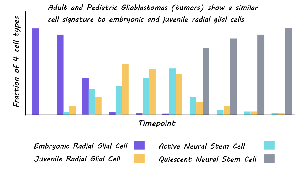
The single-cell atlas of normative brain development comprised 102,504 single cells, with an average of 3,500-13,000 unique molecular identities per cell. These cells could be clustered into three developmental stages: embryonic (mid-late gestation), juvenile (first week of life), and adult (childhood to adulthood). In the embryonic stage, cells were more likely to be undergoing neurogenesis, followed by a switch to gliogenesis which peaked in the juvenile period and continued into young adulthood. Unbiased clustering of the cells also revealed four major clusters with distinct transcriptional profiles. Embryonic radial glial precursor cells denominated the first cluster, juvenile radial glial precursor cells the second, third were glial cells also expressed in the juvenile period, and the fourth was quiescent neural stem cells expressed in the adult period. A deeper characterization of the regional distribution of these cells revealed distinct expression patterns based on the geographical location of the cells within the brain, particularly for the embryonic cells. This incredible resource is publicly available through an app developed by the authors.

The second aim of the study was to use this new atlas to classify the developmental stage of cells from both adult and pediatric brain tumors (glioblastomas). They were able to establish a reliable cross-species mapping between the mouse cells and adult and pediatric human cells, providing another useful resource for the scientific community. Cells from the cerebral gliomas were most similar to those in the embryonic and juvenile stages of the mouse atlas, rather than the adult. This was true for both adult and pediatric tumors. There was also a high similarity between the human fetal cells, cultured glioblastoma cells, and embryonic mouse cells. This both bolsters confidence in the ability of the mouse atlas to characterize cellular heterogeneity in cerebral tumors, and in the embryonic nature of the tumor cells. Finally, malignant tumor cells from the adult mouse brain tumors (from the Nestin-Cre mice with conditional loss of expression of two tumor suppressor genes) also had high similarity to embryonic and juvenile radial glial precursor cells providing further validation for their results. 
What's the impact?

This study presents the most comprehensive single-cell atlas of the developing mouse cortex. Furthermore, by achieving reliable cross-species alignment of single-cell transcriptional profiles, the authors were able to use this atlas to classify tumor cells from human adult and pediatric glioblastomas. They found that expression profiles of human tumor cells specifically overlap with embryonic radial glial precursor cells. These findings provide insight into the origins of human brain tumors and aid in our understanding of how normal cells turn into cancer cells. Further, these findings identified potential molecular targets for the treatment of cerebral tumors.   
Going Beyond the Diagnosis: What it Means to be “Transdiagnostic”

Post by Anastasia Sares

A diagnosis isn’t the whole story?

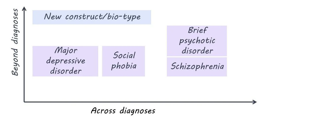
“Transdiagnostic research” is a current buzzword in psychiatry. It comes from the prefix “trans,” meaning through/across or beyond, and “diagnostic” meaning a medical category, so transdiagnostic means “across or beyond diagnosis”. The current world of psychiatry is dominated by the DSM (Diagnostic and Statistical Manual of Mental Disorders) and ICD (International Classification of Diseases), which defines the symptom criteria for conditions like depression, anxiety, post-traumatic stress, eating disorders, personality disorders, and psychotic disorders. If you’ve ever taken a psychology class, this is likely the model you’ve been taught.

However, there are some problems with this system. First, there is a high amount of variability between individuals with a given diagnosis - in extreme cases, two individuals with the same diagnosis may only share one or two symptoms. Second, some symptoms overlap between disorders, and comorbidities (when someone has 2 or more diagnoses simultaneously) are more common than we would expect. Third, our categories are based largely on symptoms, not on causes or effective treatments. So, some scientists and clinicians think we need to put the old categories aside and search for a new classification system.

What would transdiagnostic research and treatment look like?

To come up with a new psychiatric classification system, we would have to find a set of traits that span across current diagnostic categories. These new traits would need to do a better job of predicting a patient’s symptoms and/or their response to treatment.

For example, one study that looked at both schizophrenia and bipolar depression found three distinct “biotypes” that were spread across the two diagnoses. One biotype showed marked cognitive impairments and lowered sensory reactivity, another showed some cognitive impairment but heightened sensory reactivity, and a third group had cognitive and sensory features comparable to the non-patient group. Knowing someone’s diagnosis doesn’t help very much in predicting what their biotype is, so these biotypes could be considered transdiagnostic.

What’s new?

Transdiagnostic research is gaining momentum, with large groups of researchers combining their efforts to try and find a new classification system. However, some are critical of the recent flood of research. A systematic review by Fusar-Poli and colleagues found that only three out of the 111 studies they examined were truly transdiagnostic. Less than half of self-titled “transdiagnostic” studies compared their new classifications directly to the current gold standard diagnoses, and when they did, the transdiagnostic approach often performed no differently than the current diagnosis-based approach. Twenty percent of the studies were not transdiagnostic at all. The authors came up with guidelines for future transdiagnostic research to guide the growing field.

What's the bottom line?

It’s obvious that the current diagnostic system in psychiatry is imperfect, but it’s the best we have so far. While there are some good examples of transdiagnostic research out there, the field needs to be rigorous and insightful if it ever hopes to replace the DSM and ICD.

References +

Dalgleish, T., Black, M., Johnston, D., & Bevan, A. (2020). Transdiagnostic approaches to mental health problems: Current status and future directions. Journal of Consulting and Clinical Psychology, 88(3), 179–195. https://doi.org/10.1037/ccp0000482

Fusar‐Poli, P., Solmi, M., Brondino, N., Davies, C., Chae, C., Politi, P., Borgwardt, S., Lawrie, S. M., Parnas, J., & McGuire, P. (2019). Transdiagnostic psychiatry: A systematic review. World Psychiatry, 18(2), 192–207. https://doi.org/10.1002/wps.20631

Clementz, B. A., Parker, D. A., Trotti, R. L., McDowell, J. E., Keedy, S. K., Keshavan, M. S., Pearlson, G. D., Gershon, E. S., Ivleva, E. I., Huang, L.-Y., Hill, S. K., Sweeney, J. A., Thomas, O., Hudgens-Haney, M., Gibbons, R. D., & Tamminga, C. A. (2022). Psychosis Biotypes: Replication and Validation from the B-SNIP Consortium. Schizophrenia Bulletin, 48(1), 56–68. https://doi.org/10.1093/schbul/sbab090


The Role of Dopamine in Forgetting Competing Information

Post by Shannon Kelly

The takeaway

Actively forgetting memories that compete with useful memories is important for our ability to remember information that supports our goals. This study found that the neurotransmitter dopamine in the prefrontal cortex plays a critical role in the active forgetting of competing memories.

What's the science?

Unlike the passive decay of memories that are not used over time, active forgetting is a process by which distracting memories are selectively forgotten to support our ability to retrieve useful memories. Active forgetting can happen when we retrieve certain memories and must inhibit competing memories (i.e., retrieval-induced forgetting). Although the neurotransmitter dopamine is known to play an important role in skills that relate to active forgetting, such as the ability to flexibly change behaviors, its direct involvement in retrieval-induced forgetting is not well understood. This week in The Journal of Neuroscience, Gallo and colleagues found that active forgetting in rats was hindered by reducing the brain’s ability to respond to the presence of dopamine and improved by increasing dopamine-related activity in the prefrontal cortex.

How did they do it?

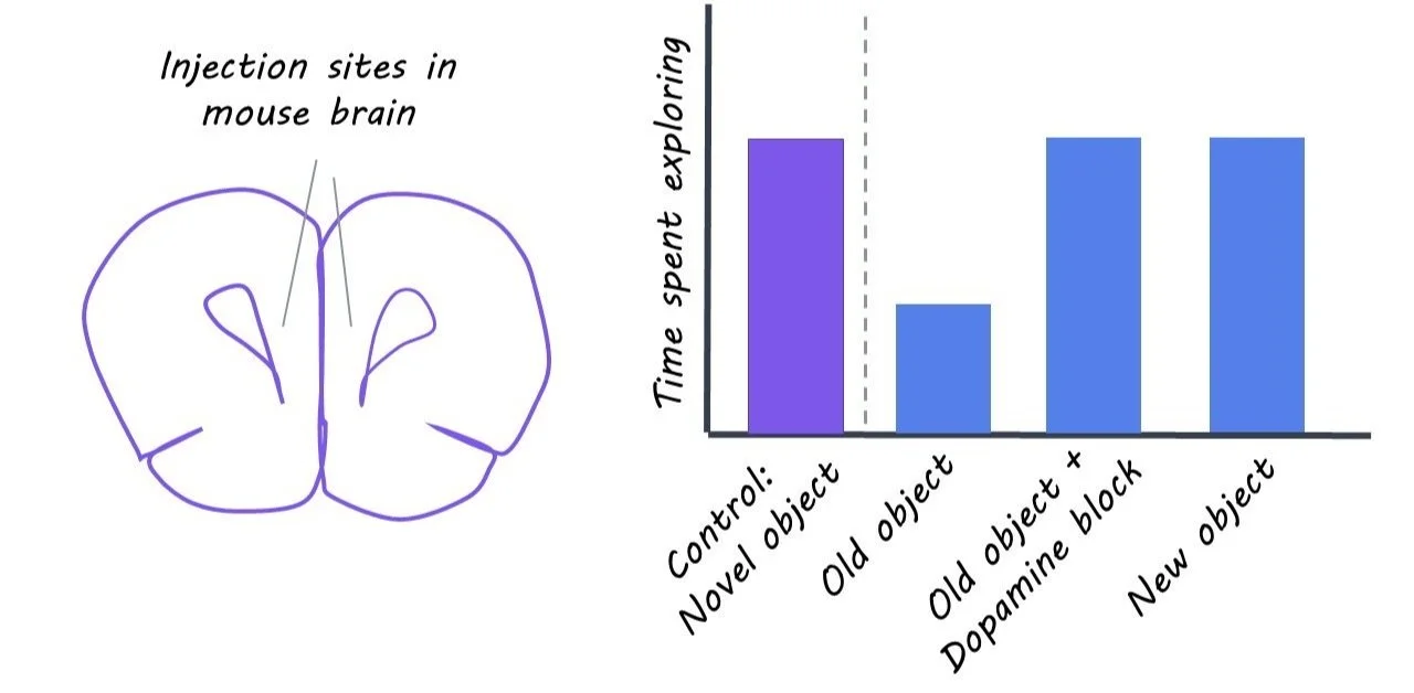
The authors tested the effects of dopamine-related drugs on rats’ memory. They first showed rats pairs of objects to remember, then injected either harmless salt water or a drug that affects dopamine-related activity in the medial prefrontal cortex (mPFC; similar to the dorsolateral prefrontal cortex in humans). In the retrieval condition, rats then were shown pairs of one previously seen object and one new object so that they used their memory to guide their exploration. In the control conditions, rats either were shown pairs of two new objects or rested for the same amount of time. Finally, they showed the rats a pair of one new and one previously seen object and measured how long the rats examined each object. Since rats are known to prefer new objects, a large difference in time spent exploring the new object compared to the old one indicated that the rats remembered the previously seen object. If the rats preferred the new object in both control conditions but showed no or less preference for the new object in the retrieval condition, this indicated that the rats showed retrieval-induced forgetting of the previously seen object. The authors tested the effects of (1) a drug that decreases reactivity to dopamine by blocking dopamine receptors, (2) a drug that inhibits dopamine release into the mPFC from the ventral tegmental area (VTA), and (3) a drug that activates dopamine receptors on retrieval-induced forgetting.

What did they find?

Rats that were injected with either the drug that blocks dopamine receptors or the drug that suppresses the release of dopamine from VTA into mPFC showed no difference in behavior between the retrieval and control conditions, indicating no retrieval-induced forgetting. These findings show that dopamine-related activity in the mPFC is necessary for active forgetting of competing memories. For rats whose dopamine release from VTA was suppressed, retrieval-induced forgetting was improved back to normal levels when given the drug that activates dopamine receptors in mPFC. Finally, rats given only the dopamine-enhancing drug showed better retrieval-induced forgetting compared to control rats during a more difficult version of the memory test. Together, these findings show that active forgetting can be causally improved or hindered by increasing or decreasing dopamine-related activity in mPFC.

What's the impact?

This study showed that the ability to selectively forget distracting information can be directly influenced by affecting the level of dopamine-related activity in the prefrontal cortex of rats. These findings clarify the mechanisms by which experience helps determine which memories are maintained and which are forgotten. Based on findings that active forgetting involves similar brain mechanisms in humans as in rats, these findings suggest that prefrontal dopamine plays a critical role in our ability to adapt our memories based on the demands of our environments.

Access the original scientific publication here.