Ripples Facilitate Memory Recall via Widespread Synchronization of Brain Activity

Post by Leanna Kalinowski

The takeaway

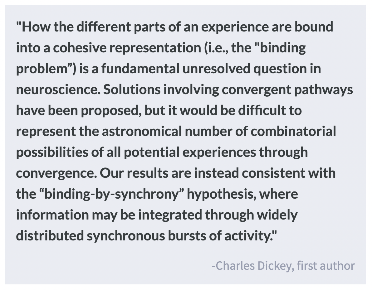
The encoding, consolidation, and retrieval of memory requires the integration of multiple regions across the cortex. High-frequency brain oscillations, called “ripples”, may help to organize activity across these regions into a coherent representation.

What's the science?

Different elements of a memory are stored in different regions across the brain. Therefore a coordination of brain activity is required whenever a memory is consolidated, encoded, or retrieved. While this coordination is not yet fully understood, it is believed that these regions become synchronized via high-frequency brain oscillations called “ripples”. This week in PNAS, Dickey and colleagues used intracranial recordings in humans to measure ripple patterns during sleep, waking, and memory recall.

How did they do it?

The researchers used data from 25 participants who already had electrodes surgically implanted into their brains for an unrelated medical issue (i.e., treatment-resistant epilepsy). This procedure, called stereoelectroencephalography (SEEG), is used to measure activity in regions deeper than what regular electroencephalography (EEG) can reach. In order to study healthy brain activity, they excluded data that was suspected to be related to underlying epilepsy. They measured ripple patterns from the cortex and hippocampus during three key periods: (1) during non-rapid eye movement (NREM) sleep, which is when the brain replays and sorts through memories for consolidation into long-term storage, (2) during waking, which is when information is transferred from the cortex to the hippocampus for memory encoding, and (3) during memory recall.

They then evaluated memory recall by having participants undergo the paired-associated memory task. In this task, participants first encoded eight pairs of words (e.g., “moon” + “mouth”) and were subsequently cued with one word from each pair (e.g., “moon”) to immediately recall the second word in the pair (e.g., “mouth”). After a 60-second delay with distraction, they were once again asked to recall the second word in the pair. This process was repeated until each participant attempted to recall about 100-160 unique word pairs in total.

What did they find?

The researchers found that ripples couple (i.e., occur within 500 ms of each other) and co-occur (i.e., have > 25 ms of activity overlap between these ~70 ms long events) across multiple lobes and both hemispheres during both NREM sleep and waking. They also found an increase in ripple co-occurrences between cortical sites and between the cortex and hippocampus during the memory recall task. These ripples phase lock with neuron firing (i.e., synchronize individual neuron activity) and phase synchronize with each other (i.e., synchronize activity across brain regions) across short and long distances.

What's the impact?

Taken together, these results demonstrate a mechanism by which the brain synchronizes activity across multiple cortical regions, which may help to integrate different components of a memory. This synchronization is not impacted by distance, including instances where both hemispheres of the brain are activated. Further work is needed to assess how ripples may help with the integration of brain activity for neural events beyond memory.

Statistical Learning From Speech

Post by Anastasia Sares

The takeaway

Spontaneous speech synchronizers are those who unconsciously align their own speech production with an external stimulus. People who do this are also better at identifying patterns in artificial babble speech, and it may be because they are using additional brain areas when processing speech in general.

What's the science?

Language learning is a complex and challenging process. When first learning to speak, or when learning a second language, we often hear strings of sounds with no obvious break between them. But through repeated listening, our brains can pick up on patterns, determining which groups of sounds occur together more often—these are more likely to be words. The process is called “statistical learning,” and it can be tested in the lab using artificial speech. Individuals vary in their statistical learning ability. In a recent study, it was discovered that some people spontaneously synchronize their own speech with external speech, and these people are also better at statistical learning.

But why would spontaneous speech synchrony make a person better at statistical learning? This week in PLoS Biology, Orpella, Assaneo and colleagues tried to see whether brain activity during statistical learning could distinguish between spontaneous synchronizers and non-synchronizers and whether this difference in brain activity could account for their enhanced statistical learning ability.

How did they do it?

The authors recruited two groups of people: one to confirm the effect of spontaneous speech synchrony on statistical learning, and another group to perform this task while undergoing magnetic resonance imaging (MRI) to understand the brain regions involved.

The first measure was spontaneous speech synchrony—participants heard a string of spoken syllables and were instructed to whisper “tah” repeatedly while listening. Synchronizers will align their speech to the syllables they hear, while non-synchronizers will not align their speech, so this task separated the participants into two groups.

In the statistical learning task, participants heard strings of nonstop syllables containing embedded “words” (three syllables that repeatedly occurred together in the same order, like “rakuso”). Afterward, they were tested by being shown two “words” and choosing which one they had heard. There were two conditions in this task—in one, participants listened to the stream of syllables normally, while in the other, they were required to say “tah” repeatedly (this is supposed to interfere with the statistical learning).

The second group of participants completed the statistical learning task while in an MRI, and afterward their brain activity was split up into networks using a technique called Independent Component Analysis (ICA). The networks were tested individually to see which ones had activity related to the task.

What did they find?

Synchronizers did better at the statistical learning task, but only if they were allowed to listen passively. When they had to say “tah” while listening, their performance decreased to the level of the non-synchronizers. Non-synchronizers did equally well whether they were saying “tah” or passively listening.

MRI revealed that synchronizers were recruiting an additional brain network to process speech during statistical learning. While non-synchronizers only used the auditory network, synchronizers also used a fronto-parietal network, which is composed of some regions in the frontal lobe and others in the parietal lobe. Previous research identifies the fronto-parietal network as being involved in salience, attention, or monitoring.

What's the impact?

This study highlights some important individual differences in language processing. Research into spontaneous synchronization and the fronto-parietal network’s role in speech might shed some light on normal and abnormal language development.

Access the original scientific publication here.

Brain Health Following Bariatric Surgery to Treat Obesity

Post by Lani Cupo

The takeaway

“Brain age” refers to how old a brain appears when compared to normative trajectories of development across the lifespan. Obesity increases brain age, however, bariatric surgeries can reduce the impact of obesity on brain age.

What's the science?

The brain age gap is defined as the difference between a person’s predicted age based on biological features (in this case brain grey matter density) and a person’s actual chronological age. Advanced brain age — when the predicted age is greater than the chronological age — in adults has been associated with various disorders and reliably predicts cognitive impairment. While obesity has been linked to increased brain age in the past, it was previously unknown whether weight-loss interventions can reduce brain age and improve brain health. Recently in NeuroImage, Zeighami and colleagues investigated brain age in individuals with obesity and the impact of bariatric surgery on brain and cardiovascular health.

How did they do it?

The authors used three different large magnetic resonance imaging (MRI) datasets for this study. They derived a measure of volume at each voxel, (like a 3D pixel) in the brain images, allowing them to examine volume across the entire brain. The first dataset (640 participants, 324 females) was used to construct a model that describes how the brain’s grey matter changes with age in healthy participants. Based on the model from the first dataset, brain age was predicted for the second data set (92 participants, 46 with obesity). In this “test” dataset, the authors compared the brain age gap for participants with and without obesity. The final dataset (87 at baseline, 65 females, 34 at last visit, 25 females) was used to examine brain age in subjects with obesity at baseline (before surgery) and then 4, 12, and 24 months after surgery. Lastly, in this dataset, brain age was also compared with improvement in “cardiometabolic metrics” including body mass index (BMI), blood pressure, triglycerides, cholesterol, glucose, and a measure of insulin resistance (HOMA-IR).

What did they find?

First, the authors found that compared with individuals who did not have obesity, those with obesity had an increased brain age gap, reflecting poorer brain health. On average, the brains of those with obesity appeared about 5 years older than those of controls. Second, the authors found that compared with baseline, participants who completed all four visits demonstrated improved brain age (reflected in a reduced brain age gap) 12 months and 24 months after the surgery. At 12 months, the decrease in brain age was about 2.9 years, and at 24 months it was about 5.9 years compared to baseline, reducing the gap almost to control levels.

In the participants who completed all four visits, the authors found an association between higher brain age and cardiometabolic metrics, including higher BMI and blood pressure, and lower HDL cholesterol (the so-called “good” cholesterol). When participants who did not complete all four visits were included, the authors found an association between higher brain age and higher BMI, blood pressure, and HOMA-IR. The discrepancy between the two samples could reflect some systematic difference between the participants who remained in the study and those who dropped out. At 24 months post-surgery, the reduction in brain age was driven by a global change across brain regions, however, the somatomotor, visual, and ventral attention networks were strong contributors to this reduction.

What's the impact?

This study is the first to show that surgeries that target weight-loss and cardiometabolic health can improve brain health and reduce the brain age gap. These results could suggest that improved brain health following bariatric surgery may result from increased cerebral blood flow and greater insulin sensitivity. Ultimately, these findings reinforce the benefits of bariatric surgeries in patients with obesity, not only in terms of cardiovascular health but for brain health as well.