Brain Health Following Bariatric Surgery to Treat Obesity

Post by Lani Cupo

The takeaway

“Brain age” refers to how old a brain appears when compared to normative trajectories of development across the lifespan. Obesity increases brain age, however, bariatric surgeries can reduce the impact of obesity on brain age.

What's the science?

The brain age gap is defined as the difference between a person’s predicted age based on biological features (in this case brain grey matter density) and a person’s actual chronological age. Advanced brain age — when the predicted age is greater than the chronological age — in adults has been associated with various disorders and reliably predicts cognitive impairment. While obesity has been linked to increased brain age in the past, it was previously unknown whether weight-loss interventions can reduce brain age and improve brain health. Recently in NeuroImage, Zeighami and colleagues investigated brain age in individuals with obesity and the impact of bariatric surgery on brain and cardiovascular health.

How did they do it?

The authors used three different large magnetic resonance imaging (MRI) datasets for this study. They derived a measure of volume at each voxel, (like a 3D pixel) in the brain images, allowing them to examine volume across the entire brain. The first dataset (640 participants, 324 females) was used to construct a model that describes how the brain’s grey matter changes with age in healthy participants. Based on the model from the first dataset, brain age was predicted for the second data set (92 participants, 46 with obesity). In this “test” dataset, the authors compared the brain age gap for participants with and without obesity. The final dataset (87 at baseline, 65 females, 34 at last visit, 25 females) was used to examine brain age in subjects with obesity at baseline (before surgery) and then 4, 12, and 24 months after surgery. Lastly, in this dataset, brain age was also compared with improvement in “cardiometabolic metrics” including body mass index (BMI), blood pressure, triglycerides, cholesterol, glucose, and a measure of insulin resistance (HOMA-IR).

What did they find?

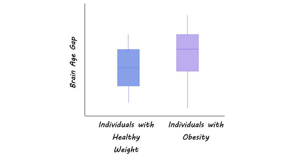
First, the authors found that compared with individuals who did not have obesity, those with obesity had an increased brain age gap, reflecting poorer brain health. On average, the brains of those with obesity appeared about 5 years older than those of controls. Second, the authors found that compared with baseline, participants who completed all four visits demonstrated improved brain age (reflected in a reduced brain age gap) 12 months and 24 months after the surgery. At 12 months, the decrease in brain age was about 2.9 years, and at 24 months it was about 5.9 years compared to baseline, reducing the gap almost to control levels.

In the participants who completed all four visits, the authors found an association between higher brain age and cardiometabolic metrics, including higher BMI and blood pressure, and lower HDL cholesterol (the so-called “good” cholesterol). When participants who did not complete all four visits were included, the authors found an association between higher brain age and higher BMI, blood pressure, and HOMA-IR. The discrepancy between the two samples could reflect some systematic difference between the participants who remained in the study and those who dropped out. At 24 months post-surgery, the reduction in brain age was driven by a global change across brain regions, however, the somatomotor, visual, and ventral attention networks were strong contributors to this reduction.
What's the impact?

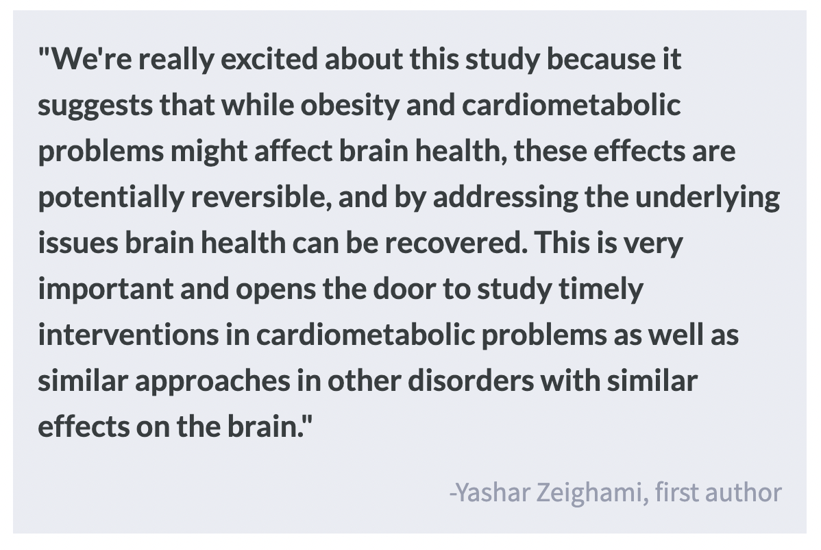
This study is the first to show that surgeries that target weight-loss and cardiometabolic health can improve brain health and reduce the brain age gap. These results could suggest that improved brain health following bariatric surgery may result from increased cerebral blood flow and greater insulin sensitivity. Ultimately, these findings reinforce the benefits of bariatric surgeries in patients with obesity, not only in terms of cardiovascular health but for brain health as well.
Specific Meningeal Layers Involved in CNS Autoimmune Responses

Post by Elisa Guma

The takeaway

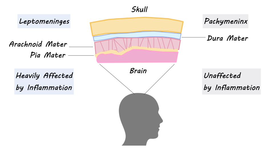
The meningeal layers of the central nervous system play distinct roles in acute and chronic CNS autoimmunity. The outermost dural layer seems to be largely unaffected by inflammation, while the internal leptomeningeal layers are heavily involved in the inflammatory response.

What's the science?

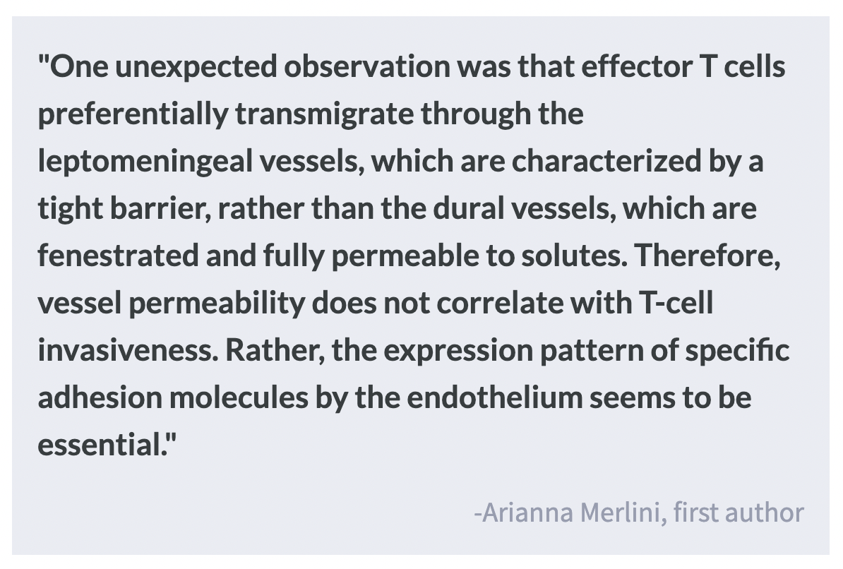
The central nervous system is surrounded and protected by a three-layered membrane called the meninges, which include the leptomeninges (including the pia layer, closest to the brain, followed by the arachnoid layer), and pachymeninx (also known as the dura mater). In addition to their physically protective role, the meningeal layers are densely populated by immune cells and are involved in the autoimmune response of the CNS, both in acute and chronic conditions (such as autoimmune diseases like multiple sclerosis). However, the distinct role of the three meningeal layers in CNS autoimmunity has yet to be fully understood. This week in Nature Neuroscience, Merlini and colleagues investigate the differential role of the meningeal layers in modulating autoimmune inflammation in an animal model of multiple sclerosis, experimental autoimmune encephalomyelitis (EAE), and human MS patients.  

How did they do it?

The authors studied the meninges (pachymeninx or dura mater vs. the leptomeninges) in both rat and mouse models of autoimmune encephalomyelitis (EAE), which exhibits signs of autoimmune inflammation, similar to those seen in human patients with multiple sclerosis. To induce meningeal inflammation, the authors gave healthy rodents T cells that specifically activate in the presence of certain proteins that exist in the brain (myelin basic protein or beta synuclein). Four to five days following exposure, rodents exhibit clinical symptoms. 

To characterize the immune response of the meninges, the authors tracked the migration of fluorescently labeled immune cells using two-photon laser scanning microscopy in the EAE model. Next, to assess the properties of the meningeal vasculature they injected rats with fluorescent tracers that rapidly leak out of vessels and measured accumulation in the perivascular space, which would indicate regions where the vasculature is leaky. 

Next, the authors investigated the stimulatory capacity of T cells present in the dural layer and the leptomeningeal compartment. They also measured the motility of T cells and if they made contact with antigen-presenting cells, a group of cells that mediate the immune response by processing and presenting antigens for recognition to certain T cells, to determine whether differences in immune response across the layers were due to reduced opportunity for inflammation. They also investigated the role of lymphatic drainage in modulating the immune response. Finally, to confirm the observations they made in the rodent autoimmune models, they studied the immune profile of post-mortem samples of the meninges of patients with chronic MS. 

What did they find?

In the rodent EAE models, the authors found evidence of massive inflammation of the leptomeninges and parenchyma but hardly any inflammation in the dura. These results were replicated in meningeal samples from human patients with chronic multiple sclerosis. 

To better understand these results, the authors studied the meningeal vasculature: they found that the dura had high vessel permeability (based on leakage of a fluorescent tracer), which indicated that a lack of permeability is not preventing the dura from accumulating inflammatory cells. Transcriptional analysis of the dural and leptomeningeal vessels identified distinct gene expression profiles, whereby genes encoding blood-brain barrier components, and molecules relevant for effector T cell adhesion to the vessels, were expressed at significantly lower levels in the dura, which may limit the ability of the T cells to adhere to the dural vessels. In inflammatory conditions, however, the dural endothelia enabled T cell adhesion. Therefore, other mechanisms should be accounting for the reduced involvement of the dura in the CNS autoimmune process. 

The authors investigated if the reduced levels of inflammation within the dura could be due to 1) T cell anergy (a state in which cells fail to produce a response), 2) to an immune suppressive environment in the dura, or 3) to the inability of the locally resident phagocytes to present antigen. However, they found that the dural T cells had the same intrinsic stimulatory capacity as the T cells in the leptomeninges and that dural antigen-presenting cells were also fully competent. What they did find was that the substrate for successful autoreactive T cell activation, namely CNS autoantigens, did not reach the dura in sufficient quantities to activate the T cells. 

Finally, the authors investigated the role of lymphatic vessels in the inflammatory response. They found that although antigens could be transported from the CNS to the dura and the deep cervical lymph nodes, there was higher uptake in the leptomeningeal cells than in the dura or deep cervical lymph nodes.  

What's the impact?

This study highlights the differential role of the meningeal layers in both acute and chronic autoimmune disease, with the dura playing a passive role relative to the leptomeninges. The findings presented here are surprising since the dura would be expected to play a more active role in these inflammatory processes, given its location, vascularity, and dense population of immune cells. Future research is needed to investigate potential immunomodulatory therapies specifically targeted at the leptomeninges. 

An Effective Treatment for Post-Traumatic Stress Headaches in Veterans

Post by Megan McCullough

The takeaway

Cognitive behavioral therapies are an effective treatment for veterans with post-traumatic headaches and post-traumatic stress disorder symptoms.

What's the science?

Veterans are more at risk for developing post-traumatic headache (PTH) associated with mild traumatic brain injuries compared to civilians. There are no current treatments for PTH, but previous research suggests that cognitive behavioral therapy (CBT) could offer a safe and effective treatment. Although previous studies have found no benefit of CBT for treating PTH in civilians, veterans could respond differently to treatment because of their higher risk of post-traumatic stress disorder (PTSD) symptoms. This week in JAMA Neurology, McGeary and colleagues tested the efficacy of CBT compared to usual treatments in veterans with mild traumatic brain injury-related headaches and symptoms of PTSD.

How did they do it?

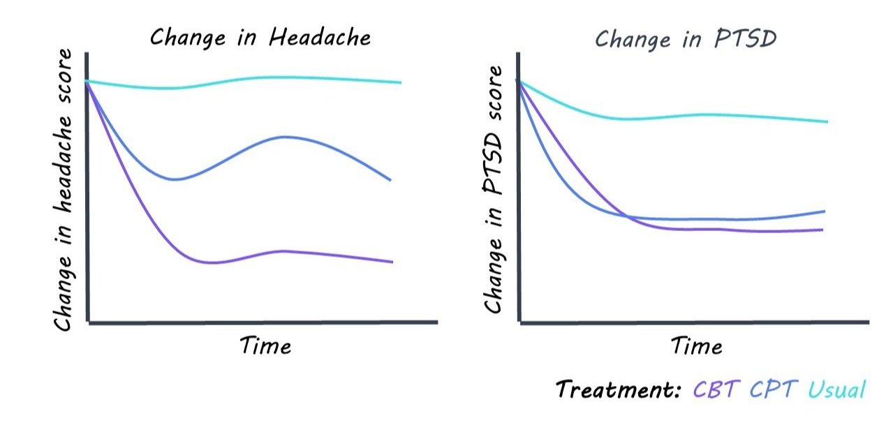
Participants included 193 combat veterans with PTH and PSTD symptoms. Participants were randomized into one of three groups: cognitive behavioral therapy for headaches, cognitive processing therapy, and usual care. Treatment was administered over the course of 6 weeks and participants completed pre-treatment assessments, post-treatment assessments, and 3 and 6-month follow-ups. The CBT treatment focused on behavioral interventions and stress management with a focus on headaches, while the cognitive processing therapy treatment focused on PTSD symptoms. The assessments administered to measure the efficacy of the therapies included self-reports of headache and PTSD symptoms. Statistical analyses were used to determine the effect of each treatment on the alleviation of headache and stress symptoms.

What did they find? 

The authors found that CBT treatment significantly improved headaches in veterans. Specifically, those in the CBT group showed significant improvement in both headache symptoms and PTSD symptoms compared to those who received usual care or cognitive processing therapy. Cognitive processing therapy, which focused on the alleviation of PTSD symptoms, did not improve headache disability but did lead to a significant decrease in the severity of PTSD symptoms.

What's the impact?

This study is the first published randomized clinical trial for PTH treatments in veterans. The data show that CBT improved both headache and PTSD symptoms, while cognitive processing therapy only improved the PTSD symptoms. This suggests that cognitive behavioral therapy can be an effective tool for veterans experiencing the ramifications of trauma and brain injuries.

Access the original scientific publication here.