How Sleep Facilitates Relational Associative Memory

Post by Andrew Vo

The takeaway

Sleep plays a critical role in our ability to make connections among indirectly learned but overlapping items in our memories. Building a theoretical neural model that can learn and simulate sleep states revealed specific mechanisms by which sleep may improve associative memory.

What's the science?

Relational memory refers to the ability to form associations between individual items. An important feature of relational memory is transitive inference, the ability to form associations between indirectly learned but overlapping items. For example, indirectly learning that A→C after directly learning that A→B and B→C. Previous research has suggested that sleep is important in forming such memories. The exact mechanisms underlying this process are not entirely clear, however. This week in Journal of Neuroscience, Tadros and Bazheno build a thalamocortical network to test how sleep might strengthen relational memories.

How did they do it?

The authors built a computer model of a thalamocortical network that could learn a relational memory task as well as simulate awake/sleep states. The network contained a cortex, composed of two layers of neurons that represented the primary visual cortex and associative cortex, and thalamus. Excitatory and inhibitory connections among neurons were randomly modelled. Network states were simulated by changing levels of neuromodulators, such as acetylcholine and GABA, until neuron firing rates became characteristic of different sleep cycles.

The relational memory task was comprised of three stages: supervised training, unsupervised training, and sleep. During supervised training, each of six individual items was stimulated in cortical layer 1 followed by layer 2, forming network pathways that represented these items. Next, during unsupervised learning, pairs of items were stimulated at the same time to induce associative learning between individual items. During a sleep phase, neuromodulator levels were changed to simulate slow oscillation neuron firing typical of slow-wave sleep and no stimulation was provided. Finally, the network’s ability to learn and recall direct and indirect relational memories following sleep was tested by stimulating each individual item in layer 1 and measuring responses in layer 2.

What did they find?

The authors found that following supervised training, stimulation of a given item in layer 1 induced corresponding activity for that item measured in layer 2. After unsupervised training, they observed an increase in direct but not indirect relational memories. Stimulation of a given item in layer 1 led to activity in layer 2 that corresponded only to directly associated items. It was only after a sleep phase that the network demonstrated increases in indirect relational memories. Stimulation of a given item in layer 1 now produced activity in layer 2 corresponding to indirectly associated items. This sleep-related improvement in relational memory was modulated by the length of training and the duration of sleep. These findings reveal that sleep is necessary for the formation of indirect relational memories.

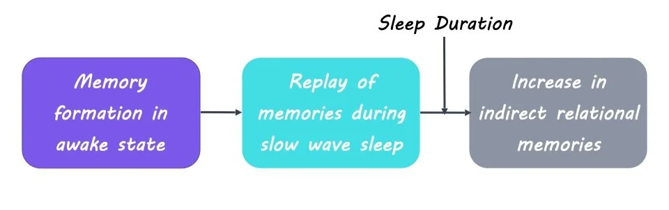
To investigate the exact mechanism by which sleep can improve relational memory, the authors looked specifically at neuron spiking events during slow oscillations that were considered replay events. These replay events are related to the reactivation and consolidation of a memory. The authors found that the number of replay events was correlated with the strengthening of neural connections, suggesting that the replay of memory traces during sleep underlies the formation of not only direct but also indirect relational memories.

What's the impact?

The present study highlights the importance of sleep in our ability to form connections between indirectly learned but overlapping memories. It also demonstrates how building models of the brain that simulate different states can allow for controlled testing of specific hypotheses about human cognition.

Access the original scientific publication here.

Two Distinct Functions of Noradrenaline

 Post by Shireen Parimoo

The takeaway

The locus coeruleus (LC) is a small brainstem structure that releases noradrenaline, which has both specific and widespread effects on cortical regions. Distinct populations of neurons in LC have distinct roles during the expression of learned behaviors, including coding for unexpected and expected rewards and punishment.

What's the science?

The locus coeruleus (LC) is a small nucleus in the brainstem that produces noradrenaline in the brain. Noradrenaline has a variety of effects on cognition, including regulating arousal, sleep, attention, and decision-making processes. LC function is difficult to study in humans because of its small size. It was previously thought to be a homogenous structure. However, new animal studies indicate that different sub-populations of LC neurons may project to different cortical regions and therefore play a role in distinct cognitive processes. This week in Nature, Breton-Provencher and colleagues used optogenetics and in vivo imaging techniques to investigate the functional organization of LC neurons.

How did they do it?

Activity from LC neurons was recorded while mice performed an auditory go/no-go task, in which they were trained to press a lever in response to “go” tones and withhold a lever press in response to “no-go” tones. The tones varied in volume, which allowed the authors to determine whether stimulus intensity influenced LC activity. Mice were rewarded on correct hit trials (i.e., when the mice pressed the lever in response to the go tone) and punished with an air puff on false alarm trials (i.e., when the mice pressed the lever in response to the no-go tone). No reinforcement was provided on missed trials (did not press the lever on go trials), but an unexpected reward was randomly delivered on a few correct rejection trials (did not press the lever on no-go trials). In addition to the hit and false alarm rates, the authors also estimated the probability of pressing the lever on a given trial.

They examined the impact of optogenetic inhibition on LC activity and behavior, both on the current trial and the subsequent trial. The authors optogenetically inhibited activity in LC neurons during tone onset, lever press, and reinforcement. Additionally, they identified populations of LC neurons that projected to the motor cortex and the dorsomedial prefrontal cortex (DMPFC) with retrograde tracing. To examine the impact of LC input to cortical regions on behavior, they inhibited LC axons specifically in motor cortex and DMPFC using optogenetics.

What did they find?

High-intensity tones improved performance on both go and no-go trials. That is, mice were more likely to press the lever when the go tone intensity was high (i.e., louder) and conversely, less likely to press the lever when the no-go tone intensity was high. In the absence of optogenetic inhibition, punishment increased the probability of lever pressing for go tones on the subsequent trial, but this effect was eliminated with LC inhibition. Expected rewards led to an increase in hit rates whereas unexpected rewards increased false alarms. However, LC inhibition during expected rewards had no impact on behavior, but it reduced the false alarm rate following unexpected reward delivery, demonstrating that the LC differentially codes for surprising compared to expected positive reinforcement.

Different subpopulations of LC neurons were active during false alarms compared to correct hits, and spike rates of LC neurons increased (1) right before mice pressed the lever, and (2) after reinforcement delivery. The increase in spiking activity prior to lever pressing was positively related to go tone intensity and did not differ between hits and false alarms. Lever pressing was associated with greater input from LC to the motor cortex. On the other hand, punishment, but not reward, led to greater activity in the LC that was distributed homogenously in both the motor cortex and DMPFC. Spiking activity also increased after the delivery of unexpected rewards, regardless of tone intensity. Photoinhibition of LC axons in the motor cortex impaired behavior in all trials, whereas inhibiting the DMPFC had no impact on behavior. Together, these results show that distinct LC neurons are activated at different time points, with differential roles in motor responding for task execution and processing reinforcement to improve performance accuracy.

What's the impact?

This study demonstrates that different sub-populations of the locus coeruleus are involved in the preparation of motor responses and in learning related to the delivery of rewards and punishment. These findings are crucial for improving our understanding of the functional organization of the locus coeruleus and its role in various cognitive processes.

Individual Differences and Similarities in How Pain is Represented in the Brain

Post by Lincoln Tracy

The takeaway

Brain regions receiving direct spinothalamic input display more consistent pain representations between different individuals. These regions have the potential to be used as targets for personalized clinical interventions.

What's the science?

Pain is a complex and multidimensional experience. Certain brain regions have consistently been reported to play a role in pain processing, while other regions have been less consistently linked. Researchers suspect pain arises from activity in a variety of brain pathways, which may differ from person to person. However, it is currently unclear as to which brain regions have more consistent versus variable representations of pain across individuals. This week in Nature Neuroscience, Kohoutová and colleagues used a personalized brain mapping approach to identify brain regions with high or low interindividual variability in their relationship to pain.

How did they do it?

The authors obtained existing functional magnetic resonance imaging data from 404 individuals who had previously participated in one of 13 experimental pain studies across two independent laboratories. They then used these data to undertake personalized brain mapping of pain through predictive modeling. From these models, they sought to identify which brain regions were important for pain prediction. They also sought to explore how much variability each of the identified brain regions (either individually or as a collective) displayed in terms of pain prediction between individual participants. Finally, the authors validated their findings by replicating the analyses in a novel dataset of 124 individuals.

What did they find?

The authors identified 21 pain-predictive brain regions, including the anterior midcingulate cortex, the dorsolateral prefrontal cortex, and the cerebellum. When they compared the individual variability of these important regions, certain regions (e.g., the ventromedial prefrontal cortex) displayed high levels of variability between individuals, while other regions (e.g., the posterior midcingulate cortex) displayed lower levels of variance. This indicates substantial variability in the role of these regions in pain between individuals. Similar results were observed when considering the brain regions as a collective (i.e., multivariable analysis) rather than individually. The ventrolateral prefrontal cortex, the vermis, and the ventromedial prefrontal cortex showed the highest individual variability. In contrast, the posterior midcingulate cortex, the supplementary motor area, and the sensorimotor cortex were the most stable regions among individuals. Importantly, these findings were replicated when the analyses were repeated in a novel and independent dataset. Taken together, these findings indicate that the relationship between brain regions and pain perception at the individual level is more complicated than how they are usually summarized at the group level.  

What's the impact?

This study found that various brain regions display differing levels of variability in pain representation between individuals. These findings enhance our understanding of how individuals process pain differently. Knowing which brain regions contribute to pain processing at the individual level could assist treatment decision-making processes for people with chronic pain.

Access the original scientific publication here.