What is Neurofeedback and How is it Used?

Post by Ewina Pun

Sensory feedback and neuroplasticity

Have you ever tried to walk in a straight line with your eyes closed, or eat a meal in complete darkness? Movement control becomes significantly harder when sensory feedback is limited. When we’re learning, we rely heavily on various forms of feedback, such as tactile, visual, and audio feedback. For some individuals, sensory feedback is lost due to injuries and neural deficits. Fortunately, our brain has the ability to quickly adapt to new circumstances - also known as neuroplasticity.

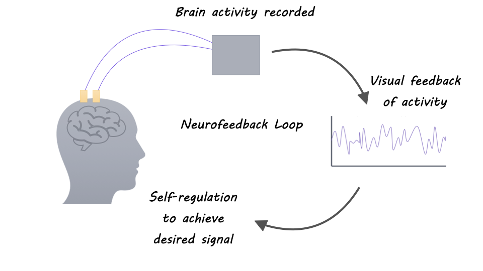
Some researchers study neuroplasticity with neurofeedback: a form of biofeedback that provides a representation of the recorded neural activity as a visual, auditory, or other signal back to the individual in real-time to facilitate self-regulation. In other words, one can modulate their brain activity by integrating information about one’s own brain activity to elicit a different behavior or pathology. Neuroplasticity not only enables cognitive and perceptual learning but also forms the basis of clinical neurorehabilitation. And researchers have begun to investigate the use of neurofeedback for treatments of brain and behavioral disorders.

Neurofeedback to study brain networks

Research suggests that there is a specific network of brain regions involved in self-regulation. Changes in brain activity during neurofeedback can be seen via changes in electroencephalography (EEG) amplitudes and blood oxygen level-dependent signals in functional magnetic resonance imaging (fMRI) in brain areas such as the anterior insular cortex, anterior cingulate cortex, dorsal lateral prefrontal cortex, inferior parietal lobule, basal ganglia, and thalamus. This indicates that neurofeedback involves networks involved in reward processing, cognitive control, and learning and memory.

Neurofeedback for clinical application

Neurofeedback has been extensively studied to treat attention deficit hyperactivity disorder (ADHD). For instance, neurofeedback therapy can provide information about the patient’s brain state to allow the patient to consciously match to and maintain the desired brain state through reinforcement. EEG-based Neurofeedback helps reduce the elevated low-frequency (theta/delta) synchronization observed in children with ADHD and improved ADHD symptoms.

Studies have also found several benefits of neurofeedback for stroke recovery. One study showed that severely impaired, chronic stroke patients learned to upregulate ipsilesional sensorimotor rhythm (SMR) through controlling a brain-computer-interface (BCI) with a hand orthosis, and the neurofeedback training also improved their upper limb functions. In addition, virtual reality in combination with EEG-BCI can offer a more immersive representation of neurofeedback for stroke rehabilitation and increase the perceived embodiment (sense of control as their own). However, more robust evidence is needed to support the efficacy of neurofeedback therapies for ADHD and stroke rehabilitation.

Neurofeedback for BCI motor control

The use of neurofeedback is not exclusive to self-regulate one’s neural state. It is also widely used in BCI for closed-loop motor control. In motor BCI, individuals can directly manipulate an external device with their neural signals being translated into action commands. For example, after training, a BCI maps neural activity patterns to control commands in real-time and provides visual feedback of the current position of the external device being controlled. Such real-time feedback allows users to reevaluate, refine and correct their control. With trial-and-error, people with paralysis can decide which set of mental motor imagery (e.g., imagining controlling a joystick or a computer mouse) was most effective for control. With minimal practice using an intracortical BCI, people with paralysis were able to coordinate movements of a seven degrees-of-freedom robotic arm, control a computer cursor, or functionally stimulate muscles for movement restoration.

Bidirectional closed-loop neurofeedback

In addition to providing just visual feedback, researchers try to directly restore tactile and cutaneous sensations in arm and hand function, which is important during grasping or manipulation of objects. A bidirectional BCI for motor control refers to a system that (1) decodes neural signals in the motor cortex into commands to control a device and (2) provides somatosensory feedback by delivering electric stimulation patterns to the primary somatosensory cortex (S1) or the spinal cord. Microstimulation of the cortical surface of M1 and S1 using high-density electrocorticography (ECoG) has provided tactile sensations such as “buzzing”, “tingling”, “brushing”, “light tapping,” or a “feeling of movement” to participants with paralysis. An adaptive deep brain stimulation framework targeted at the thalamus is capable of concurrent biomimicry stimulation and sensing for better closed-loop therapies for psychiatric disorders, epilepsy, or chronic pain. More research is needed to quantify perceptual qualities and improve naturalistic sensations to provide functional benefits for BCI control.

What’s next?

Neurofeedback is a novel and valuable way to study brain function and neuroplasticity. Further, neurofeedback has exciting potential as a therapeutic tool. Although researchers have begun to understand some of the mechanisms underlying neurofeedback, future research will likely further clarify the psychological and neural underpinnings of self-regulation, which will help to design more-effective neurofeedback technologies for treating a variety of diseases and conditions.

References +

Sitaram et al. Closed-loop brain training: the science of neurofeedback. Nature. (2016)

Saha et al. Progress in Brain Computer Interface: Challenges and Opportunities. Front. Syst. Neurosci. (2021)

Ramos-Murguialday, et al. Brain–machine interface in chronic stroke rehabilitation: a controlled study. Ann. Neurol. (2013).

Zotev et al. Self-regulation of human brain activity using simultaneous real-time fMRI and EEG neurofeedback. Neuroimage (2014).

Collinger et al. High-performance neuroprosthetic control by an individual with tetraplegia Lancet (2013).

Hochberg et al. Neuronal ensemble control of prosthetic devices by a human with tetraplegia. Nature (2006).

Hughes et al. Bidirectional brain-computer interfaces. Handbook of Clinical Neurology (2020).

Ansó et al, Concurrent stimulation and sensing in bi-directional brain interfaces: a multi-site translational experience. Journal of Neural Engineering (2022).

Predicting and Tracking Hallucinations

Post by Leanna Kalinowski

The takeaway

Hallucinations, a common symptom in disorders like schizophrenia, have traditionally been difficult to study given that they cannot be directly observed. Scientists have successfully applied a computational framework to screen for and track hallucinations.

What's the science?

Hallucinations, which are perceptions that occur in the absence of a stimulus, are a hallmark sign of several psychosis-spectrum disorders, such as schizophrenia. Traditionally, hallucinations have been difficult to study given that scientists cannot directly observe them. However, the rise of the field of computational psychiatry field now allows scientists to use mathematical frameworks to better understand the neurological underpinnings of psychosis-spectrum disorders.

One such framework, predictive processing theory, shows promise as a tool for better understanding hallucinations. In this framework, “perception” is described as the process of determining the cause of one’s sensations by considering (1) one’s internal expectations of their surroundings based on prior knowledge (called “priors”) and (2) the available sensory evidence that is weighted by the participant’s certainty in the source of the information. Evidence suggests that hallucinations arise when the priors are over-weighted compared to incoming sensory evidence, but this exact relationship is unclear. This week in Biological Psychiatry, Kafadar, Fisher, and colleagues used mathematical modeling to determine the relationship between these over-weighted priors and susceptibility to hallucinations.

How did they do it?

First, 458 participants were screened for the presence of auditory hallucinations and separated into two groups: hallucinators and non-hallucinators. Then, they completed the Auditory Conditioned Hallucinations task, where participants are first trained to associate a visual pattern with an auditory tone. Once the association is learned, the researchers then recorded the conditioned hallucination rate, which is the proportion of times that the participants reported hearing the tone when the visual pattern was displayed, without the tone. Finally, a subset of the hallucinators group was invited back to the lab 6-12 months later to determine whether performance on this task is related to changes in symptom severity.

What did they find?

The researchers found that conditioned hallucination rates were a predictor of the frequency of self-reported hallucinations. These rates were sensitive to hallucination state and the over-weighting of priors compared to incoming sensory evidence. They also found that conditioned hallucination rates and prior weighting are higher in the hallucinator group. Changes in these rates were further associated with changes in the frequency of reported hallucinations at the follow-up test, suggesting that this approach may inform future clinical screening tools.

What's the impact?

Taken together, these results indicate that conditioned hallucination rates and over-weighting of priors can be used as markers of hallucination status. This can be useful when tracking the development, trajectory, and treatment response of psychosis-spectrum disorders.

How Gut Microbiota Affect Brain Health

Post by Elisa Guma

What is your gut microbiome?

Living inside (and on the skin) of every person are trillions of bacteria, viruses, fungi, and other organisms that collectively make up our microbiome. These microorganisms can coexist with their human hosts, causing no harm, they can have mutualistic relationships with their hosts, providing them benefits, or they can be harmful, producing unwanted metabolites. Many of these metabolites can influence brain structure and function. Although many organs have their own distinct microbial colonies, the gut microbiome has attracted a great deal of attention, particularly because there is bidirectional communication between the gut, its microbiome, and the brain. Our first big dose of microbiota comes from birth (via the vaginal canal and breastfeeding), but its composition continues to evolve throughout the lifespan, influenced by our environment and diet.

The gut-brain axis

Your gut and your brain are in an ongoing dance of bidirectional communication, forming a circuit often referred to as the gut-brain axis. Our central nervous system (i.e., our brain and spinal cord) can influence the composition and function of the gut microbiota via the autonomic nervous system, regulating gastrointestinal motility, mucus secretion and permeability, and luminal release of neurotransmitters. In turn, microbiota in the gut can affect the permeability of the gut and blood-brain-barrier, as well as shape brain development, behaviour, and mood. This bidirectional communication can be neural, i.e. through the vagus nerve. It can also be neuroendocrine or immune, via metabolites and neurotransmitters produced in the gut. Microbiota can produce these signaling molecules from the food we ingest (carbohydrates, amino acids), from our bodily secretions (estrogens), or from chemical substances to which we are exposed (pesticides or medications). Some of these metabolites include (or are precursors for the production of) short-chain fatty acids (for example, butyrate), neurotransmitters (serotonin or γ-aminobutyric acid (GABA)), hormones (for example, cortisol), and immune system modulators (for example, quinolinic acid).

How can diet impact the microbiome?

Diet plays a critical role in shaping the diversity and proportions of microorganisms in our gut. This in turn can modulate brain structure and function through the communication channels discussed above: neuroendocrine, neural, and immune. Importantly, diet intervention has been found to alter both microbiome diversity and inflammatory markers in humans. A recent randomized controlled study found that individuals who ingested diets rich in fermented foods, compared to those who ingested diets rich in fiber, had increased microbiota diversity and decreased inflammation. Although those ingesting high-fiber diets experienced positive effects from their microbiota as well, increased diversity and decreased inflammation were not observed. Thus, fermented foods may be valuable dietary additions, particularly for those dealing with increased inflammation, or decreased microbial diversity (for example, if following a course of antibiotic treatment). In contrast, rodent and human studies have shown that diets rich in high-sugar and high-fat foods can change the bacterial content of the gut rapidly, decreasing diversity, and increasing inflammatory markers.

The gut microbiome and mental health

From anecdotal observations in patients, the association between altered gut-to-brain signaling and anxiety, depression, and autism spectrum disorder (ASD) was first established. Often, these psychiatric conditions were comorbid with another diagnosis of a digestive problem, such as irritable bowel syndrome. Post-mortem studies have also identified increased intestinal permeability and heightened inflammation in individuals with ASD, suggesting a potential link between gut health and inflammation. 

Studies in animal models have shown that the composition of the gut microbiome can modulate the central nervous system and central nervous system-driven behaviours. Initial studies comparing mice with and without microbes in their gut found that the former displayed increased motor activity, decreased anxiety, and altered genes associated with synaptic function in the brain. 

Dysbiosis of the gut has also been identified in patients with major depressive disorder (compared to healthy controls). Microbiome transfer from depressed human individuals into healthy rodents has been found to induce depressive-like behaviours in those mice, which suggests a potential causal role between the microbiota and the depressive symptoms. It is unclear whether these are indirectly mediated through other factors like increased inflammation, which has also been associated with the pathology of numerous psychiatric conditions.

Important links with neurodegenerative diseases have also been made. A strain of Escherichia coli in the gut has been shown to make a protein that is similar to the misfolded alpha-synuclein protein associated with disease progression in Parkinson’s disease. Some researchers hypothesize that these misfolded proteins may travel up the vagus nerve to the brain, providing a “template” for misfolding to the alpha-synuclein protein.

Therapeutic microbes to tackle disease

Given the intriguing interactions between gut microbiota and psychiatric symptoms, many groups are investigating putative therapies aimed at altering the composition of these microbes. Microbial transfer therapy (or fecal transplants) has been successfully used to recolonize the gut of individuals suffering from severe gastrointestinal distress or following complications from antibiotic therapy. Some are starting to investigate its utility in the treatment of autism spectrum disorder. In a recent study, children with autism spectrum disorder who received a microbial transfer from the gut of healthy individuals showed a decreased severity of autistic and gastrointestinal symptoms. In contrast, probiotic treatment of individuals with major depressive disorder or schizophrenia has shown mixed findings, with some individuals showing improvements and others no changes. While targeting gut microbiota in the treatment of mental illness shows great promise, there is much more research to be done to understand the gut-brain axis, and how best to develop therapies to effectively modify the gut microbiome.

References +

Horn et al. Role of diet and its effects on the gut microbiome in the pathophysiology of mental disorders. Translational psychiatry (2022).

Neufeld et al. Effects of intestinal microbiota on anxiety-like behaviour. Communicative & Integrative Biology (2011).

Parker et al. Gut microbes and metabolites as modulators of blood-brain-barrier integrity and brain health. Gut Microbes (2020).

Sgritta et al. Mechanisms underlying microbial-mediated changes in social behaviour in mouse models of autism spectrum disorder. Neuron (2019).

Shoubridge et al. The gut microbiome and mental health: advances in research and emerging priorities. Molecular Psychiatry (2022).

Wastyk et al. Gut-microbiota-targeted diets modulate human immune status. Cell (2021).

Willyard. How gut bacteria alter the brain. Nature (2021). Zhu et al. The progress of gut microbiome research related to brain disorders. Journal of Neuroinflammation (2020),