Predicting and Tracking Hallucinations

Post by Leanna Kalinowski

The takeaway

Hallucinations, a common symptom in disorders like schizophrenia, have traditionally been difficult to study given that they cannot be directly observed. Scientists have successfully applied a computational framework to screen for and track hallucinations.

What's the science?

Hallucinations, which are perceptions that occur in the absence of a stimulus, are a hallmark sign of several psychosis-spectrum disorders, such as schizophrenia. Traditionally, hallucinations have been difficult to study given that scientists cannot directly observe them. However, the rise of the field of computational psychiatry field now allows scientists to use mathematical frameworks to better understand the neurological underpinnings of psychosis-spectrum disorders.

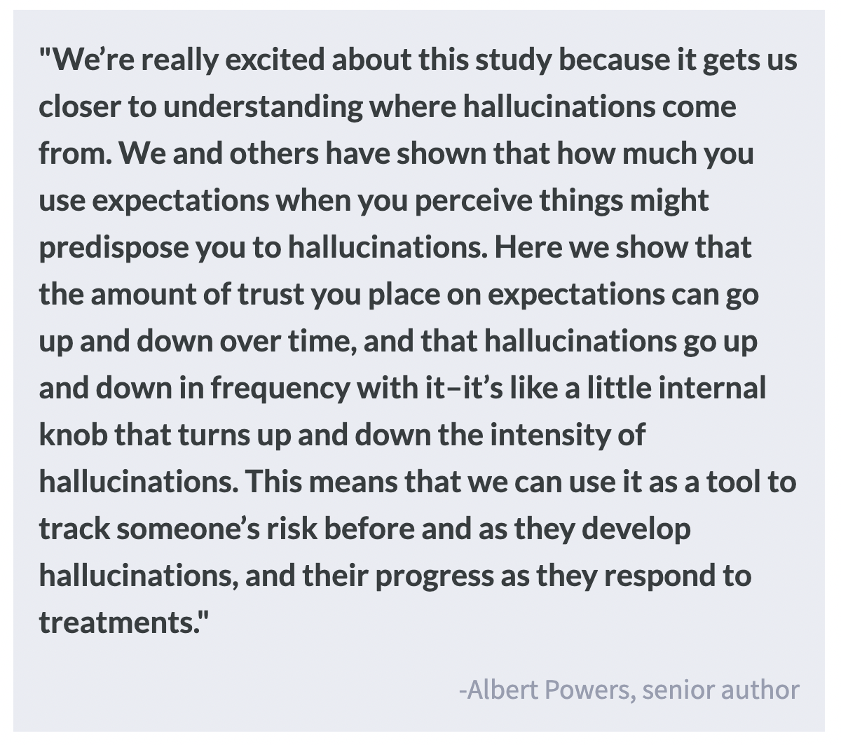
One such framework, predictive processing theory, shows promise as a tool for better understanding hallucinations. In this framework, “perception” is described as the process of determining the cause of one’s sensations by considering (1) one’s internal expectations of their surroundings based on prior knowledge (called “priors”) and (2) the available sensory evidence that is weighted by the participant’s certainty in the source of the information. Evidence suggests that hallucinations arise when the priors are over-weighted compared to incoming sensory evidence, but this exact relationship is unclear. This week in Biological Psychiatry, Kafadar, Fisher, and colleagues used mathematical modeling to determine the relationship between these over-weighted priors and susceptibility to hallucinations.

How did they do it?

First, 458 participants were screened for the presence of auditory hallucinations and separated into two groups: hallucinators and non-hallucinators. Then, they completed the Auditory Conditioned Hallucinations task, where participants are first trained to associate a visual pattern with an auditory tone. Once the association is learned, the researchers then recorded the conditioned hallucination rate, which is the proportion of times that the participants reported hearing the tone when the visual pattern was displayed, without the tone. Finally, a subset of the hallucinators group was invited back to the lab 6-12 months later to determine whether performance on this task is related to changes in symptom severity.

What did they find?

The researchers found that conditioned hallucination rates were a predictor of the frequency of self-reported hallucinations. These rates were sensitive to hallucination state and the over-weighting of priors compared to incoming sensory evidence. They also found that conditioned hallucination rates and prior weighting are higher in the hallucinator group. Changes in these rates were further associated with changes in the frequency of reported hallucinations at the follow-up test, suggesting that this approach may inform future clinical screening tools.

What's the impact?

Taken together, these results indicate that conditioned hallucination rates and over-weighting of priors can be used as markers of hallucination status. This can be useful when tracking the development, trajectory, and treatment response of psychosis-spectrum disorders.

How Gut Microbiota Affect Brain Health

Post by Elisa Guma

What is your gut microbiome?

Living inside (and on the skin) of every person are trillions of bacteria, viruses, fungi, and other organisms that collectively make up our microbiome. These microorganisms can coexist with their human hosts, causing no harm, they can have mutualistic relationships with their hosts, providing them benefits, or they can be harmful, producing unwanted metabolites. Many of these metabolites can influence brain structure and function. Although many organs have their own distinct microbial colonies, the gut microbiome has attracted a great deal of attention, particularly because there is bidirectional communication between the gut, its microbiome, and the brain. Our first big dose of microbiota comes from birth (via the vaginal canal and breastfeeding), but its composition continues to evolve throughout the lifespan, influenced by our environment and diet.

The gut-brain axis

Your gut and your brain are in an ongoing dance of bidirectional communication, forming a circuit often referred to as the gut-brain axis. Our central nervous system (i.e., our brain and spinal cord) can influence the composition and function of the gut microbiota via the autonomic nervous system, regulating gastrointestinal motility, mucus secretion and permeability, and luminal release of neurotransmitters. In turn, microbiota in the gut can affect the permeability of the gut and blood-brain-barrier, as well as shape brain development, behaviour, and mood. This bidirectional communication can be neural, i.e. through the vagus nerve. It can also be neuroendocrine or immune, via metabolites and neurotransmitters produced in the gut. Microbiota can produce these signaling molecules from the food we ingest (carbohydrates, amino acids), from our bodily secretions (estrogens), or from chemical substances to which we are exposed (pesticides or medications). Some of these metabolites include (or are precursors for the production of) short-chain fatty acids (for example, butyrate), neurotransmitters (serotonin or γ-aminobutyric acid (GABA)), hormones (for example, cortisol), and immune system modulators (for example, quinolinic acid).

How can diet impact the microbiome?

Diet plays a critical role in shaping the diversity and proportions of microorganisms in our gut. This in turn can modulate brain structure and function through the communication channels discussed above: neuroendocrine, neural, and immune. Importantly, diet intervention has been found to alter both microbiome diversity and inflammatory markers in humans. A recent randomized controlled study found that individuals who ingested diets rich in fermented foods, compared to those who ingested diets rich in fiber, had increased microbiota diversity and decreased inflammation. Although those ingesting high-fiber diets experienced positive effects from their microbiota as well, increased diversity and decreased inflammation were not observed. Thus, fermented foods may be valuable dietary additions, particularly for those dealing with increased inflammation, or decreased microbial diversity (for example, if following a course of antibiotic treatment). In contrast, rodent and human studies have shown that diets rich in high-sugar and high-fat foods can change the bacterial content of the gut rapidly, decreasing diversity, and increasing inflammatory markers.

The gut microbiome and mental health

From anecdotal observations in patients, the association between altered gut-to-brain signaling and anxiety, depression, and autism spectrum disorder (ASD) was first established. Often, these psychiatric conditions were comorbid with another diagnosis of a digestive problem, such as irritable bowel syndrome. Post-mortem studies have also identified increased intestinal permeability and heightened inflammation in individuals with ASD, suggesting a potential link between gut health and inflammation. 

Studies in animal models have shown that the composition of the gut microbiome can modulate the central nervous system and central nervous system-driven behaviours. Initial studies comparing mice with and without microbes in their gut found that the former displayed increased motor activity, decreased anxiety, and altered genes associated with synaptic function in the brain. 

Dysbiosis of the gut has also been identified in patients with major depressive disorder (compared to healthy controls). Microbiome transfer from depressed human individuals into healthy rodents has been found to induce depressive-like behaviours in those mice, which suggests a potential causal role between the microbiota and the depressive symptoms. It is unclear whether these are indirectly mediated through other factors like increased inflammation, which has also been associated with the pathology of numerous psychiatric conditions.

Important links with neurodegenerative diseases have also been made. A strain of Escherichia coli in the gut has been shown to make a protein that is similar to the misfolded alpha-synuclein protein associated with disease progression in Parkinson’s disease. Some researchers hypothesize that these misfolded proteins may travel up the vagus nerve to the brain, providing a “template” for misfolding to the alpha-synuclein protein.

Therapeutic microbes to tackle disease

Given the intriguing interactions between gut microbiota and psychiatric symptoms, many groups are investigating putative therapies aimed at altering the composition of these microbes. Microbial transfer therapy (or fecal transplants) has been successfully used to recolonize the gut of individuals suffering from severe gastrointestinal distress or following complications from antibiotic therapy. Some are starting to investigate its utility in the treatment of autism spectrum disorder. In a recent study, children with autism spectrum disorder who received a microbial transfer from the gut of healthy individuals showed a decreased severity of autistic and gastrointestinal symptoms. In contrast, probiotic treatment of individuals with major depressive disorder or schizophrenia has shown mixed findings, with some individuals showing improvements and others no changes. While targeting gut microbiota in the treatment of mental illness shows great promise, there is much more research to be done to understand the gut-brain axis, and how best to develop therapies to effectively modify the gut microbiome.

References +

Horn et al. Role of diet and its effects on the gut microbiome in the pathophysiology of mental disorders. Translational psychiatry (2022).

Neufeld et al. Effects of intestinal microbiota on anxiety-like behaviour. Communicative & Integrative Biology (2011).

Parker et al. Gut microbes and metabolites as modulators of blood-brain-barrier integrity and brain health. Gut Microbes (2020).

Sgritta et al. Mechanisms underlying microbial-mediated changes in social behaviour in mouse models of autism spectrum disorder. Neuron (2019).

Shoubridge et al. The gut microbiome and mental health: advances in research and emerging priorities. Molecular Psychiatry (2022).

Wastyk et al. Gut-microbiota-targeted diets modulate human immune status. Cell (2021).

Willyard. How gut bacteria alter the brain. Nature (2021). Zhu et al. The progress of gut microbiome research related to brain disorders. Journal of Neuroinflammation (2020),

A Subtype of Dopamine Neurons is Vulnerable to Neurodegeneration in Parkinson’s Disease

Post by Shireen Parimoo

The takeaway

Parkinson’s disease is characterized by the loss of dopamine (DA) neurons, with some DA neurons more vulnerable than others. One subtype of DA neurons that strongly express genes that have previously been linked to Parkinson’s disease is most susceptible to degeneration.

What's the science?

Parkinson’s disease (PD) is characterized by the loss of dopamine (DA) neurons in the substantia nigra pars compacta (SNpc). However, neuronal degeneration is not uniform across all subtypes of DA neurons, as some survive late into disease progression while others die early on. The genetic and molecular characteristics of the different DA neuron subtypes that make them selectively vulnerable to PD-related degeneration are not currently known. This week in Nature Neuroscience, Kamath and colleagues performed molecular profiling of DA neurons in the SNpc to identify the subtypes most at risk for degeneration in PD.

How did they do it?

The authors first identified DA and non-DA cells in post-mortem human midbrain slices, as well as the rodent and macaque midbrain, using single-nucleus RNA sequencing. They performed a clustering analysis and compared the presence of certain genes against an existing database to identify different cell types within the midbrain tissue. Within the DA neurons, they quantified the presence of certain molecular biomarkers, such as transcription factors and regulons (set of genes whose expression is controlled by a common regulatory region), to further distinguish between the different DA neuron subtypes. Next, they spatially localized the DA neuron subtypes within the macaque SNpc using Slide-seq (a spatial phenotyping technique) and in the human SNpc using fluorescence imaging. To determine which DA neuron subtypes were more vulnerable to degeneration, the authors compared post-mortem tissue of PD patients and controls. Lastly, they examined the presence of genetic and molecular markers associated with familial and sporadic PD in the different DA neuron subtypes.

What did they find?

Midbrain cells were grouped into seven categories (e.g., astrocytes, DA neurons, microglia, etc.), within which DA neurons were differentiated from non-DA neurons based on the presence of the Nr4a2 gene. There were two broad classes of DA neurons in the human and macaque midbrains: those that strongly expressed the SOX6 genes and those that strongly expressed the CALB1 genes. The SOX6-expressing neurons – particularly the subtype containing the AGTR1 gene – were primarily localized to the ventral SNpc while the CALB1-expressing neurons were predominantly in the dorsal SNpc. Within the DA neuron subtypes, there was considerable heterogeneity in the expression of transcription factors and regulon activity. This means that different subtypes of DA neurons in both the human and macaque midbrain can be distinguished from one another based on the expression of specific transcription factors.

In PD, SOX6-expressing DA neurons in the ventral SNpc were more vulnerable to neurodegeneration, especially the AGTR1 subtype, compared to CALB1-expressing neurons in the dorsal SNpc. Moreover, the AGTR1 subtype was strongly enriched for genes previously linked to PD by genome-wide association studies. Additionally, this subtype was found to have upregulated genes linked to neurodegeneration in mouse models of PD, which explains their selective vulnerability to PD-related degeneration. Together, these results implicate the SOX-AGTR1 subtype of DA neurons in PD due to the marked presence of genetic markers associated with neurodegeneration.

What's the impact?

The authors profiled DA neurons in the midbrain using single-nucleus RNA-sequencing, which allowed the authors to identify and spatially localize the DA neurons within the substantia nigra pars compacta that are selectively vulnerable to PD-related degeneration. These findings not only strengthen our understanding of the biomarkers associated with PD, but the identification of transcription factors that drive neurodegeneration in PD can additionally inform efforts to develop targeted treatments and therapies.

Access the original scientific publication here.