Psilocybin Reduces Depressive Symptoms by Reorganizing Functional Brain Networks

Post by Shireen Parimoo

The takeaway

Psilocybin, a psychedelic drug commonly known as magic mushrooms, is increasingly viewed as a viable treatment option for psychiatric conditions like major depression. It reduces the severity of depressive symptoms by reorganizing the functional connectivity of brain networks.

What's the science?

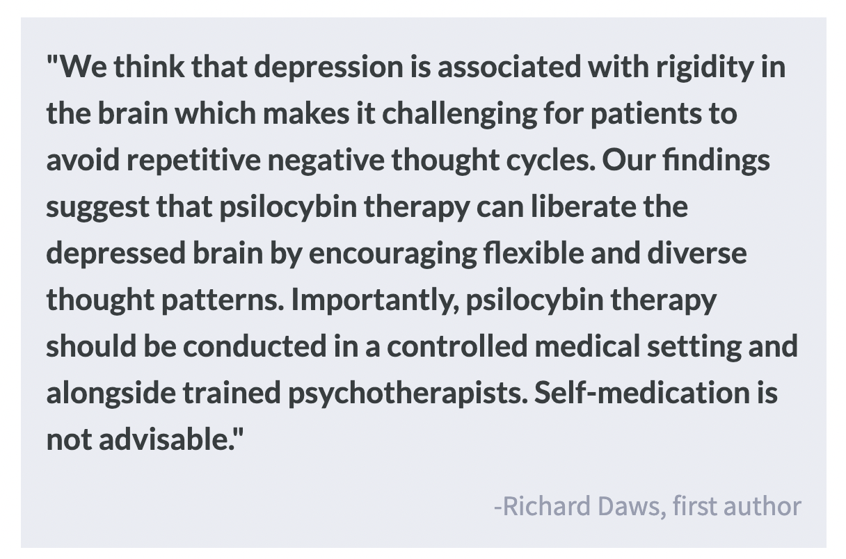
Major depressive disorder is commonly treated using serotonin-selective reuptake inhibitors (SSRIs) that increase serotonin concentrations in the brain. Psychedelic drugs like psilocybin are now being considered viable options for treating major depression as they also increase serotonin levels. In the brain, serotonin receptors are present in many brain regions, including brain regions that comprise the default mode (DMN) and executive control/salience networks (E/SN). The DMN is generally active during self-referential thinking and is over-activated in individuals with depression, while the E/SN are associated with attentional processes that are impaired in depression. One possibility is that psilocybin alters the functional properties of these networks by acting on serotonin receptors in the brain. This week in Nature Medicine, Daws and colleagues used clinical and resting-state fMRI (rs-fMRI) data from two clinical trials to investigate the effects of psilocybin treatment on brain network organization and depressive symptoms.

How did they do it?

The authors used data from two clinical trials on psilocybin treatment for major depressive disorder:  an open-label trial with 16 treatment-resistant patients who knew they would receive psilocybin treatment, and a double-blind randomized-controlled trial (DB-RCT) with patients who either received psilocybin or an SSRI. Although the patients in the DB-RCT did not know which treatment they received, they were informed that they would either receive a high dose or a negligible dose of psilocybin. In both trials, participants underwent rs-fMRI scanning and completed the Beck Depression Inventory I for an assessment of their depressive symptoms. The authors used functional connectivity analyses to identify the extent to which brain networks were integrated or segregated from each other. Lastly, they estimated the dynamic flexibility of the networks (i.e., how often different brain regions switched network membership over time) in the DB-RCT.

In the open-label trial, participants were given one low dose of psilocybin and a higher dose one week later. A clinical assessment and rs-fMRI scan were performed at baseline (prior to the first dose) and one day after the second dose of psilocybin. Clinical follow ups were also conducted three and six months after the second dose to examine the long-term effect of psilocybin treatment on depression. In the DB-RCT, participants underwent clinical assessments and scanning followed by psilocybin or SSRI treatment at baseline and three weeks later. They also took a daily capsule with psilocybin, placebo, or the SSRI for six weeks following the first dose. Finally, clinical follow-ups were conducted two weeks, four weeks, and six weeks after the first dose.

What did they find?

Psilocybin treatment reduced depressive symptom severity in both the short-term (1-6 weeks) and in the long-term (3-6 months) across both clinical trials. Moreover, in the DB-RCT, psilocybin was found to be more effective than the SSRI treatment. In both trials, there was an increase in the global integration of the brain's functional networks that followed psilocybin therapy. The increase in connectivity between the brain's networks correlated with improvements in the long-term well-being of patients. There were no observed changes in the global integration of the brain networks in patients who received the SSRI treatment. 

Digging deeper, the brain's higher-order networks that are associated with cognition and the flexible adaptation of behaviour showed significant changes after psilocybin. This was expected because these networks house the highest densities of serotonin receptors and function abnormally in depression. In the open-label trial, the DMN was less segregated and more integrated with the E/SN one day after treatment. This means that psilocybin reduced the extent to which DMN regions were connected to each other and increased their connectivity with regions of the E/SN. In the DB-RCT, greater dynamic flexibility of the E/SN was associated with greater symptom improvements after receiving psilocybin. Again, these changes were not observed in patients who received the SSRI treatment.

What's the impact?

This study shows that the rapid and sustained antidepressant effect of psilocybin works differently from a conventional SSRI. The brains of patients with depression that received psilocybin became more integrated and flexible, in a manner that is conducive to better mental health and well-being. This work is particularly exciting because the effects of psilocybin on clinical symptoms lasted for up to 6 months in patients who were previously resistant to SSRIs, making it a viable alternative to conventional treatments.

Amyloid Versus Tau Proteins in the Path to Alzheimer’s Disease

Post by Anastasia Sares

The takeaway

The two main hallmarks of Alzheimer’s disease in the brain are plaques, made from proteins called amyloid-beta, and neurofibrillary tangles, made from proteins called tau. Brain activity and performance on memory tasks depend on the levels of both of these proteins in the brain, supporting the idea that amyloid is related to disease onset, while tau is related to disease progression.

What's the science?

In recent years, the field of Alzheimer’s research has hit a wall. Both amyloid and tau proteins are correlated with having the disease, but many researchers had guessed that amyloid proteins were the key, and that tau tangles were just a downstream effect. Some animal models of Alzheimer’s were created so that they naturally over-produced amyloid, which seemed to recreate the disease state. From work on these animals, treatments were developed that targeted only amyloid, but when these treatments were tried on humans in clinical trials, they didn’t work as well as people hoped. Now, researchers are re-examining the role of tau proteins in Alzheimer’s. This week in Brain, Düzel and colleagues showed how amyloid and tau status can interact in humans without Alzheimer’s dementia, affecting memory and brain activity. 

How did they do it?

Participants gave samples of cerebrospinal fluid (the fluid that bathes the brain and central nervous system) so that the researchers could determine the amount of amyloid and tau proteins present in each person. Their brain activity was also recorded with MRI while they performed a memory test about recognizing familiar scenes. There were three groups of participants: people with mild cognitive impairment but without Alzheimer’s disease, people who complained of cognitive decline but nevertheless had a good memory, and healthy controls. With this sample, the researchers were able to get a variety of levels of amyloid and tau, both in individuals with cognitive decline and those without.

What did they find?

The researchers found that both memory performance and brain activity were predicted by an interaction between amyloid and tau. In “amyloid-negative” groups, tau levels were not related to memory performance, while in “amyloid-positive” groups, tau levels were related to memory performance. This result was also consistent with a paper-and-pencil test of memory recall and held true even with different statistical corrections (for example, accounting for group, age, sex, site of testing, etc.). This same interaction also showed up in brain activity during the tasks, specifically in the hippocampus and surrounding entorhinal cortex, key brain regions for forming new memories and recognizing what is familiar versus what is new. Individuals with a well-known genetic variant (APOE4) related to Alzheimer’s did not significantly differ from those without this variant.

What's the impact?

The findings of this study will help researchers in the field of Alzheimer’s to decide between competing theories of the disease, one of which is an “amyloid x tau” model. Once we have a correct understanding of the cause of Alzheimer’s in humans, we can look for treatments much more effectively.

The Brain Dynamically Changes Size Throughout Life

Post by D. Chloe Chung

The takeaway

Researchers have created a growth chart of the human brain, reflecting how the brain changes size throughout the lifespan. This chart can be used as a reference tool for the neuroimaging and clinical communities.

What's the science?

The advancement of neuroimaging techniques such as magnetic resonance imaging (MRI) has helped many clinicians, patients, and researchers over the past decades. However, unlike how we understand the change of height and weight throughout our lives, there has been no standard reference of what the brain looks like at a certain age. An inclusive map that describes developmental milestones and aging-related changes of the brain will benefit both researchers and clinicians. This week in Nature, Bethlehem and colleagues comprehensively examined how our brain dynamically changes its size throughout our lifespan by analyzing more than 100,000 MRI scans of the brain.

How did they do it?

The authors collected 123,984 MRI scans from 101,457 human participants (with or without medical conditions), aged from 16 weeks after conception to 100 years old. These brain scans were obtained from both primary studies and publicly available open databases. To create the brain size chart over the lifespan, the scans were quantified for structural changes in the brain and how fast these changes occur through aging. For this analysis, the authors adapted a modeling approach recommended by the World Health Organization that can help neutralize differences in measurement derived from diverse techniques and machines used across studies. The final brain chart was made into an interactive tool that can be used to analyze additional MRI datasets generated by tool users in the future.

What did they find?

The authors found that, up to 6 years of age, grey matter rapidly increases in its volume and thickness. The white matter volume also showed strong growth during early childhood but in a more delayed fashion than grey matter, peaking in size at around 30 years of age. The authors noted that these changes early in brain development highlight grey/white matter volume differentiation. After their respective peaks, both grey and white matter volume began to decrease over the rest of the lifespan. In contrast to these early developmental milestones, the authors found that the amount of cerebrospinal fluid in the brain ventricles that maintains its plateau throughout life starts to exponentially increase from around 60 years of age. In addition to defining developmental milestones using the brain chart, the authors demonstrated the utility of their brain chart in studying brain-related conditions. For example, the authors observed a faster decrease of the grey matter volume in Alzheimer’s disease patients, especially those who are biologically female, compared to non-patients of the same age.

What’s the impact?

This study generated a comprehensive growth chart of the human brain by examining the largest collection of MRI brain scans to date covering a 100-year age range. This brain chart will serve as a highly useful, standardized reference for neuroimaging in the future. The authors pointed out that even this brain chart is not inclusive enough as it covers mostly European and North American populations because neuroimaging tools are not as readily available to all global communities. Future studies will hopefully improve demographic and socioeconomic diversity in MRI research.