Brain Activity in the Mesolimbic Network is Related to Affective Behaviour

Post by Negar Mazloum-Farzaghi 

The takeaway 

The brain mechanisms underlying affective behaviours like smiling, laughing, or expressing discomfort are critical to everyday life. This research shows that there is a distributed network of brain regions associated with different affective behaviors, and it's possible to differentiate these behaviors from neutral ones using brain activity recordings.

What's the science? 

Affective states and our associated behaviors are an essential part of daily life. However, the underlying neurological mechanism of affective behaviours remains unclear. Previous studies have found that different affective behaviours are related to distinct patterns of spatial brain activity in the mesolimbic network, with certain brain regions playing a more critical role in some affective behaviours compared to others. Moreover, it remains unclear whether different spectral patterns (frequency bands) of activity in the mesolimbic network can distinguish one affective state from another. This week in Nature Human Behaviour, Bijanzadeh and colleagues examined the brain mechanisms underlying naturalistic affective behaviours from epilepsy patients who had intracranial EEG (iEEG) electrodes implanted in their mesolimbic network. 

How did they do it?

The authors aimed to investigate whether changes in specific frequency bands (i.e., spectral features) in specific mesolimbic network brain regions (i.e., spatial features) would create ‘spectro-spatial’ patterns across the mesolimbic network, which would ultimately allow for the distinction between naturalistic affective positive and negative behaviours. To investigate this, the authors obtained 24-h audiovisual recordings and continuous iEEG data from 11 hospitalized participants with epilepsy. They analyzed 116 hours of behavioural and neural data from these participants who had electrodes placed in at least three mesolimbic structures (insula, anterior cingulate cortex (ACC), orbitofrontal cortex (OFC), amygdala, and hippocampus). Behaviours were categorized as positive affective behaviours (smiling, laughing, positive verbalizations), negative affective behaviours (pain-discomfort, negative verbalization), and neutral behaviours (control condition; minimum 10-minute periods where the participants showed neither negative nor positive affective behaviours).

The authors aligned each participants’ neural and behavioural data and extracted the spectral power in brain frequency bands from the electrodes in their mesolimbic structures. The average power in each frequency band (spectral features) for each electrode (spatial features) was computed and the two sets of features combined were referred to as the spectro-spatial features. Next, using statistics and machine learning methods, the authors trained models (binary random forest models) on the spectro-spatial features for the behaviours for each participant to determine whether models could distinguish between affective behaviours and neutral behaviours. Then, the authors examined which mesolimbic spatial features influenced the performance of the models. 

What did they find?

The authors found that, at the individual level, models were able to successfully decode positive (up to 93% accuracy) and negative affective (up to 78% accuracy) behaviours from neutral behaviours significantly greater than chance, and the group-level analysis replicated those results. Moreover, it was found that affective behaviours were associated with changes in activity in the mesolimbic network. That is, affective behaviours were related to increased high-frequency (gamma) and decreased low frequency bands (theta, alpha, beta).

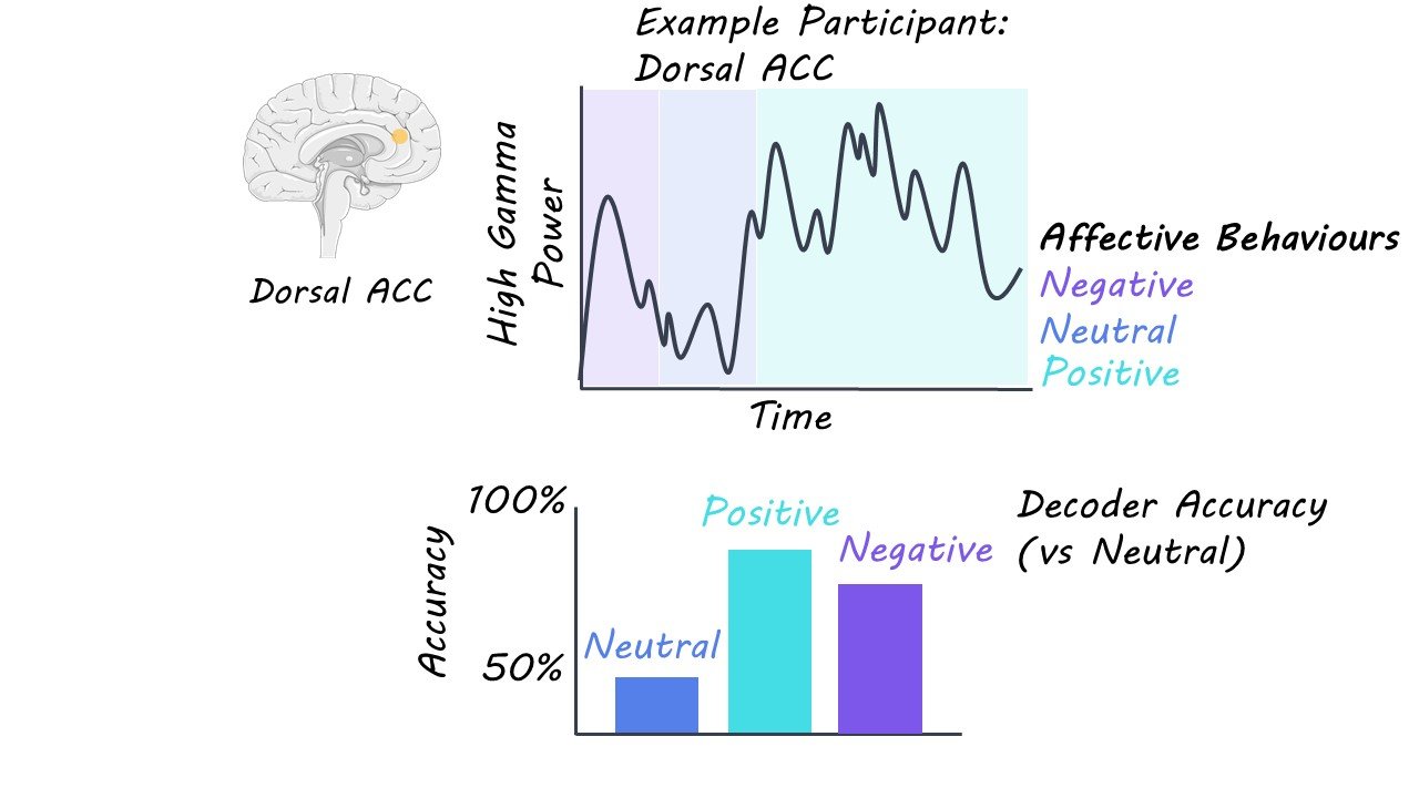
Certain regions of the mesolimbic system (insula, ACC, hippocampus, and amygdala) were found to contribute more strongly to both positive and negative affective behaviours, compared to other regions (OFC). This finding suggests that increased gamma activity in these brain regions during both positive and negative affective behaviours may reflect emotional arousal in general. However, the results also revealed that distinct structures of the mesolimbic system may contribute to positive and negative affective behaviours in different ways. For example, increased gamma activity in the ventral ACC, dorsal ACC, and hippocampus was related to positive affective behaviours. In contrast, increased gamma activity in the amygdala was related to negative affective behaviours. Thus, this research suggests that there is a distributed network of brain regions that are associated with different affective behaviours in the mesolimbic system.

What's the impact?

Using statistical and machine learning methods, this study found that spectro-spatial features of brain activity in the mesolimbic network are related to naturalistic affective behaviours. This study further elucidates the neural mechanisms at play in the mesolimbic network. Advancing decoding models to be able to relate neural signals to more complex emotions will allow for more refined brain models of affective behaviours which may be used to inform treatments for mental health disorders.

Access the original scientific publication here.

The Deep Wiring of Speech in the Human Brain

Post by Anastasia Sares

The takeaway

Compared to other animals, humans have a highly developed capacity for speech. This study showed that human speech areas have high-fidelity and fast connections to deep brain nuclei—meaning that we may indeed be hard-wired to learn language.

What's the science?

The basal ganglia are a set of structures deep in the brain that help to regulate almost all other activity: they form circuits with other brain areas, regulating them and deciding whether to perform an action, or when to stop. One of these circuits is called the hyperdirect pathway; this pathway puts the brakes on an action or a process. In rodents, it was discovered by injecting special viral proteins and dyes that can climb along neural pathways. This was not ethically possible to do in humans, so it was unclear whether our brain architecture was similar. Scientists came one step closer in the 2010s, when the hyperdirect pathway was found in primates. In 2018, evidence of this pathway in humans was observed by careful electrical recordings during surgery. This confirmed that the hyperdirect pathway exists in humans, and it links to many areas of the cortex. This week in Cell Reports, Jorge and colleagues used a similar electrical stimulation and recording technique to look at speech-related areas of the brain. They concluded from the timing of the electrical responses that there is a hyperdirect pathway that connects areas associated with speech to the deep parts of the brain, perhaps explaining humans’ unique ability with language.

How did they do it?

The study recruited patients with Parkinson’s disease who were already being implanted with electrodes in a procedure known as “deep brain stimulation.” In this procedure, a long, thin electrode is inserted into the interior of the brain and is connected to an exterior pacemaker-like device that can deliver pulses of electricity directly to stimulate that brain region.

For Parkinson’s, the target of these stimulations is the subthalamic nucleus: one of the brain’s deep nuclei that works to inhibit actions that are not necessary. Stimulating the subthalamic nucleus helps to suppress the resting tremors that are associated with Parkinson’s disease. The subthalamic nucleus also happens to be the first stop of the hyperdirect pathway, and when stimulated, electrical activity can actually travel backwards to the cortex (at least, this is true in animal models). So, the researchers were able to take advantage of some electrodes placed on the surface of the brain during the surgery to measure and map out these backward-traveling signals. They would stimulate the subthalamic nucleus, then time how long it took for the signal to reach cortex. If the timing was short (< 10 milliseconds), then it was likely that the two regions were directly connected.

What did they find?

First, the authors confirmed the 2018 finding: they were able to measure electrical signals traveling backward from the subthalamic nucleus to the cortex. These signals arrived quickly, under 10 milliseconds, which means the connections were likely only a single neuron in length.

They then looked at how the placement of the stimulating electrode affected the activity in the cortex. Stimulating areas closer to the midline of the brain generated stronger signals in the parts of cortex that control movement while stimulating further from the midline generated signals in parts of the cortex that deal with sensory perception and forming associations. Many areas known for processing speech were affected by subthalamic stimulation, including the inferior frontal gyrus (classically known as Broca’s area), the auditory cortex, and association areas in the temporal cortex (roughly equivalent to the classical Wernicke’s area).

What's the impact?

This study demonstrates that speech areas of our brain are just a single neuron away from the deep inner nuclei of the brain. This super speedy pathway may contribute to our extraordinary capacity for speech, and help us understand what makes humans unique. Many current models of speech skip or gloss over the role of these deep brain loops in speech, and therefore these models may need to be updated to reflect the importance of these pathways. 

Access the original scientific publication here

Angiotensin-Converting Enzyme Inhibitors Modulate Brain Opioids

Post by Shireen Parimoo

The takeaway

Angiotensin-converting enzyme (ACE), an enzyme that normally regulates blood pressure in the body, is also important for reward processing in the brain. Inhibition of ACE in the nucleus accumbens may be a viable option for treating addiction and substance use disorders.

What's the science?

The nucleus accumbens (NAc) is a key region of the reward circuit in the brain and is implicated in addiction. Two types of NAc neurons – DS1 and DS2 neurons – are important in reward processing but are difficult to study separately because they receive similar inputs and express many of the same genes. This, in turn, makes it difficult to develop pharmacological treatments for addiction that specifically target DS1 or DS2 neurons. Interestingly, only DS1 neurons express angiotensin-converting enzyme (ACE), which is thought to modulate excitatory transmission within the NAc by acting on opioid receptors and may be important in reward processing. This week in Science, Trieu and colleagues sought to identify the exact mechanism by which ACE regulates synaptic transmission in the NAc and to elucidate its role in the reward pathway.

How did they do it?

The authors conducted a series of ex vivo and in vivo experiments. First, they quantified ACE expression in slices of NAc neurons and applied captopril (an ACE inhibitor) and naloxone (an opioid receptor antagonist). Recording mEPSPs (miniature excitatory postsynaptic potentials) from both DS1 and DS2 neurons in those slices allowed them to determine whether ACE affected synaptic transmission in the NAc through opioid signaling. Next, they used liquid chromatography-tandem mass spectrometry to examine the effect of NAc stimulation on the concentration of different enkephalins (e.g., Leu-enkephalins, MERF, etc.). Enkephalins are opioid peptides released by DS2 neurons and except for MERF, most enkephalins can be degraded by ACE. The authors assessed how ACE inhibition and optogenetic stimulation affected enkephalin levels, as well as the subsequent impact of elevated enkephalin levels on synaptic transmission in the NAc. They then applied captopril and MERF to NAc slices along with opioid receptor antagonists to identify the specific opioid receptors that are involved in ACE inhibition.

The authors also investigated the effect of ACE inhibition on excitatory transmission in vivo by administering captopril and optogenetically stimulating medial prefrontal neurons that provide excitatory input to the NAc. Finally, they used a place conditioning paradigm to study the impact of ACE inhibition on reward learning in mice. In this paradigm, mice typically prefer a context that is paired with a reward compared to a control context. The authors examined the impact of captopril on preference for a context paired with fentanyl, an opioid drug that normally has an excitatory effect on NAc neurons.

What did they find?

There was a higher concentration of ACE in DS1 compared to DS2 neurons in the NAc. Inhibition of ACE led to long-term depression (a form of synaptic plasticity) in the DS1 neurons but had no impact on synaptic transmission in DS2 neurons. Naloxone – an opioid receptor antagonist – prevented long-term depression in DS1 neurons when it was applied together with captopril. However, applying naloxone after captopril did not reverse LTD. These findings demonstrate that ACE inhibition triggers synaptic plasticity by acting on opioid receptors in the NAc.

Extracellular concentrations of all enkephalins increased following NAc stimulation, including MERF. However, inhibiting ACE increased MERF but had no impact on other enkephalins. Moreover, stimulating DS2 neurons increased MERF levels even in the presence of captopril, which indicates that MERFs are released by DS2 neurons. Applying enkephalins to both DS1 and DS2 neurons decreased the frequency (but not amplitude) of mEPSPs in the NAc, with the strongest effects resulting from MERF application. In DS1 neurons, captopril and MERF alone did not affect mEPSPs but applying them together reduced mEPSP frequency. Additionally blocking the mu opioid receptor prevented the reduction in mEPSP frequency, which suggests that ACE inhibition triggers synaptic plasticity changes in DS1 neurons by acting on the mu opioid receptors. Finally, medial prefrontal input to the NAc typically increases excitatory transmission in DS1 neurons, but this sensitivity was reduced in the presence of captopril. Similarly, mice who underwent place conditioning showed a strong preference for the fentanyl-associated context, but this preference was smaller when captopril was administered. Thus, ACE inhibition reduced NAc activity in vivo and modulated reward learning in mice by dampening the impact of reward at the neural level.

What's the impact?

This study is the first to demonstrate how ACE inhibitors modulate synaptic transmission in specific types of neurons in the nucleus accumbens. Given the crucial role of the nucleus accumbens in reward processing, ACE inhibitors hold considerable potential for the development of targeted drugs for treating addiction and substance use disorders.

Access the original scientific publication here.