Online Single-Session Interventions Can Help Depressed Teenagers

Post by D. Chloe Chung

The takeaway

Accessible, online single-session interventions that teach coping skills can effectively improve symptoms related to depression in teenagers, especially during the COVID-19 pandemic with heightened teen depression.

What's the science?

Rates of depression among teenagers soared during the COVID-19 pandemic as they faced social isolation due to school closure and financial difficulties. While teen depression is the major health risk for young people, more than half of depressed teenagers lacked access to professional help even before the pandemic. As many teenagers find it challenging to seek mental healthcare services due to stigma from family or financial reasons, it is important to create effective and accessible platforms to help those in need. Recently in Nature Human Behaviour, Schleider and colleagues showed that accessible, online single-session interventions that teach coping skills can reduce depression in teenagers.

How did they do it?

The authors used Instagram advertisements to recruit a diverse group of teenagers (13-16 years old) in the United States who were experiencing depressive symptoms. Recruited teenagers were informed that they would be rewarded up to $20 by participating in the minimal-risk, free, confidential online psychology study. Since depressed teenagers often feel uncomfortable telling their guardians about their mental health problems, the study was approved to waive consent from parents for participation. A total of 2,452 eligible teenagers who completed the baseline survey were randomly assigned with one of three web-based, single-session interventions designed by the authors. The first active intervention (“growth mindset”) teaches the participants that symptoms and personal traits can change. The second active intervention (“behavioral activation”) teaches participants how to adapt behaviors to experience positive sensations such as happiness and accomplishment. The third intervention (“supportive therapy”), that encourages sharing emotions with others, acts a placebo as it does not teach specific coping skills. Each of the self-guided interventions included peer narratives and writing activities taking 20-30 minutes to complete. The participants were asked to complete the follow-up survey three months after the interventions. Both pre- and post-intervention surveys assessed a range of symptoms including depression, anxiety, COVID-19-related trauma, hopelessness, eating behaviors, and perceived sense of agency.

What did they find?

The authors compared the depressive symptoms measured during the baseline survey and the follow-up survey. From this analysis, they found that both “growth mindset” and “behavioral activation” interventions substantially decreased depressive symptoms compared to the placebo session. The degree of reduction in depressive symptoms was similar between groups of each active intervention. Both active interventions also improved other aspects in depressed teenagers. Specifically, three months after these interventions, participants reported decreased hopelessness, decreased restrictive eating, and increased sense of agency. However, when it comes to reducing trauma-related to COVID-19 and anxiety, only the “growth mindset” intervention and not the “behavioral activation” intervention was effective.

What's the impact?

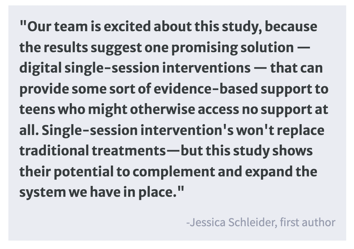
This study has shown that even a single online intervention session can help depressed teenagers who may otherwise have difficulties in seeking accessible professional help. Study results were promising as the access to mental healthcare services is even more limited during the global pandemic. Since the reduction in depressive and other symptoms by these active interventions was modest, depressed teenagers should be provided with more intense and long-term help for sustainable care. Still, this non-traditional, highly accessible mental healthcare service can support teenagers who might otherwise have trouble getting appropriate help.

Memory Strategies Shift How Information is Represented in Prefrontal Cortex Neurons

Post by Lani Cupo

The takeaway

Brain regions encode information differently depending on the memory strategy used. In the lateral prefrontal cortex of monkeys, neural activity shifts in a strategy-dependent manner between individual neurons and populations of neurons.

What's the science?

Humans and other animals use strategies to organize information in order to counteract the limited capacity of working memory and execute complex cognitive processes, however, how this information is represented in neural mechanisms is still poorly understood. These processes rely on the prefrontal cortex, an area that is flexible enough to adapt to the demands of different tasks. However, information can either be represented at the single-neuron level or at the population level, and it can dynamically respond to employed strategies. This week in Neuron, Chiang and colleagues investigated how working memory strategies employed by monkeys impacted neuronal ensemble coding in the lateral prefrontal cortex (LPFC).

How did they do it?

Previous research established that, like humans, monkeys use strategies to exceed the natural limits to working memory (WM) (usually around 4 items). In the present study, two monkeys were presented with a visual task where six identical, colored circles were presented on a screen and the monkeys had to make a saccade (eye movement) to each one only once, returning their eyes to a central point between each selection, remembering which targets they had already visited. The task forced them to remember up to 6 targets in each trial, exceeding the typical capacity of WM and engaging additional mnemonic strategies. Microelectrodes were implanted in the bilateral LPFC of the monkeys to record neuronal activity from groups of about 40 neurons simultaneously during the task, allowing the authors to link neuronal activity to the task performance. To examine the representation of three variables (location of targets, order of saccades, and color of targets), the authors applied a technique known as linear discriminant analysis (LDA) to the neural activity data, allowing them to separate neuronal representations of each variable. In order to understand how each neuron contributed to the population representation of the overall ensemble code, the authors employed a procedure where they removed each unit from their LDA model in turn to see how the overall pattern changed, identifying that neurons contributed differently to encoding at a population level. To examine the impact of sequencing strategies on task performance and the underlying neuronal activity, the authors examined whether the monkeys were likely to visit targets in a similar order across a block (set of trials). When monkeys were more likely to fixate on circles in a specific pattern, the block was given a high stereotyped index (SI), and when there was more diversity in the pattern it was given a low SI. The categorization allowed them to assess whether monkeys were employing a sequencing strategy (high SI blocks), and how this strategy impacted task performance and neuronal firing.

What did they find?

First, reaction times increased across saccades (on later target selection), and monkeys were more likely to fail (look at a target they had already looked at before), suggesting that selections became more difficult, and working memory was being taxed. However, the monkeys performed better on blocks with a higher SI, implying the mnemonic strategy helped compensate for limits to working memory. One of the main findings was that stereotyped behaviors, representing sequencing strategies, were associated with more distributed neuronal encoding in the LPFC. As behavior became more stereotyped, individual neurons contributed less to the ensemble. Overall, when strategies were used and behavior became more routine (associated with better performance on the task), more neurons were recruited, with smaller individual contributions, whereas when behavior was more flexible, fewer neurons were recruited, each contributing more to the signal.

What's the impact?

This study found that using mnemonic strategies improved task performance and altered the underlying representation of the behavior, shifting towards a more distributed pattern of activity with more neurons contributing less individually. These findings provide a new perspective on how information is represented differently on a neuronal level dependent on cognitive strategies employed. This study represents a step for investigations that seek to further uncover the neural mechanisms underlying higher-order cognitive abilities.

Creative Fluency is Driven by Default Network Activity

Post by Lincoln Tracy

The takeaway

Creative thinking is a complex and crucial part of the human experience that cannot be linked to one specific area of the brain. Stimulating the brain’s default network - a brain network that is active during activities like daydreaming or mind-wandering -  limits creative thinking.  

What's the science?

Creative thinking—the ability to produce novel and useful ideas—has been a key evolutionary mechanism underlying the rapid advancement of humans as a species. While certain senses or processes are linked to a specific area of the brain (such as vision with the visual cortex), creative thinking cannot be pinned down to just one area. Previous evidence suggests that connectivity between brain regions associated with the default network may contribute to creative thinking, but no causal relationship has been identified. This week in Molecular Psychiatry, Shofty and colleagues utilized a creative thinking task in the unique environment of awake brain surgery to explore the effects of default network stimulation on creativity.

How did they do it?

The authors recruited 13 patients (three women, age range 19-69 years) with gliomas, a common type of brain cancer. All patients were scheduled for awake brain surgery to remove their tumor. At the beginning of surgery, the left default network was electrically stimulated while patients completed an alternate uses task (AUT), a task commonly used to assess creativity. In this task, patients were presented with a series of everyday objects and asked to list possible alternative uses. For example, a newspaper can be used to swat files, start a fire, or be used for a ransom note. Patients were scored on fluency (the number of alternative uses for each object they could think of) and originality (how many other people suggested the same use). Patients underwent a pre-surgery functional magnetic resonance imaging (fMRI) scan to map their own (individualized) default mode network, which was then stimulated during surgery.

What did they find?

First, the authors sought to validate the AUT in an awake brain surgery setting. Correlating baseline AUT scores with a marker of default network integrity (obtained from the pre-surgery fMRI) revealed a positive correlation for fluency but no correlation with originality. When the authors examined the effect of stimulation on creative thinking during awake brain surgery, they found creative fluency was reduced during default network stimulation. No effect of stimulation on originality was observed. The authors then investigated how stimulating regions associated with the default network affected creativity. Changes in fluency were observed by stimulating the parietal, frontal, and temporal regions of the default network; no effects were observed for originality. Taken together, these findings mean that cortical stimulation impacts creative fluency—but not originality—and that stimulating areas more connected to the default network results in a greater impact.  

What's the impact?

This study found a causal link between the default mode network and creative fluency, where direct stimulation disrupted creative fluency. These results imply that different aspects of creativity are controlled by specialized parts of the default network. These findings hint at the possibility of using such a technique to identify and preserve creativity and other cognitive functions of patients undergoing brain surgery.