A Step Towards Personalized Classification in Epilepsy

Post by Andrew Vo

The takeaway

Treating patients with temporal lobe epilepsy is challenging due to the high degree of individual variability along the disease spectrum. Using machine learning and data-driven analysis of disease factors may allow for individualized patient diagnosis and care.

What's the science?

Despite sharing a common diagnostic label, patients with drug-resistant temporal lobe epilepsy (TLE) often display widespread and non-overlapping brain changes. Large individual variation along the disease spectrum has made it difficult to accurately characterize TLE patients and predict treatment responses using traditional “one-size-fits-all” group-level analyses. This week in Brain, Lee et al. applied machine learning to brain imaging measures to estimate the expression of TLE “disease factors” and tested how well these factors predicted clinical outcomes in TLE.

How did they do it?

The authors first obtained magnetic resonance imaging (MRI) of the brains of a large group of TLE patients. These non-invasive MRI measures described different structural and microstructural properties of the brain, including cortical thickness, myelin (white matter) changes, and gliosis (scarring in response to neuronal damage). They then applied machine learning to these data. Latent Dirichlet allocation is an unsupervised machine learning technique that estimates the co-expression of disease factors (i.e., patterns of disease-related brain changes) in each patient. Unlike more traditional approaches that aim to classify patients into a single subtype, the analysis technique used here allows multiple disease factors to be expressed to varying degrees in each patient. The authors determined the specificity of identified disease factors by estimating their expression in groups of healthy and other disease (i.e., frontal lobe epilepsy) control groups. Finally, they tested how well these disease factors could predict individual patient drug response and seizure outcomes after surgery, as well as the degree of cognitive dysfunction.

What did they find?

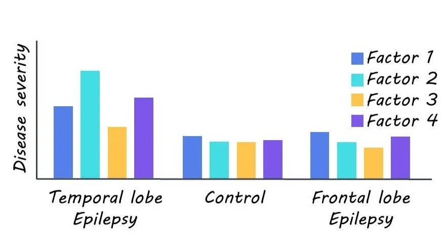
Four latent disease factors were identified. All factors were expressed to varying degrees in each TLE patient, not at all in healthy controls, and negligibly in frontal lobe epilepsy, thus highlighting the specificity of their findings. Factor 1 was characterized by microstructural changes, myelin loss, and atrophy largely in the hippocampus. Factor 2 was predominately marked by gliosis in paralimbic and hippocampal regions. Factor 3 was distinguished by bilateral neocortical thinning. Lastly, Factor 4 was largely marked with bilateral microstructural changes and minimal hippocampal changes.

The identified disease factors were then used to train classifier models, which predicted individual patient drug response and surgical outcome with 76% and 88% accuracy. Notably, classifiers trained on these disease factors out-performed untrained classifiers. Similarly, these disease factor-trained models could more accurately predict individual cognitive dysfunction (assessed in terms of verbal IQ, memory, and visuomotor learning) than baseline models.

What's the impact?

This study illustrated how using a machine learning approach can improve the characterization and prediction of clinical outcomes in TLE. Rather than classifying patients into a specific subtype, the strategy used here allows individuals to be represented along multiple dimensions that better capture their complex underlying pathology. Considering individual variability along the disease continuum will allow for personalized care and improved prognosis not just in TLE but other heterogeneous disease groups.

Access the original scientific publication here.

The Impact of Running on Brain Function and Mental Health

Post by Megan McCullough

The takeaway

A 10-minute running session of moderate intensity was shown to increase mood and executive function through the bilateral activation of the prefrontal cortex

What's the science?

Previous research has shown that physical exercise can lead to an increase in mood and executive function through the activation of the dorsolateral prefrontal cortex. These studies have predominantly measured the effects of exercise on the brain by using pedaling as the form of exercise. Since running uses different muscles and parts of the body, it may have different effects on the brain than other forms of exercise. This week in Scientific Reports, Damrongthai and colleagues studied the effects of running on mood, executive function, and the prefrontal cortex.

How did they do it?

The participants consisted of 26 healthy individuals that completed both a 10 minute run at moderate intensity and a control resting period in a randomized order. After both activities were completed, executive function was then evaluated using the colour-word matching Stroop task. This task involves a list of names of colours, written in different colours; participants are tasked with naming the colour the word is written in and not the word itself. The Stroop test was used to test executive function because it measures the ability of participants to control their responses despite external lures. Mood was also measured before and after exercise using a mood scale the states of arousal and pleasure the participant was in. Finally, functional near-infrared spectroscopy was used to measure blood flow and thus activation in the prefrontal cortex.

What did they find?

The authors found higher blood flow in the prefrontal cortex after running trials compared to after the resting control trials. This suggests that running increased bilateral activation in the prefrontal cortex, an area associated with cognition and mood. The authors also found that participants performed better at the Stroop test after the moderate exercise compared to control trials. This shows the positive effect of running on cognition. Finally, the authors found that running led to an increase in mood, in particular an increase in pleasure level that has never been found in their previous pedaling studies. Together, these results suggest that a moderately intense running session can lead to improvements in cognition and mood through bilateral activation of the prefrontal cortex.

What's the impact?

This study adds to the growing body of research showing that physical activity benefits mental health. Critically, this research shows that running ⁠— a whole-body locomotion exercise ⁠—  is also an effective way to achieve improvements in mood, especially pleasure level, which can benefit exercise adherence and ​executive function.  

The Effects of Traumatic Brain Injury in the Presence of Aß Pathology

Post by Negar Mazloum-Farzaghi

The takeaway

There is a link between traumatic brain injury and the development of dementia. The double burden of traumatic brain injury and Aß deposits in the aging brain can cause alterations in cellular processes that lead to neuron death.

What's the science?

Previous research has established an association between traumatic brain injury (TBI) sustained in early adulthood and the risk of developing dementia later in life. It is less clear whether TBI sustained as an elderly individual (geriatric TBI) increases the risk of developing dementia, and what the cellular mechanisms would be involved. The aging brain’s response to TBI may be influenced by several factors, such as increased inflammation, increased amyloid beta load (Aß; a hallmark of dementia), and decreased autophagic activity (important process for degrading unnecessary or dysfunctional intracellular macromolecules). This week in Scientific reports, Streubel-Gallasch and colleagues used an in vitro model, designed to simulate the conditions of the aging brain, to investigate cellular responses to TBI in the presence of Aß pathology.

How did they do it?

In order to investigate the effects of physical injury caused by TBI in the presence of Aß deposits, the authors exposed co-cultures of astrocytes (specialized glial cells fundamental to maintaining homeostasis and cellular function) and neurons to Aß protofibrils. After the exposure, they subjected the cultures to a “scratch injury” using a scalpel in order to mimic TBI. There were four experimental groups: untreated (control), scratch injury (TBI), Aß protofibril exposure, and the combination of both TBI and Aß.

The authors performed immunocytochemistry to examine the effect of TBI and Aß protofibril exposure on neuronal survival. They employed the same method to examine how TBI and Aß protofibril exposure affects astrocytes. To do this, they used two well characterized astrocytic markers, Glial fibrillary acidic protein (GFAP) and calcium-binding protein (S100ß). They used transmission electron microscopy to further assess changes to cellular homeostasis caused by TBI and Aß protofibril exposure by examining the function of the mitochondrial network of astrocytes. Finally, using both immunocytochemistry and immunoblotting, they also examined whether autophagic activity was impaired in response to TBI and Aß protofibril exposure.

What did they find?

The number of neurons in the experimental TBI group, Aß protofibril-exposure group, and control group remained stable. However, in the experimental group with the combination of Aß and TBI, the percentage of neurons decreased significantly. One explanation for this finding may be that the double burden of Aß deposits and TBI hindered astrocytic functions, which threw their ability to maintain homeostasis off balance, making it more difficult to clear debris and protect neurons.

Aß protofibril-exposed astrocytes displayed higher GFAP levels. In contrast, astrocytes subjected to both Aß and TBI displayed lower GFAP levels. This was a surprising finding as GFAP usually upregulates in response to TBI. The other astrocytic protein, S100ß, showed increased levels in astrocytes in all three experimental groups compared to the control group. This is in line with previous work that has shown that S100ß upregulates in response to trauma and has increased levels in the tissue cells and astrocytes of dementia patients. However, it remains to be determined whether these increased levels have positive or negative effects in pathological conditions.

Aß pathology and TBI caused mitochondrial damage in astrocytes (but no clear additive effect of the two), which may have negative consequences for vital astrocytic functions thereby disturbing homeostasis. There was an increase in autophagic activity in the Aß protofibrils exposure group and TBI group. Interestingly, when cells were exposed to a combination of Aß protofibrils and TBI, autophagy failed to upregulate, which may be indicative of an inefficient compensatory mechanism when faced with this double burden.

What's the impact?

This is the first study to show that the double burden of Aß deposits and TBI result in neuronal loss, altered astrocytic responses as indicated by changes in the expression of two key astrocytic proteins, mitochondrial disturbances, and abnormal autophagic activity. The findings of this study demonstrate the underlying cellular mechanisms involved in the relationship between TBI and the development of dementia. Overall, this study advances our understanding of geriatric TBI on the cellular level.